http://www.inosr.net/inosr experimental sciences/ Ikuomola et al
INOSR Experimental Sciences 8(1): 84 94, 2022.
©INOSR PUBLICATIONS
International Network Organization for Scientific Research
ISSN: 2705 1692 Evaluation of the effect of secnidazole on the histology of the testes and epididymis of male Wistar rats 1,2 E.O. Ikuomola , 1O.S Akinsonmisoye, 1R.O. Owolabi and 3M. B. Okon
1Department of Physiological Sciences, Faculty of Basic Medical Sciences, Obafemi Awolowo University, Ile-Ife, Osun State, Nigeria;
2Department of Physiological Sciences, Kampala International University, Western Campus, Uganda.
3Department of Biochemistry, Kampala International University, Western Campus, Uganda.
ABSTRACT
The effect of secnidazole on the histology of the testes and epididymis of male Wistar rats was evaluated. Thirty five male rats were grouped into 7 groups containing 5rats each. Group A1 (n=5) were given distilled water (2ml/kg) which was the vehicle used to dissolve the drug. A2 (n=5) were given distilled water and allowed to recover for another 8 weeks. Group B (n = 5) received 14.3mg/kg of secnidazole (low dosage) for 8 weeks only. Group C (n = 5) Received 28.6mg/kg of secnidazole (medium dosage) for 8 weeks only. Group D (n = 5) Received 57.2mg/kg of secnidazole (high dosage) for 8 weeks only. Group E (n = 5) Received 14.3mg/kg of secnidazole (low dosage) for 8 weeks once daily and allowed to recover for another 8 weeks. Group F (n = 5) Received 28.6mg/kg of secnidazole (medium dosage) for 8 weeks once daily and allowed to recover for another 8 weeks. Group G (n=5) Received 57.2mg/kg of secnidazole (high dosage) for 8 weeks once daily and allowed to recover for another 8 weeks. All administrations were done orally with the aid of an oral cannula. Group A1 showed compactly arranged seminiferous tubule with small interstitial space with scanty Leydig cells, intact germinal epithelium (GE) with longer spermatids lying at the luminal surface (L). Group B showed Focal exfoliation of germinal epithelium (GE) and minimally enlarged interstitial space (I). Group C and D shows relatively large interstitial space with scanty Leydig cells, germinal epithelium appear intact (GE). Group A2 showed compactly arranged seminiferous tubule with small interstitial space with scanty Leydig cells, intact germinal epithelium (GE) with elongating spermatids lying at the luminal surface (L). Group E showed compact tubular arrangement, intact germinal epithelium (GE), small interstitial space with scanty Leydig cells. Group F and G showed relatively wide interstitial space (I) with few interstitial Leydig cells. Group A1 and A2 showed intact seminiferous tubule epithelium with normal arrangement of spermatogenic cells and Sertoli cells (evident by their nuclei) in the tubule epithelium (GE). Spermatogonia is distinctly evident on the basement membrane of the tubule (S) and the most mature germ cells localized to the tubule lumen (L). Leydig cells nuclei are evident between the tubules, in the interstitium (LC). Group B showedsloughing of germinal epithelium and the associated spermatogenic cells (asterisks). Group C and D shows germinal epithelium distortion (GE), aberrant appearance of germ cells, vacuolar degeneration in the germinal epithelium partial depletion of elongating spermatids in the lumen (L) when compared with the control. Group E and F showed germinal epithelium is relatively better than in Group B and C, although there are region of exfoliated germ cells (asterisks) in Group F Group G showed germinal epithelium sloughing (asterisks) and depletion of elongated spermatids in the lumen. Group A1 and A2, the epithelium appeared normal the principal and basal cells that constitute the pseudo stratified columnar epithelium are not distinguishable at this magnification.Group D epithelium appears normal, spermatozoa appeared less dense in some of the lumen, the ducts appeared compact with less connective tissue stroma (CT) Group E and F epithelium showed normal appearance with compact arrangement of the ducts and less dense spermatozoa within the lumen. Group G duct appeared less dense, epithelium appear thin/attenuated with less connective tissue stroma and dense spermatozoa. In conclusion, histological observation of testicular sections subjected to hematoxylin and eosin stain in the rats subjected to secnidazole treatments groups showed alterations in the germinal epithelium, seminiferous tubules and the depletion of spermatogonic cells, large interstitium, partial depletion of elongated spermatids in the lumen, when compared with the control.
Keywords: secnidazole, histology, testes, epididymis and Wistar rats
http://www.inosr.net/inosr experimental sciences/ Ikuomola et al
INOSR Experimental Sciences 8(1): 84 94, 2022.
INTRODUCTION
Infertility has become an ominous problem in Africa on the average, about 10% of all couples face difficulty in startingafamilyandthiscreatesafeeling ofgreatpersonalfailure[1] Many factors both extrinsic andenvironmental factors including the increased use of antibiotics and anti effective drugs have been implicated as potential causes of male infertility. Studies have shown that antimicrobial combination therapy such as metronidazole, quinolones, tetracycline, ketoconazole, fluconazole and other imidazole group of antibiotic drugs are among the most prescribed classes of drugs in medicine. There is a high possibility that some of the couples presenting with history of infertility or inability to conceive may be due to these groups of drugs [2] Nitro imidazole, an antimicrobial drug is among the most clinically prescribed classes of drugs for couples presenting with infertility in the clinics. However, these classes of drug have been shown to possess mutagenic activities in bacterial assay and reproductive toxicity including inhibition of spermatogenesis in rats [3; 2] Studies have also shown that some nitro imidazole class of drugs such as metronidazole and ketoconazole may have being responsible for some reproductive toxicity which causes inhibition of spermatogenesis in rats earlierreported by [4] Secnidazole is one of the representatives of nitro imidazole group of drugs. It is highly effective and differs from other compounds in this group by a prolonged serum half life of about 17 29 hours [5]. The prescription of secnidazole have increased due to its pharmacological advantages in treatment of adults and children with intestinal amoebiasis, urinary tract infection (UTI), pelvic inflammatory disease (PID) and vaginal infectionsmostly of mild or moderate severity [5]. Secnidazole is an anti biotic with distinct pharmacological properties that are different from other
imidazole group of drugs, giving it a relative pharmacological advantage [6]. The frequency of prescription of secnidazole has increased tremendously in recent years due to its pharmacological advantages [5; 6] The tolerability profile of secnidazole does not differ markedly from other 5 nitroimidazoles. The most commonly reported adverse events in clinical trials involved the gastrointestinal tract (nausea, vomiting, glossitis, anorexia, epigastric pain and a metallic taste) and occurred in 2 to 10% of patients. Headacheanddizzinesswereexperienced by about 2% of patients. Secnidazole was reportedtobewelltoleratedinadultsand children, and no adverse event required therapeutic intervention or treatment withdrawal [7] Secnidazole, from its pharmacological properties has a mutagenic effect in bacterial assay. This mutagenic effect has been proved to reduce a nitro group and attacks the nucleic acid of the micro organism. Furthermore, it inhibits further DNA synthesis and causes degradation of already existing DNA [8]. The effect of secnidazole on reproductive system was studied because reports from previous studieshave shown increasing number of people using secnidazole in clinical settings especially among the young ones andthoseinthereproductiveagebracket. Also, the pharmacological properties of secnidazole have been predicted to be reprotoxic.Secnidzole isoneofthenitro imidazole drugs mostly prescribed in the clinical settings because of it increase in patient’s compliance and most of the infections that it is used to treat are infections of the reproductive system suchasurinarytraitinfection(UTI),Pelvic inflammatory disease (PID), Vaginal infections.
Objective of the study
The specific objective of the study is to determine the effect of secnidazole on the histology of the testes and epididymis of male Wistar rats.
http://www.inosr.net/inosr experimental sciences/ Ikuomola et al
INOSR Experimental Sciences 8(1): 84 94, 2022.
METHODOLGY
Animals
FortyadultmaleWistarratsweighing150 200g were used for this study. The animals were obtained from the Animal House,CollegeofHealthScience,Obafemi Awolowo University, IleIfe, Nigeria. The rats were housed in the Animal House, Faculty of Basic Medical Sciences, Obafemi Awolowo University, Ile Ife, Nigeria.Theratswerekeptincagesunder normal environmental conditions and given free access to standard pellet diet and water. They were allowed to acclimatizetothelaboratoryenvironment
for two weeks before the commencement oftheexperiment.
Drugs
Secnidazole was procuredbyMay &Baker Company, SangtoOtta, Ogun State, Nigeria.
Ethical Clearance
Ethical clearance for this study was obtained from the Health Research Ethics Committee (HREC) of the Institute of Public Health, College of Health Sciences, ObafemiAwolowoUniversity,IleIfe,Osun State Nigeria. (HREC Assigned Number: IPHOAU/12/1624).
Experimental Design
GROUPS NUMBER OF RATS TREATMENTS
NUMBER OF DAYS
GROUP A1 A2 5 5 2mls of distilled Water (control) 8 Weeks treated 8 Weeks Recovery
GROUP B 5 14.3mg secnidazole (low dose) 8 Weeks
GROUP C 5 28.6mg secnidazole (medium dose) 8 Weeks
GROUP D 5 57.2mg secnidazole (high dose) 8 Weeks
GROUP E 5 14.3mg secnidazole (low dose)+ Recovery
GROUP F 5 28.6mg secnidazole (medium dose) + Recovery
GROUP G 5 57.2mg secnidazole (high dose)+Recovery
Secnidazole Drug Administration
Rats in group 1 received 2 ml/kg of distilled water only, group 2 rats received 14.3mg/kgofsecnidazole(lowdosage)for 8 weeks; group 3 rats received 28.6 mg/kg of secnidazole (normal dosage) for 8 weeks; group 4 rats received 57.2 mg/kg secnidazole (high dosage) for 8 weeks; Rats in group 5, 6 and 7 received 14.3 mg/kg, 28.6 mg/kg, 57.2 mg/kg of secnidazole respectively for 8 weeks and they were allowed to recover from treatmentforanother8weeks.
Stock Solution Preparation
Astocksolutionof500mgwaspreparedin 20mls of distilled water and from this a dosage of 2mls/kg was administered. All administration was done orally with the aidofanoralcannula.
8 Weeks treated + 8 Weeks Recovery
8 Weeks treated + 8 Weeks Recovery
8 Weeks treated + 8 Weeks Recovery
Animal Groupings and Administration
Thirty five male rats were grouped into 7 groupscontaining5ratseach.
Group A1 (n=5) were given distilled water (2ml/kg) which was the vehicle usedtodissolvethedrug.
A2 (n=5) were given distilled water and allowed to recover for another 8weeks
Group B (n = 5) received 14.3mg/kg of secnidazole(lowdosage)for 8weeksonly [5]
Group C (n = 5) Received 28.6mg/kg of secnidazole (medium dosage) for 8 weeks only[5].
Group D (n = 5) Received 57.2mg/kg of secnidazole (high dosage) for 8 weeks only[5]
http://www.inosr.net/inosr experimental sciences/ Ikuomola et al
INOSR Experimental Sciences 8(1): 84 94, 2022.
Group E (n = 5) Received 14.3mg/kg of secnidazole(lowdosage)for8weeksonce dailyandallowedtorecoverforanother8 weeks[5].
Group F (n = 5) Received 28.6mg/kg of secnidazole (medium dosage) for 8 weeks once daily and allowed to recover for another8weeks[5]
Group G (n = 5) Received 57.2mg/kg of secnidazole (high dosage) for 8 weeks once daily and allowed to recover for another 8 weeks [5]. All administrations were done orally with the aid of an oral cannula.
Mode of Sacrifice and Organ Collection
After eight weeks of administration of secnidazole, the rats were sacrificed by cervical dislocation. Their blood was collected through cardiac puncture. The testis and epididymis were harvested and weighed using digital weighing scale. The right testis of each rat was immediately fixed in Bouin’s fluid for histological processing. The semen for sperm parameters was obtained from the caudal epididymis and their sperm parameters were assessed under the microscope. The same sacrificial procedure was repeated forratsintherecoverygroups.
Histological Analysis
Tissue Processing
Immediatelyaftertheratsweresacrificed, their right testes and epididymis were carefullyexcisedforconsistencyandthen weighed.Theorgans werefixedinBouin’s fluid to prevent putrefaction and autolysis and were further taken from the tissuesandprocessedasfollows:
Dehydration: After fixation (48 hours), the tissues were transferred into ascending grades of alcohol for dehydration at room temperature as follows: 50 % alcohol, 70 % alcohol, 90% alcohol, absolute alcohol I, absolute alcoholIIanhoureach.
Clearing:Dehydratedtissueswerecleared at room temperature in two changes of xylene to replace the alcohol for 1 2 hours.
Infiltration: The tissues were then infiltrated in two changes of molten paraffinwaxat600 Cfor30minuteseach.
Embedding: Infiltrated tissues were embedded in paraffin wax (at 600 C for 40 minutes) to provide rigid support for microtomy. Sections of 5 μm thickness were produced from the tissue using a Leica rotary microtome (Bright B5143 Huntington, England). These sections were floated in a water bath (45oC) to allow spreading of the folded sections, mounted on clean glass slides and dried at40oConhotplatetoenhanceadherence ofthesectionstotheslide.
Following fixation, the tissues were processed for paraffin wax embedding. This is the most commonly used embedding medium in both normal and pathological histology. The recommended procedure of [9] was adopted. All tissues were dehydrated through ascending grades of ethanol by immersion as follows:
50%alcoholI .. 1hr 50%alcoholII 1hr 70%alcoholI 1hr 70%alcoholII 1hr 95%alcoholI 1hr 95%alcoholII .. 1hr AbsolutealcoholI .. 1hr AbsolutealcoholII .. 1hr
Dehydratedtissues wereclearedinxylene asfollows: 1:1absolutealcoholandxylene 1hr XyleneI 1hr XyleneII .. 1hr
The tissues were infiltrated in two changes of molten paraffin wax at56°C in the oven for one hour each and finally embedded in paraffin wax using cassettes.
Paraffin blocked tissues were trimmed and made ready for sectioning proper. The rotary microtome was set to 5µm thickness and at this, thin sections were obtained. The sections were spread in warm bath and collected on clean glass slides smeared with egg albumen. The slides were dried on a drying plate at a temperature of 40°C to ensure adherence, and stored in slide racks until they were readyforstaining.
http://www.inosr.net/inosr experimental sciences/ Ikuomola et al
INOSR Experimental Sciences 8(1): 84 94, 2022.
Hematoxylin and Eosin Staining Procedure:
Haematoxylin and Eosin method [9] was used to demonstrate the histology and morphology of testis. Paraffin wax is poorly permeable to some fluids and stains, so sections were dewaxed in two changes of xylene for three minutes each. Xylene again is removed because it is not miscible with aqueous solution and low gradesofalcohol.Sectionswerepassedin absolute alcohol in two changes for two minutes each. To avoid the possibility of diffusion current causing damage and perhaps detachment of sections, sections were rehydrated through 95%, 90%, 70%, and 50% ethanol for about two minutes each, double change and then brought to water.
Hematoxylin Preparation
Working solution A: Hematoxylin 6g Absolutealcohol 60ml The solid was dissolved in the liquid and allowedtostay: Working Solution B: Ammoniumorpotassium 120g Doubledistilledwater 1200ml
The two ingredients were mixed and thesolutionwasbroughttoboilingstate.
1. When solution B was build, solution A was added to the flask containing B and these were brought to boilquicklyandremovedfromheat.
2. Flask containing both solutions was placed in cold water and 3g of mercuricoxidewasaddedgradually.
3. The flask was kept in cold water until mixture develops a dark purple color.
4. When it was cold, 48ml of glacial acetic acid was added to increase selectivityofstainfornucleus.
5. Allow stain to mature or ripe for oneweekbeforeusing.
Effect of secnidazole on histology of the testes x100 magnification
Hematoxylin and Eosin Staining Protocol
1. Sections were dewaxed in 2 changesofxylenefor2minuteseach.
2. Rehydrated in descending grades ofalcohol:95%,90%,70%and50%ethanol for2minutes each.
3. Rinsedindistilledwater.
4. Stained in hematoxylin for 10 15 minutes
5. Wash well in running tap water for 23 minutes and examined microscopically to confirm sufficient degreeofstaining.
6. Excess stain was removed or differentiated in 1% acid in 70% alcohol for few seconds as the acid breaks the mordantdyelinkages
7. The sections were washed in running tap water for 10 15 minutes to regainthebluecolorasobservedwiththe nakedeye.
8. Stain in 1% aqueous eosin for about35minutes
9. Surplus stain was rinsed off in running water and examined with a microscope
10. Dehydrate rapidly in ascending grades of ethanol. The over staining with eosin was removed by low grades of ethanol.
11. Mounted in distrene plasticizer xylene(DPX)usingcleanglasscoverslips.
Photomicrography:
SlideswereviewedundertheLeicaDM750 microscope and digital photomicrographs takenbyanattachedLeicaICC50camera.
Statistical analysis: Data were analyzed by using one way analysis of variance (ANOVA) followed by Students Neuman Keuls (SNK) and Turkey test for multiple comparisons. Results wereexpressedasmean±S.E.M.,p<0.05 was taken as accepted level of significant difference.
RESULTS
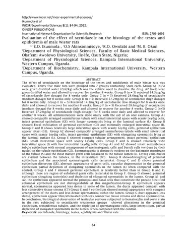
Group A1 showed compactly arranged seminiferoustubule withsmall interstitial space with scanty Leydig cells, intact germinal epithelium (GE) with longer spermatids lying at the luminal surface
(L). Group B showed Focal exfoliation of germinal epithelium (GE) and minimally enlargedinterstitialspace(I).GroupCand D shows relatively large interstitial space with scanty Leydig cells, germinal
http://www.inosr.net/inosr experimental sciences/ Ikuomola et al
INOSR Experimental Sciences 8(1): 84 94, 2022.
epithelium appear intact (GE). Group A2 showed compactly arranged seminiferoustubule withsmall interstitial space with scanty Leydig cells, intact germinal epithelium (GE) with elongating spermatids lying at the luminal surface
(L). Group E showed compact tubular arrangement, intact germinal epithelium (GE), small interstitial space with scanty Leydig cells. Group F and G showed relatively wide interstitial space (I) with fewinterstitialLeydigcells.(Plate1)
Plate 1: Effect of Secnidazole on histology of the testes
Representative light micrographs of sections of male rat testes subjected to H&E stain x100 Magnification.
LEGEND, A1 2ml/kg distilled water only, B 14.3mg/kg secnidazole, C 28.6 mg/kg secnidazole,D 57.2mg/kgsecnidazole,A2 Recoverycontrol,E,FandG 14.3mg/kg,28.6 mg/kg,57.2mg/kg+8weekswithdrawalofsecnidazoleadministration
KEYS: L = Luminal surface I = Interstitial space GE = Germinal epithelium
http://www.inosr.net/inosr experimental sciences/ Ikuomola et al
INOSR Experimental Sciences 8(1): 84 94, 2022.
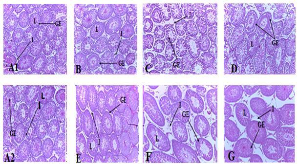
Effect of secnidazole on histology of the testes x400 magnification
Group A1 and A2 showed intact seminiferous tubule epithelium with normal arrangement of spermatogenic cells and Sertoli cells (evident by their nuclei) in thetubuleepithelium (GE). Spermatogoniaisdistinctlyevidentonthe basement membrane of the tubule (S) and the most mature germ cells localized to the tubule lumen (L).Leydig cells nuclei are evident between the tubules, in the interstitium (LC). Group B showedsloughing of germinal epithelium and the associated spermatogenic cells
(asterisks).GroupCandDshowsgerminal epithelium distortion (GE), aberrant appearance of germ cells, vacuolar degeneration in the germinal epithelium partialdepletionofelongatingspermatids in the lumen (L) when compared with the control. Group E and F showed germinal epithelium is relatively better than in Group B and C, although there are region of exfoliated germ cells (asterisks) in Group F. Group G showed germinal epithelium sloughing (asterisks) and depletion of elongated spermatids in the lumen(Plate2)
LEGEND, A1 2ml/kgdistilledwateronly, B 14.3mg/kg secnidazole, C 28.6 mg/kg secnidazole, D 57.2 mg/kg secnidazole,A2 Recoverycontrol,E,Fand G 14.3 mg/kg, 28.6 mg/kg, 57.2 mg/kg
+ 8 weeks withdrawal of secnidazole administration
KEYS: S = Spermatogonia L = Lumen
http://www.inosr.net/inosr experimental sciences/ Ikuomola et al
INOSR Experimental Sciences 8(1): 84 94, 2022.
* = Exfoliated germs
GE = Germinal epithelium
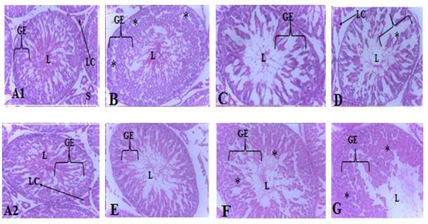
Effect of secnidazole on histology of the epididymis x100 magnification
Group A1 and A2, the epithelium appeared normal the principal and basal cells that constitute the pseudo stratified columnar epithelium are not distinguishable at this magnification. The surrounding fibro muscular stroma appeared much (CT), the duct circumscribing, peristalticcontraction aiding smooth muscle attached firmly around the duct and spermatozoa appeared dense within the lumen. Group B and C showed Epithelial lining of the
LC= Leydig cell
epididymis appeared attenuated (ET), spermatozoa appeared dense within the lumen (S). Group D epithelium appears normal, spermatozoa appearedless dense in some of the lumen, the ducts appeared compact with less connective tissue stroma (CT) Group E and F epithelium showed normal appearance with compact arrangement of the ducts and less dense spermatozoa within the lumen. Group G duct appeared less dense, epithelium appear thin/attenuated with less connective tissue stroma and dense spermatozoa(Plate3)
Plate 3: Effect of secnidazole on histology of the epididymis
Representative light micrographs of cross sections of male rat epididymis subjected to H&E stain x 100 magnification.

http://www.inosr.net/inosr experimental sciences/ Ikuomola et al
INOSR Experimental Sciences 8(1): 84 94, 2022.
LEGEND, A1 2ml/kg distilled water only, B 14.3mg/kg secnidazole, C 28.6 mg/kg secnidazole,D 57.2mg/kgsecnidazole,A2 Recoverycontrol,E,FandG 14.3mg/kg,28.6 mg/kg,57.2mg/kg+8weekswithdrawalofsecnidazoleadministration
KEYS
CT = Connective tissues (Fibro muscular stromal tissue)
S= Spermatozoa
Ep= Epithelium
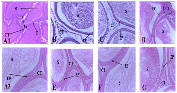
Effect of secnidazole on histology of the epididymis x400 magnification
GroupA1andA2showedtubularsections of the ductus epididymis are seen, with tall principal cells nuclei (P) and shorter basal cells nuclei (bs) that constitute the pseudo stratified columnar epithelium of this organ revealed (giving the appearance of two rows of nuclei) surrounded by a fibro muscular stroma (CT). Spermatozoa appeared dense within thelumen.GroupBandCEpitheliallining of the epididymis appeared breached and
vacuolated (ET). The nuclei of the columnar cells and basal cells are not distinguishable. Spermatozoa appear less dense within the lumen. Group D Epithelium appeared normal with the nuclei of the columnar cells located at moderate distance from the nuclei of the basal cell. Group E, F, and G epithelium showed normal appearance, the nuclei of the columnar and basal cells are not distinguishable. There are also normal circular arrangements of smooth muscle tissue unsheathing the tubules across thesegroups.(Plate4)
Plate 4: Effect of secnidazole on histology of the epididymis
Representative light micrographs of cross sections of male rat epididymis subjected to H&E stain x 400 magnification.
http://www.inosr.net/inosr experimental sciences/ Ikuomola et al
INOSR Experimental Sciences 8(1): 84 94, 2022.
LEGEND: A1 2ml/kg distilled water only, B 14.3mg/kg secnidazole, C 28.6 mg/kg secnidazole,D 57.2mg/kgsecnidazole,A2 Recoverycontrol,E,FandG 14.3mg/kg,28.6 mg/kg,57.2mg/kg+8weekswithdrawalofsecnidazoleadministration.
KEYS:
CT = connective tissue (Fibro muscular stromal tissue)
S = Spermatozoa bs = Basal Cells
P = Principal cells
DISCUSSION
This study evaluated the effect of secnidazole on the histology of the testes and epididymis of male Wistar rats. Histopathological evaluation of animal reproductive tissue is useful in male reproductive risk assessment. The testis is regarded as the most sensitive end point in the evaluation of male reproductive toxicity. In this study, histological observation of testicular sections subjected to hematoxylin and eosin stain in the rats subjected to secnidazole treatments groups showed alterations in the germinal epithelium, seminiferous tubules and the depletion of spermatogonic cells, large interstitium, partial depletion of elongated spermatids in the lumen, when compared with the control. The drug treatment was highly effective in the rats treated with medium dose(C) and high dose(D), however the alterations observedin histo architectural structure of the testis was reversedin the recovery groups after the rats were allowed to recovered for 8weeks. The
degenerated histo architectural structure observed in group F and G were partially reversed in the recovery group when compared with the control. This study is in line with that reported by [10], who reported sloughing of germinal epithelium, seminiferous tubules and the spermatogonic cells, large interstitium, partial depletion of elongated spermatids inthelumen in male white mice, treated with 0.37mg/kg/mouse/30 days of metronidazole(a member of nitro imidazole group). This might be due to the fact that secnidazole breaches the blood testis barrier and gains access to the germ cells and seminiferous tubules. Whereas, there was no damage recorded in the epididymis for both drugtreated andrecoverygroups whencomparedwith the control groups. This shows secnidazole does not affect the epididymis and suggested secnidazole to be better than other nitroimidazole groupofdrugs.
CONCLUSION
In conclusion, histological observation of testicular sections subjected to hematoxylin and eosin stain in the rats subjected to secnidazole treatments groups showed alterations in the germinal epithelium, seminiferous
tubules and the depletion of spermatogonic cells, large interstitium, partial depletion of elongated spermatids in the lumen, when compared with the control.
REFERENCES
1. Bayasgalan, G., Naranbat, D. and Radnaabazar, J. (2004) Male infertility: Risk factors in Mongolian men. Asian Journal Androl6:305311.
2. Tenaw, A. and Tsige, G. M. (2004) SelfMedication Practices of Drug Consumers. Ethiopian Journal of HealthSciences,14,111.
3. Groover, V. A., M. H., Stolt, M. H., Genthner, A. and Daniels, W. L.
(2001). Spatial Variability in Palustrine Wetlands.
https://doi.org/10.2136/sssaj2001 .652527x
http://www.inosr.net/inosr experimental sciences/ Ikuomola et al
INOSR Experimental Sciences 8(1): 84 94, 2022.
4. Sohrabi, M., Tavakoli, A. andKargari, A. (2007). A review of methods for synthesis of nanostructured metals with emphasis on iron compounds. JournalChemical Papers https://doi.org/10.2478/s11696 00700147
5. Gillis, J. C and Wiseman, L. R. (1996).Secnidazole.Areviewofits antimicrobial activity, pharmacokinetic properties and therapeutic use in themanagement of protozoal infections and bacterial vaginosis Drugs. 51(4), 621 638.
6. Newark, N. J. (2017). Centers for Disease Control and Prevention (CDC) Division of Adolescent and School Health (DASH) Youth Online.
7. Edwards,S (1993) Openness, Trade Liberalization, and Growth in Developing Countries. Journal of Economic Literature, Semtembre,volXXXI,p1358 1393.
8. Eisenstein, B. I. and Schaechter, M. (2007).DNA and Chromosome Mechanics. In: Schaechter's MechanismsofMicrobialDisease.
9. Drury,R.A.andWallington,E.A. (1980) Carleton'sHistological Techniques.5thEdition,Oxford UniversityPress,NewYork,195.
10.Saad,J.F.,Griffiths,K.R.,Kohn,M. R., Clarke, S., Williams, L. M. and Korgaonkar, M. S. (2017). Regional brain network organization distinguishes the combined and inattentive subtypes of attention deficit hyperactivity disorder.Neuroimage Clin.15, 383 390. doi: 10.1016/j.nicl.2017.05.016.
11.Ugwu Okechukwu, P. C., Okpo, F. A., Onyeke, S. C. and Okon, M. B. (2022). The effect of Ethanol Extract of Rauwolfia vomitoria on Hematological Parameters of Chloroform Intoxicated Albino Wistar Rats. IAA Journal of BiologicalSciences8(1),119127