
www.idosr.org
©IDOSR PUBLICATIONS
International Digital Organization for Scientific Research
IDOSR JOURNAL OF EXPERIMENTAL SCIENCES 9(2) 115-129, 2023. https://doi.org/10.59298/IDOSR/JES/101.1.7009
Akindele
ISSN: 2579-0781
Antibiotic resistance and Virulence Genes of Pseudomonas aeruginosa isolates in southwest, Nigeria
Akeem Abiodun Akindele1, Victoria Olanike, Omoloye1, Richard Olulowo Ojedele2 , *Emmanuel Ifeanyi Obeagu3 and Abdulwasiu Oladele Hassan4
1Department of Medical Laboratory Science, Ladoke Akintola University of Technology, Ogbomosho,OyoState,Nigeria.
2Genomics and Sequencing Unit, National Reference Laboratory, Nigeria Centre for Disease ControlandPrevention(NCDC),Nigeria.
3DepartmentofMedicalLaboratoryScience,KampalaInternationalUniversity,Uganda.
4DepartmentofMedicalLaboratoryScience,AchieversUniversity,Owo,OndoState,Nigeria.
ABSTRACT
Pseudomonas aeruginosa is a ubiquitous bacterium that causes various hospital- acquired and community-acquired infections. It has been reported that the clinical isolates of P. aeruginosa aredifficulttotreatbecauseoftheirvirulencefactorsandantibioticsresistances. Theaimofpresentstudywastoscreentheantibioticresistancepatternsandtheprevalence of virulence factor genes in a setof Pseudomonas aeruginosa isolatedfromOgbomoso,and to determine whether a correlation exists between the prevalence of virulence factors and antibiotic resistance of P. aeruginosa. A total of 100 P. aeruginosa isolates were collected fromvarioustypesofclinicalspecimens.Antimicrobialsusceptibilitytestingwasperformed usingtheKirby-bauermethod.Inaddition,PCRassayswereusedforscreeningfourvirulence encoding genes (OPRL, LasB,PLCH andToxA). The results showed thatOPRL (79%) andLasB (62%)werethemostfrequentvirulencegenesin P.aeruginosa strains,followedbyPLCH(41%) and ToxA (35%). The highest resistance was detected towards Piperacillin (42%) and Tetracycline (42%). Moderate rate of resistance (12-39%) were detected towards the other antibiotics. The virulent factors identified in this study provide valuable information regardingtheprevalenceofresistancegenesof P. aeruginosa isolatesinOgbomoso,Nigeria andtheirpotentialimpactontreatmentsthatexploittheuniquephysiologyofthepathogen. This will be useful for the health workers to improve infection control measures and to establishasurveillancesystem.
Keywords:antibioticresistance,virulencegenes, Pseudomonas Aeruginosa
INTRODUCTION
Pseudomonas aeruginosa is a ubiquitous gram-negative bacillus which is responsiblefordifferenthospital-acquired and community-acquired infections. This bacterium is considered an opportunistic pathogen that affects the health of immunocompromised individuals such as thosewithdiabetes,cancer,cysticfibrosis, advanced HIV infections, severely burned patients and those that underwent major surgeries [1]. Pseudomonas aeruginosa is responsible for several nosocomial infections like pneumonia, urinary tract infections, surgical site infections, and
some of the community-acquired infectionssuchasotitisexterna,ulcerative keratitis, and soft tissue infections. P. aeruginosa is associated with high morbidity and mortality and it has been reportedas an acute infection in over 70% of cases [2]. As a pathogen, Pseudomonas aeruginosa is of increasing clinical importance because of its innate resistancetomultipleagentsanditsability to develophigh-level multidrug resistance (MDR) due to the presence of several virulence factors encoded in its genome [3].Oneofthesevirulencefactorsthatplay
a main role in tissue lysis and bacterial invasion is exotoxin A (exoA). The hemolysin phospholipase H (plcH) act to destroy lipids and lecithin contributing to tissue invasion. P. aeruginosa also producesexoenzymeS(exoS),whichisthe cytotoxinresponsible fordamage to many typesofhostcellsandelastaseB(lasB)that play an important role during the acute infection [3-4]. Some strains produce alginate that forms the matrix of the biofilm which protects bacteria from the hostdefence during the chronicinfection. The GDP-mannose 6-dehydrogenase (algD) isoneofthreeproteinsthatareimplicated in the production of alginate [5-6]. These toxins are thought to promote the organism,diffusioninthesiteofinfection and the organism invade to host immune system and inhibits DNA synthesis, so leads to host cell damage and death [7]. Severalstudieshaveshownthatthisinnate resistance is directly associated with the expression of bacterial efflux pumps and porins such as the Resistance-Nodulationcell-Division (RND)-type efflux pump,
MexAB-OprM, MexC–MexD–OprJ, MexE–MexF–OprN, and MexXY-OprM. [8]. These important RND-type efflux pumps are constitutively expressed in wild-type cells and are responsible for the intrinsic resistancetomostantimicrobialagents[9]. Under iron-limiting conditions, P. aeruginosa produces two main siderophores, pyoverdine and pyochelin, which scavenge iron from host proteins, contributingtoitsvirulence[10].Theferripyoverdine complex uptake is carried out by TonB-dependent receptors with the help of the transporter FpvB, resulting in its internalization into the periplasm. In addition to its molecular function, the pyoverdine receptor genes have also been used to genotype several strains of P. aeruginosa. Therefore, the characterization of B-lactamases from drug-resistant P. aeruginosa can greatly contribute to the understanding of mechanisms of molecular pathogenesis, antibiotic resistance and virulence in bacteria[11].
MATERIALS AND METHODOLOGY MATERIALS
The materials used include disposable protective latex gloves, cotton wool, immersion oil, normal saline, glass slides, coverslips,Pasteurpipettesandwireloop.
STUDY AREA
The study areas are UNIOSUN Teaching Hospital, Osogbo, Osun State; OAU Teaching Hospital, Ile-Ife, Osun State;
University College Hospital, Ibadan, Oyo State; and LAUTECH Teaching Hospital, Ogbomoso,OsunState.
ETHICAL APPROVAL
Ethicalapprovalwasobtainedfrom Ethical Review committee of UNIOSUN Teaching Hospital, Osogbo,Osunstate.
INCLUSION CRITERIA
All Pseudomonas aeruginosa isolates obtained from clinical samples from patients of all age-group, gender and ethnicitywereincluded.
EXCLUSION CRITERIA
Pseudomonas aeruginosa isolatesobtained from non-clinical samples (such as animal samples,watersamples,foodsamples,etc. wereexcluded.
LABORATORY ANALYSIS
Isolation Identification of Pseudomonas Aeruginosa
From September 2021 to December 2021, one hundred P. aeruginosa isolates from wound swab, urine, stool, sputum, blood cultures, cerebrospinal fluid, and catheter tip samples obtained from different patients were collected from the Medical microbiologylaboratoriesoffourhospitals located at Ibadan, Ogbomoso, Osogbo and Ile-Ife.
Preliminary identification of bacteria was based on colony characteristics of the organisms including beta hemolysis on blood agar, non-lactose fermentation and pigment production (greenish yellow and bluish green pigments) on Mac-Conkey agar. The isolates that showed these characteristics were then sub-cultured ontobloodagartoobtainapureculture.
Gram Staining and Microscopy
Gram staining was performed on colonies from subcultures for the identification of their gram reaction. Specimens were smeared onto clean grease-free glass slides, air dried, heat fixed and gram
stained.Thestainedslideswereidentified asgram-negativeif theydidnotretain the purple stain of crystal violet and were counter-stainedpinkbysafranin.
Biochemical Tests
Oxidase Test
A colony of organisms was smeared on a filter paper soaked with a drop of oxidase reagent.Itwasobservedforcolourchange.
A purple colouration indicated an oxidase producing organism (Pseudomonas aeruginosa)
Catalase Test
A drop of Hydrogen peroxide was placed onaglassslide,andacolonyoforganisms was emulsified in it and observed for gas bubble production. The production of gas
bubbles signified the presence of catalase producing bacteria (Pseudomonas aeruginosa).
ANTIMICROBIAL SUSCEPTIBILITY TESTS
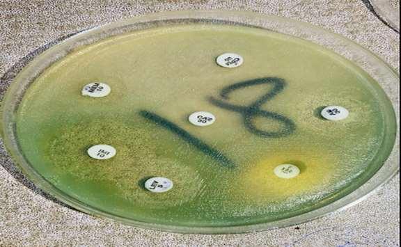
Disc Diffusion
The susceptibility testing was determined by the disc diffusion methodaccording to the European Committee on Antimicrobial Susceptibility Testing (EUCAST) recommendations. Inhibition zone diameters were evaluated according to EUCAST guidelines [12] The antimicrobial agents assayed in this study include Ceftazidime (CAZ), Meropenem (MEM), Imipenem (IPM),Cefoxitin(FOX),Cefepime (FEP), Cefotaxime (CT),aztreonam (ATM), ticarcillin (TIC), Ciprofloxacin (CIP), Amikacin (AK), Ofloxacin (OFX), Cefalexin (CN), Tetracycline (TE), Tobramycin (TOB), Piperacillin (PRL), Ceftriaxone (CRO), Trimethoprim-sulfamethoxazole (SXT),
Levofloxacin (LEV), Ticarcillinclavulanic acid (TIM), Piperacillintazobactam (TZP). The minimum inhibitory concentrations (MICs) of imipenem were determined for all strains that displayed resistance or intermediate resistance to imipenem, by using the broth microdilution method as described elsewhere. The results were interpreted based on the EUCAST breakpoints [12]. The multidrug-resistant (MDR) phenotypes are defined as resistancetoatleastthreeormoreclasses of antimicrobial agents. P. aeruginosa ATCC 27853 was employed as a control strain.
GENOTYPIC ANALYSIS
DNA Extraction for Pseudomonas aeruginosa
The isolates were plated out on Mueller Hinton agar and incubated at 37°C for 18 to 24 hours. The DNA of the isolates were extracted by suspending 4-5 bacteria coloniesin200µlofmoleculargradewater inEppendorftubesappropriatelylabelled. Themixturewerevortexedvigorously.The cells were boiled at 100oc for 10 minutes
and were cooled rapidly on ice for 30 minutes. The cell lysate were centrifuged briefly at high speed (13, 400 rpm for 3 min), and the supernatants containing the genomic DNA were transferred into fresh sterile Eppendorf tubes. The extracted DNAs were stored at -21°C until required forPCR.
PCR Procedure for Gene Amplification
A20µlreactioncontaining4µlof 5Xready to load mastermix, 0.5µl of forward primer, 0.5µl of reverse primer, 2 µl of DNAlysateand13µlofnuclease-freewater was used for PCR. Amplifications were subjected to initial denaturation at 95OC for 5min, followed by 35 cycles of
denaturation at 95OC for 1 min, annealing at 550C (OPRL), 56OC (Tox A), 56OC (PLCH), and 56OC (LasB) for 1 min respectively, extension at 72OC for 1 min and final extension procedure was carried out at 72OCfor10min[13].
Gel Electrophoresis
Aftertheamplification,PCRproductswere resolved on l.5% agarose gel prepared by dissolving 1.5g of agarose powder in 100 ml of 1X Tris-borate-EDTA (TBE) buffer solution inside a clean conical flask. The 1.5% agarose solution was heated in a microwave oven for 2-3 minutes and was observed for clarity which was an indication of complete dissolution. The mixture was then allowedto cool to about 50 0C after which 0.5 µl of ethidium bromidewasthenadded.Itwasallowedto cool further and then poured into a tray sealedatbothendswithsupporttoforma mould with special combs placed in it to create wells. The comb was carefully
removedafterthegelhadsetandtheplate wasplacedinsidetheelectrophoresistank which contained 1X TBE solution, 5µl of the amplicon was mixed with 1µl of loadingbufferandthemixturewasloaded to the wells of the agarose gel. The power supply was adjusted to 100 volts for 25 minutes. For each run, a 100 base-pair molecular weight DNA standard (size marker) was used to determine the size of each PCR product. The DNA bands were then visualized with a short wave ultraviolet trans-illuminator and photographedusingagenegelbioimaging system. The PCR product was then analyzed.
RESULTS
Table 1: Antibiotic susceptibility pattern of the isolated P. aeruginosa in the study populationn(%)
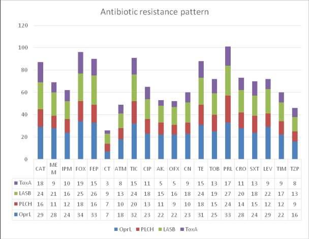
Table 1 shows the rate of sensitivity, intermediary and resistance of 100 different P. aeruginosa isolates to each of the twenty (20) antibiotics used. This result shows that a higher percentage of the organisms are sensitive to the antibiotics used with a range of 48-88% sensitivity and 12-42% resistance. In the susceptibility tests, 36 (36%) strains showedresistancetoCeftazidime,34(34%) strains showed resistance to Meropenem, 35 (35%) strains showed resistance to Imipenem, 38 (38%) strains showed resistance to Cefoxitin, 38 (38%) strains showed resistance to Cefipime, 12 (12%) strainsshowedresistancetoCefotetan, 25 (25%) strains showed resistance to Aztreonam, 39 (39%) strains showed resistance to Ticarcillin, 28 (28%) strains
showed resistance to Ciprofloxacin, 29 (29%) strains showed resistance to Amikacin, 29 (29%) strains showed resistance to Ofloxacin, 28 (28%) strains showedresistancetoGentamicin,42(42%) strains showed resistance to Tetracycline, 36 (36%) strains showed resistance to Tobramycin, 42 (42%) strains showed resistance to Piperacillin, 33 (33%) strains showedresistancetoCeftriaxone,28(28%) strains showed resistance to Trimethoprim/Sulfamethoxazole, 39 (39%) strains showed resistance to Levofloxacin, 28 (28%) strains showed resistance to Ticarcilline+Clavulanic acid,and20(20%) strains showed resistance to Piperacillin/Tazobactam
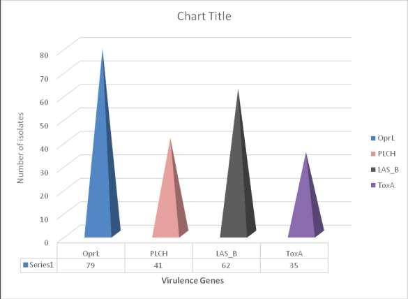
FromTable2, theresultshighlightedthatOPRL(79%)and LasB (62%) were the most frequent virulence genes in P. aeruginosa strains, followed by PLCH (41%), while the least commonly detected virulence factor gene
was ToxA (35%). Showing the distribution ofthevirulencefactorsinthesamples,the results showed that, isolates from ear swabs had the highest frequency of virulencefactors.
www.idosr.org
Table 3: showing the association between the virulence factors and their resistance to antibiotics
N.B: p-value ≤ 0.05 is taken to be statistically significant.
FromFigure2, the results showed that OPRL (79%) and LasB (62%) were the most frequent virulence genes in P. aeruginosa strains,followedbyPLCH
(41%), while the least commonly detected virulence factor gene was ToxA(35%).

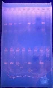
(1) Amplification of the OPRL gene, (2) amplification of the LasB gene, (3) amplification of the PLCH gene and (4)

amplification of the ToxA gene. L=DNA ladder, C=Positive control, N=Negative control.


Pseudomonas aeruginosa is widely known to significantly cause nosocomial infections with high mortality and morbidity rate, especially in immunocompromised patients [14]. Findings from this study have critically highlighted important potential clinical sources of this bacterium which include cathetertip(5%),earswab(24%),eyeswab (8%), sputum (14%), urine (11%), vaginal swab (21%) and wound swab (17%). Earlier reports fromNigeriaon theisolationrates of this organism from clinical samples include those of Odunsanya [15] who reported 4.6% for urine and 16.3% for wounds, and from Ogbolu et al. [16] who reported 41.9% and 39.35% for ear and wound swab respectively. However, from thisstudymajorityoftheisolateusedwere taken from ear swabs as it accounts for 24%ofthetotalisolate.
It is interesting to note that Amikacin which shares the same aminoglycoside classasgentamicinisalsoveryeffectiveas 62% of the isolates were found to be susceptible to it. However, this is lesser than the 99.4% reported by Elmouaden et al.[17]andthatofMohammadzadeh et al. [18]. In the present study, 50% of the isolates were sensitive to ticarcillin as opposed to the 46% reported in Sarwat et al. [19]whichwereresistant.
It was found that the rates of antibiotic resistance of P. aeruginosa were (25%) to aztreonam, (28%) to ciprofloxacin, and (36%)toceftazidime,whereasBadamchi et al [20] reported that (27.1%) of P. aeruginosa were resistant to ciprofloxacin and(25.9%)toceftazidime.Severalsurveys from developed and developing countries admit the direct link between irrational antibiotics use and the emergence of resistant strains. To reduce this problem, it is important to implement infection control measures such as good hand hygiene and proper use of antimicrobial agents[20]
Furthermore, our results revealed that the majority of the isolates are susceptible to Carbapenems, (58%) to Imipenem and (56%) to Meropenem. These findings are not in agreement with the previous result of a study done by Gierhart et al. [21] in
which all strains exhibited significant decreases in susceptibility to imipenem. However, it is in accordance with that of Kireçci et al. [22] in which the majority of the isolates were susceptible to imipenem andmeropenem.
AsopposedtothestudyofLi etal. [23]and Isichei-Ukah and Enabulele [24] the findings of this studyshows that majority of the isolates were susceptible to Ofloxacin (62%), Levofloxacin (53%), Tobramycin (57%), Trimethoprim/ Sulfamethoxazole (65%), Piperacillin/Tazobactam(70),Ticarcilline+ Clavulanic(65%).
In accounting for the susceptibility of the isolates to cephalosporins, they were foundtobesusceptibletoCefotetan(88%), Ceftriaxone (60%), Ceftazidime (54%), Cefoxitin (51%), and Cefipime (53%). This does not agree with the study conducted by Jazani et al. [25] in which there was 75.4% resistance to Cefepime and that of Prakash et al [26] who reported 65.26% resistance. This could have resulted from adherence to the control programs organizedtocurbantibioticresistancethat must have included the proper usage ofantibiotics. Moreover, the high sensitivity of the isolates to Ceftazidime (54%) is congruent with the study of Oladipo et al [27] who reported 76% sensitivity, Kechrid and Hassen [28] who foundsensitivitytobe97%.
PCR assays were used for screening four virulence encoding genes present in a variableamountintheisolatesengagedfor the study, the genes and their level of occurrenceareasfollows; Oprl (79%), plcH (41%), lasB (62%)and ToxA (35%).Ofall the four genes identified in the isolates, Oprl occurred the most as it was identified in 79%oftheisolates.
The LasB gene which is one of the most important proteases of P.aeruginosa has a 62%occurrenceintheisolatesusedforthis study, this is lesser than the 98.7% occurrence reported in the study by Elmouaden et al. [17].
The41%occurrenceoftheplcHgeneinthe isolates is dissimilar to that reported by Elmonaden et al. [17] where almost all (96.1%) harboured the plcH gene and that
www.idosr.org
of Lanotte et al., 2004 where all strains possessed the gene. Although the ToxA gene has the lowest (35%) level of occurrence in the isolates, it, however, sponsors the majority of the statistically significant resistance of the organism to antibiotics such as in resistance to CAZ (p=0.018), FOX (p=0.021), OFX (p=0.017), and AK (p=0.028). Its occurrence in the present study is opposed to the 69.4% reported in the study of Badamchi et al.[20].
The divergences in the distribution of virulence factor genes in the different populations might be due to the probabilitythatsome P aeruginosa strains arebetteradaptedtotheconditionsfound ininfectious sites thatmaybe returnedto the diverse geographical and environmental sources. The prevalence of P. aeruginosa and its virulence genes depends on various causes consisting of the nature of places, degree of
Akindele et al
contaminationandtype,immunestatusof individual patients, and virulence of strains.
The main advantage of this investigation was the collection of a set of strains isolated from various samples to determine the antibiotic susceptibility patterns and the prevalence of virulence factor genes, and although most previous studies have focused separately on virulence or resistance, this current study assessed the potential relationship between the distribution of virulence factor genes and antibiotic resistance profiles in P. aeruginosa strains. This survey provides data about phenotypic and genotypic characteristics of P. aeruginosa that emerged in Southwest, Nigeria, which could be useful for the health workers to improve infection control measures and establish a surveillancesystem.
CONCLUSION
Thepresentstudyprovidesaninsightinto antimicrobial resistance and virulence factors of P. aeruginosa isolates in Southwest, Nigeria. Our finding highlighted a moderate rate of resistance to antibiotics. The virulent factors
identified in this study provide valuable information regarding the prevalence of resistancegenesandtheirpotentialimpact on treatments that exploit the unique physiologyofthepathogen.
REFERENCES
1. PirnayJP,BilocqF,PotB,Cornelis P, Zizi M, Eldere JV, Deschaght P, Vaneechoutte M, Jennes S, Pitt T, Devos D. Pseudomonas aeruginosa population structure revisited.PLoSOne.2009; 4:1-17.
32.
2. SchulertGS,FeltmanH,RabinSD, et al. Secretion of the toxin ExoU is a marker for highly virulent Pseudomonasaeruginosaisolates obtained from patients with hospital-acquired pneumonia. Journal of Infectious Diseases. 2003;188:1695-1706
3. Idris SN, Desa MN, Aziz MN, Taib NM. Antimicrobial susceptibility pattern and distribution of Exo u and Exo s in clinical isolates of Pseudomonas aeruginosa at a Malaysian hospital, Southeast Asian. J Trop Med Public Health. 2012; 43:116-123.
4. Montero JG. Pseudomonas aeruginosa.In Vincent JL, Hal JB, editors.Encyclopediaofintensive care medicine. Berlin: Springer. 2012;1868-1872.
5. Cotar A, Chifiriuc M, Dinu S, Bucur M. Screening of molecular virulence markers in Staphylococcus aureus and Pseudomonas aeruginosa strains isolated from clinical infections. IntJMolSci.2010;11:5273-5291.
6. GholamiS,Tabatabaei M,Sohrabi N. Comparison of biofilm formation and antibiotic resistance pattern of Pseudomonas aeruginosa in human and environmental isolates. MicrobPathog. 2017; 109:94-98.
7. Jabalameli F, Mirsalehian A, Khoramian B et al. Evaluation of biofilm production and
www.idosr.org
characterization of genes encoding type III secretion system among Pseudomonas aeruginosa isolated from burn patients. Burns. 2012; 38: 11921197.
8. Livermore DM. Of Pseudomonas, Porins,Pumps andCarbapenems. Journal of Antimicrobial Chemother.2001;47:247–50.
9. Strateva T, Yordanov D. Pseudomonas aeruginosa - a phenomenon of bacterial resistance. Journal of Medical Microbiology.2009;58:1133–48.
10.Meyer J, Neely A, Stintzi A, Georges C, Holder I. Pyoverdin is Essential for Virulence of Pseudomonas aeruginosa. Infection and Immunity. 1996;64:518–23.
11.Sambrano H, et al. Prevalence of antibiotic resistance and virulent factors in nosocomial clinical isolates of Pseudomonas aeruginosafromPanamá.
12.European Committee for Antimicrobial Susceptibility Testing (EUCAST) (http://www.eucast.org/ fileadmin/src/media/PDFs/EUCA ST files/Disk test documents/ 2020 manuals/Manual v 8.0 EUCASTDiskTest2020.pdf).
13.Auda A, khdier A, Hashim KN, Abdul-LatifBelal S. Occurrence of MexAB-OprMEffluxPumpOperon on Septicemic Pseudomonas aeruginosa Chromosome. Iraqi Postgraduate Med Journal. 2012;11:97–102.
14.Kerr KG, Snelling AM. Pseudomonas aeruginosa: a formidable and ever-present adversary. Journal of Hospital Infection. 2009; 73:338–344.
15.Odusanya OO. Antibiotic susceptibility of microorganisms at a General Hospital in Lagos, Nigeria. Journal of the National Medical Association. 2002; 94:994 – 998.
16.OgboluDO,OgunledunA,Adebiyi DE,DainiOA,TerryAO.Antibiotic
Akindele et al
sensitivity pattern of Pseudomonas aeruginosa to available anti-pseudomonal drugs in Ibadan, Nigeria. African Journal of Medicine and Medical Sciences, 2008; 37(3):339-344.
17.Elmouaden et al. Virulence genes and antibiotic resistance of Pseudomonas aeruginosa isolated from patients in the NorthwesternofMorocco. J Infect DevCtries. 2019;13(10):892-898.
18.Mohammadzadeh A, Mardaneh J, Ahmadi R, Adabi J. Evaluation of the virulence features and antibiotic resistance patterns of pathogenic Pseudomonas aeruginosa strains isolated from hospitalizedpatientsinGonabad, Iran. Arch Pediatr Infect Dis. 2017;5:1-8.
19.Sarwat T, Rashid M, Rastogi V, Chander Y. Original research article a comparative study of antibiogram of Pseudomonas aeruginosa in hospital and community-acquired infections. IntJCurrMicrobiolAppSci.2015; 1:286-291.
20.Badamchi A, Masoumi H, Javadinia S, Asgarian R, Tabatabaee A. Microbial pathogenesismoleculardetection of six virulence genes in Pseudomonas aeruginosa isolates detected in children with urinary tract infection. MicrobPathog. 2017;107:44-47.
21.Gierhart S, Chukwuma U. Annual surveillance summary: Pseudomonas aeruginosa infections in the military health system. Mil Heal Syst. 2015; 195:1-22.
22.Kireçci E, Kareem R. Antibiotic sensitivity patterns of Pseudomonas aeruginosa strains isolated from various clinical specimens. Sky J Microbiol Res 2014;2:13-17.
23.LiD, YuT,Zhang Y, Yang M,LiZ, Liu M, Qi R. Antibiotic resistance characteristics of environmental bacteria from an oxytetracycline
www.idosr.org Akindele et al
production wastewater treatment plant and the receiving river. Applied and Environmental Microbiology. 2010; 76:3444–3451.
24.Isichei-Ukah,O.B.andEnabulele, O.I.Prevalenceandantimicrobial resistance of pseudomonas aeruginosa recovered from environmental and clinical sourcesin BeninCity,Nigeria. Ife Journal of Science. 2018;20(3).
25.Jazani NH, Babazade H, Sabahi Z, Zartoshti M. The evaluation of antibiotic resistance to Cefepime in hospital isolates of Pseudomonas aeruginosa.Journal of Medicine and Biomedical Sciences.2010;34:2073-78.
26.Prakash V, Mishra PP, Premi HK, Walia A, Dhawan S, Kumar A.
Increasing incidence of multidrug-resistant Pseudomonas aeruginosa in inpatients of a tertiary care hospital. International Journal of Research in Medical Sciences. 2014; 2:1302-1306.
27.Oladipo EK, Oloke JK, Omomowo IO, Oyeniran AO, Awoyelu EH, Ogundele S.O. Incidence of cephalosporin resistance among clinical isolates of Pseudomonas aeruginosa in Ibadan, SouthWesternNigeria.Int.J.Med. Biomed.Res.2015; 4:135-141
28.Kechrid A, Hassen B. (Pseudomonas aeruginosa: a multicentric study of antibiotic resistance. Tunis Med Habib Bourguiba Hospital Tunis. 2000; 82:1070-4.
Akeem Abiodun Akindele, Victoria Olanike, Omoloye, Richard Olulowo Ojedele, *Emmanuel Ifeanyi Obeagu and Abdulwasiu Oladele Hassan (2023). IDOSR Journal of Experimental Sciences 9(2) 115-129, 2023. https://doi.org/10.59298/IDOSR/JES/101.1.7009