CONTRIBUTORS
RalphGreen,MD,PhD,FRCPath ProfessorofPathologyandMedicine DepartmentofPathologyandLaboratoryMedicine UniversityofCalifornia,Davis Sacramento,California
XylinaT.Gregg,MD UtahCancerSpecialists SaltLakeCity,Utah
MichaelR.Grever,MD ProfessorEmeritus DivisionofHematology DepartmentofInternalMedicine TheOhioStateUniversity Columbus,Ohio
AmelHamdi,PhD DepartmentofPhysiology LadyDavisInstitute McGillUniversity Montreal,Quebec,Canada
XiangrongHe,MD ClinicalFellow LaboratoryMedicineandPathology MayoClinic Rochester,Minnesota
JeanneE.Hendrickson,MD Professor DepartmentsofLaboratoryMedicineandPediatrics YaleUniversitySchoolofMedicine NewHaven,Connecticut
PaulC.Herrmann,MD,PhD ProfessorandChair DepartmentofPathologyandHumanAnatomy LomaLindaUniversitySchoolofMedicine LomaLinda,California
AchilleIolascon,MD,PhD ProfessorofMedicalGenetics DepartmentofMolecularMedicineandMedicalBiotechnology UniversityofNaplesFedericoII Naples,Italy
RamiKhoriaty,MD AssistantProfessor,DepartmentofInternalMedicine AssistantProfessor,DepartmentofCellandDevelopmentalBiology SectionHead,ClassicalHematology CoreMember,RogelCancerCenter UniversityofMichigan AnnArbor,Michigan
Contributors x
AbdullahKutlar,MD ProfessorofMedicine AugustaUniversity Augusta,Georgia
MarshallA.Lichtman,MD,MACP ProfessorEmeritusofMedicineandofBiochemistryandBiophysics DeanEmeritus,SchoolofMedicineandDentistry JamesP.WilmotCancerInstitute UniversityofRochesterMedicalCenter Rochester,NewYork
ChristineLomas-Francis,MSc,FIBMS ImmunohematologyandGenomics NewYorkBloodCenter LongIslandCity,NewYork
GerardLozanski,MD ProfessorofPathologyClinical DepartmentofPathology TheOhioStateUniversity Columbus,Ohio
NaomiL.C.Luban,MD ProfessorofPediatricsandPathology SchoolofMedicineandHealthSciences GeorgeWashingtonUniversity; MedicalDirector,OfficeofHumanSubjectsProtection SeniorHematologist Children’sNationalHospital Washington,DC
JeffreyMcCullough,MD GlobalBloodAdvisor Edina,Minnesota; EmeritusProfessor LaboratoryMedicineandPathology UniversityofMinnesota Minneapolis,Minnesota
AnanyaDattaMitra,MD SectionofHematopathology DepartmentofPathologyandLaboratoryMedicine UniversityofCalifornia,DavisHealth,SchoolofMedicine Sacramento,California
JoelMoake,MD ProfessorofMedicineEmeritus BaylorCollegeofMedicine SeniorResearchScientist DepartmentofBioengineering RiceUniversity Houston,Texas
MohandasNarla,DSc LaboratoryofRedCellPhysiology NewYorkBloodCenter NewYork,NewYork
DianaMorlote,MD AssistantProfessor HematopathologyandMolecularGeneticsPathology DivisionofGenomicsandBioinformatics DepartmentofPathology TheUniversityofAlabamaatBirmingham Birmingham,Alabama
SrikanthNagalla,MBBS,MS ChiefofBenignHematology MiamiCancerInstitute Miami,Florida
CharlesH.Packman,MD ProfessorofMedicine DepartmentofHematologicOncologyandBloodDisorders LevineCancerInstitute UniversityofNorthCarolinaSchoolofMedicine Charlotte,NorthCarolina
CharlesJ.Parker,MD ProfessorofMedicine DepartmentofMedicine DivisionofHematologyandHematologicMalignancies UniversityofUtahSchoolofMedicine SaltLakeCity,Utah
JohnD.Phillips,PhD DivisionofHematology DepartmentofMedicine UniversityofUtahSchoolofMedicine SaltLakeCity,Utah
JosefT.Prchal,MD ProfessorofHematologyandMalignantHematology AdjunctinGeneticsandPathology UniversityofUtah&HuntsmanCancerInstitute SaltLakeCity,Utah 1.interníklinikaVFNaÚstavpatologickéfyziologie,1.LFSchoolof Medicine UniversitaKarlova,Prague,CzechRepublic
VishnuV.B.Reddy,MD SectionHead,UABHospitalHematologyBoneMarrowLab Director,HematopathologyFellowshipProgram DivisionofLaboratoryMedicine Professor,DepartmentofPathology TheUniversityofAlabamaatBirmingham Birmingham,Alabama
RobertaRusso,PhD AssistantProfessorofMedicalGenetics DepartmentofMolecularMedicineandMedicalBiotechnology CEINGE
BiotecnologieAvanzate UniversityofNaplesFedericoII Naples,Italy
GeorgeB.Segel,MD
EmeritusProfessorofPediatric ProfessorofMedicine
JamesP.WilmotCancerInstitute UniversityofRochesterMedicalCenter Rochester,NewYork
VivienA.Sheehan,MD,PhD AssistantProfessorofPediatrics
BaylorCollegeofMedicine Houston,Texas
SujitSheth,MD DepartmentofPediatrics
WeillCornellMedicine NewYork,NewYork
SweeLayThein,MD
NationalHeart,Lung,andBloodInstitute TheNationalInstitutesofHealth Bethesda,Maryland
PerumalThiagarajan,MD ProfessorofMedicineandPathology
BaylorCollegeofMedicine DirectorofTransfusionMedicineandHematologyLaboratory MichaelE.DeBakeyVAMedicalCenter Houston,Texas
EduardJ.vanBeers,MD,PhD Hematologist VanCreveldkliniek UniversityMedicalCenterUtrecht UtrechtUniversity Utrecht,TheNetherlands
RichardvanWijk,PhD AssociateProfessor CentralDiagnosticLaboratory UniversityMedicalCenterUtrecht UtrechtUniversity Utrecht,TheNetherlands
NealS.Young,MD Chief,HematologyBranch NationalHeart,Lung,andBloodInstitute MarkHatfieldClinicalResearchCenter NationalInstitutesofHealth Bethesda,Maryland
PREFACE
Thediscoveryoftheruddyglobules(redcells)isattributedtoJan Swammerdam(1637-1680)inAmsterdam;but,itwasAntonjvan Leeuwenhoek(1632-1723)ofDelft,whoasaresultofhisabilitytogrind lenseswithgreatermagnifyingpower(x275),madeamoredetailed descriptionofredcells,delineatingtheirgrossstructure.
Thebiochemistry,physiology,andbiophysicsoftheredcellhave beenstudiedintensivelyoverthreecenturiesand,althoughconsidered a“simple”structure,sinceitisanucleateandafteronedayinthecirculationhasnocytoplasmicorganelles,itsmysterieshavebeenslow tobeunraveled.Theprocessofenucleationoftheerythroblastinthe hematopoieticspaceandthemovementoftheanucleatecellfromthe hematopoieticspacetothemarrowsinusandfromtheretothesystemiccirculation,accomplishedbyacellwithoutanintrinsicapparatus tosupportamoeboidmotility,andthedeterminantsofitsaveragelife spanofapproximately120daysarestillbeingelucidated.Itsstructural andbiophysicalproperties,biochemicalpathways,andtherelationship amongthosefeatureshavebeenofcontinuedinteresttoscientists.Its absenceofinterferinggranules,containingproteolyticenzymes,organelles,andothercomplexitieshaveallowedtheisolationofhighlypurifiedredcellmembranesandtheearlyexplorationofthebiochemical andbiophysicalfeaturesofcellmembranes,applicabletoothercells, includingthecharacteristicsofmembranetransportofvariousmolecules.Thenatureofthestructureandfunctionofhemoglobinandthe explorationoftheglycolyticpathway,thehexosemonophosphateshunt, andtheLuebering-Rapoportpathwaywereotherrewardsreapedfrom thestudyofredcells.
Muchisknown,butasourmentor,friend,andcolleague,Ernest Beutler,cautionedPh.D.graduatesataScrippsInstitutedoctoralgraduation,oneshouldnotassumethatourunderstandingofthebiomedical sciencesissoprofoundthatwhatisleftforusistofillinsomegaps. Hearguedthatmuchfundamentalbiomedicalknowledgewasstill undiscoveredandwaitingtobeilluminated.Amonghismanycontributionstothepathogenesisofdiseaseandapplicationoftherapy,his contributionstounderstandingtheredcellandanemiawerenotable. Theseobservationsincludedaclassicseriesofpapersdescribingthe effectsofoxidantstressonindividualswithredcellglucose-6-phosphate dehydrogenasedeficiencyandalife-longinterestintheenzyme’svariantsandepidemiology.Hismonographonmethodsformeasuringred cellenzymeswasanearlycontributiontoenhancingthespecificityof thediagnosisofhemolyticanemia.Publishedoverfivedecadesago, itremainsanunsurpassedsourceofmethodsfortheassayofredcell enzymes.Beutler,also,usedredcellenzymemeasurementasasurrogate fordiagnosisofsystemic,untilthendifficulttodiagnosediseases,such asgalactosemia,glycogenstoragedisorders,andothers.Hefoundthat redcellglucose-6-phosphatedehydrogenasedeficiencywasinherited asanXchromosome-linkeddisorderanddescribedthemosaicismof normalanddeficientredcellsinheterozygousfemales.Thisfindingof mosaicismprovidedthebasisforanintellectualjumptothehypothesis ofXchromosomeinactivationinhumans,coincidentwithMaryLyon’s descriptionofthephenomenoninmice.He,also,madeseminalcontributionstounderstandingtheeffectsofirondeficiencyinnon-anemic womenandtheexpressionofironoverloadinthosehomozygousfor theHFEmutationandthevalueofadditivesforprolongedstorageof redcells,stillincurrentuse.
WithnoDNAorRNAsynthesis,nomitochondriaandtheir relatedenzymaticbiochemicalenergygeneratingpathways,andwith arelativelyshortlifespan,thisamitoticcellissustainedatanormal
concentrationinthebloodbyarobustdailyproductionofnewcellsin themarrow,theprocessoferythropoiesis.Thisprocessdeliverstwoto threemillionnewredcellstothebloodpersecond.Althoughremarkable,itisalsoavulnerabilityshouldredcellproductionbedampenedby diseaseorsubstrateinsufficiency:thelatter,aprincipalcauseofanemia.
In1929,3yearsafterobtaininghisM.D.degreeattheUniversityof Manitoba,hisfamilyhavingimmigratedtoCanadafromAustria,Maxwell MyerWintrobe,obtainedhisPh.D.atTulaneUniversity,hisdoctoral thesisentitled“TheErythrocyteinMan.”Wintrobeisconsideredthe fatherofclinicalhematologyhavingpublishedthefirstcomprehensive textintheEnglishlanguage,ClinicalHematology,in1942.Heintroducedthetechniqueofthehematocritdevicetomeasurethepacked redcellvolumeatatimewhenhemoglobinandredcellcountmeasurementswereneitheraccuratenorreproducible.Theword“hematocrit”wassoappealingthatitbecameasynonymforthepackedred cellvolumeratherthantheinstrumentofmeasurementasintendedby Wintrobe.Initially,the“Wintrobe”tube,asitbecameknown,wasfilled bypipettewithbloodtothe1mLmarketchedonthetubeandthegradationsonthetubeallowedonetoreadthefractionofbloodthatwas composedofredcellsaftercentrifugation.Later,themicrohematocrit centrifuge,whichreachedG-forcesthatremovedplasmatrappingasa significantconsiderationinthemeasurementincapillarytubesfilled withblood,couldbefoundoneverywardandclinicallaboratoryasthe principalmeanstomeasurethepackedredcellvolumeand,thereby, identifyanemiaorerythrocytosis.Achartallowedthedetermination ofthepackedcellvolumewhenthecapillarytube,regardlessofthe volumeofblooditcontained,wasplacedagainstitsscales.Wintrobe institutionalizedtheredcellindices,meancellvolume(MCV),mean cellhemoglobin(MCH),andmeancellhemoglobinconcentration (MCHC)andshowedintwoclassicpaperin1930and1934thatone couldclassifytheanemiasfordiagnosticpurposesbydistinguishing amongmacrocytic,normocytic,simplemicrocytic,andhypochromic microcyticanemias,amethodofdifferentialdiagnosisstillusedtoday. AftermovingtotheUniversityofUtahfromJohnsHopkinsUniversity, Wintrobeestablishedoneofthemostesteemedhematologyclinicaland researchtrainingprogramsintheworld.Healsodescribedalongwith hiscolleagueGeorgeCartwrightthattheaveragehematocritandhemoglobinconcentrationwashigherinresidentsofSaltLakeCity(elevation4300feet)thanthevalueobservedatJohnsHopkinsinBaltimore (elevation480feet).Hededucedfromthatprescientobservationthat hypoxia,inthatinstancefromhigheraltitude,isaprincipalregulatorof normalerythropoiesis.
In1953,F.WilliamSundermanandcolleaguesenhancedtheaccuracyofbloodhemoglobinmeasurementbyintroducingthecyanmethemoglobinmethod.In1956,WallaceCoulterintroducedhishigh-speed, automaticbloodcellcountermakingbloodcellcountingaccurate, reproducible,andcapableofmeetingthedemandsofabusyclinicand hospitalenvironment.The“CoulterPrinciple”heldthatcellsarepoor conductorsofelectricityinasaltsolution.Thus,whencellsarediluted insalineandaredrawnthroughatinyaperturecarryingacurrent,each cellproducesaslightimpedancetocurrentflowasitpassesthroughthe narrowaperture.Thepulsecreatedbythisimpedancecanbeamplifiedandcounted.Moreover,thesizeofthepulseisproportionaltocell volume.Thus,thenumberandvolumedistributionofredcellsina measuredvolumeofsolutioncanbeconvertedtoredcellcountand volumeelectronically.Theirproduct,redcellcountandredcellvolume, providedthehematocrit,nowaderivedvalue.Thousandsofcellscan

becountedpersecond.Sincetheredcells,leukocytes,andplateletsare sufficientlydifferentinsize,theycanbediscriminated.Theelectronic particlecounter’sderivativetechnologyofcellflowanalysis,dependent onlaserlight,providedoneofthemostpowerfuldiagnostictechnologiesinmedicine,capableofmeasuringcellDNAcontentorthesurface antigenarrayofaspecificcelltype.Onecouldusethedevicetoisolate purified,specificcellpopulationsforanalysis.TheCoulterPrinciple anditsderivativetechnologiesrevolutionizeddiagnosticmedicine,biomedical,andindustrialresearchand,morespecifically,thediagnosis andmanagementofredcelldiseases.
Agiantofstudiesoftheredcell,perhapslittleknowntoyounger scientists,wasEricPonder(d.1970),anoriginalmemberoftheRed CellClub(seefurther),whosetreatiseHemolysisandRelatedPhenomenain1948,reissuedin1971byGruneandStrattonwithaforwardby RobertI.Weed,isanextraordinarycompilationofhisresearchonthis cell.Manyofhisstudiesarestillrelevant.Allscientistinterestedinthe redcellshouldbefamiliarwiththiswork.Weed,anothergiftedcontributortoourunderstandingoftheredcell,diedprematurelyin1976, attheageof48years,ofaglioblastoma.Hewaslargelyresponsiblefor convincingtheNationalInstitutesofHealthtoexpandthedesignation oftheHeartandLungInstitutetotheHeart,LungandBloodInstitute in1976,facilitatingresearchsupportforbloodcells,especiallyredcell research.In1976,inrecognitionofhisleadershipinthatinitiativeand hiscontributionstoresearchontheredcell,hewasnamedthethird recipientoftheWilliamDameshekAwardoftheAmericanSocietyof Hematology.Atthetime,theSocietyhadtwoprizes,TheHenryStratton LectureandTheWilliamDameshekPrize.StrattonandDameshekwere veryclosefriends.DameshekwasamongtheverytopacademicclinicalhematologistsintheUnitedStatesandStrattonwastheco-owner ofGruneandStrattonPublishers.Theyweretheprimemoversofthe establishmentoftheAmericanSocietyofHematologyandstartedBlood in1946.DameshekwasthefoundingeditorandGruneandStrattonthe publisher.UnderDameshek’seditorshipBloodbecamethemostprestigiousjournalofclinicalandresearchhematologyintheworld.In1976, thejournalbecametheofficialpublicationoftheAmericanSocietyof Hematology;however,thepublisherstillownedthetitleand,technically,editorialcontrol,butsomeofitwascededtotheSociety.In1989, theAmericanSocietyofHematologyboughtthetitletoBloodfromits thenpublisherSaunders,Inc.anditbecameBlood,TheJournalofthe AmericanSocietyofHematology.Thepurchaseoftitlewasaninitiative ledbyH.FranklinBunn,adistinguishedhematologistatHarvardUniversityandaworld’sauthorityonthestructureandfunctionofhemoglobin.ThepurchaseoftheJournalhasprovidedtheSocietywithan enormouslysuccessfuleconomicenginetosupportitseducationaland researchprograms,fullcontrolofitseditorialpolicies,andanoutletfor themostimpactfulresearchinthefield,includingthatoftheredcell anditsdiseases.
BobWeed’sclosecolleaguesattheUniversityofRochester,Claude ReedandScottSwisher,werepioneersinforecastingthekeyroleof amembraneproteinabnormalityastheprimarylesioninhereditary spherocytosis,whereasothersweredistractedbyepiphenomena,such assubstratetransport.Theyshowedthatthemembranelipidcompositionofredcellsinhereditaryspherocytosiswasnormalbutafter 24hoursofincubation,lipids(cholesterolandphospholipids)werelost tothemediumintheirexactmolarproportionasintheredcellmembraneandthisphenomenoncouldbedecreasedbyaddingglucoseto themedium.Thisfindingstronglysuggestedthatthelossofsurfacearea oftheredcellsandthedisctospheretransformationdecreasingtheir surfaceareatovolumeratioandmovingtowardtheircriticalhemolytic volumewasrelatedtolossofpiecesofmembrane.Thisworkpublished in1966waswellbeforemethodsformembraneproteinanalysiswere available.Later,theabilitytocharacterizetheproteincompositionof
“pure”redcellmembranes(ghosts)incasesofspecificdisordersofthe redcell(eg,hereditaryspherocytosisversushereditaryelliptocytosis) allowedtheassignmentoffunctionalcharacteristicstothemissingor mutantproteins.Redcellghostsareapreparationofredcellmembranes freedoftheirinternalcontents,notablehemoglobinandenzymesand substratesandcolorless(ghostlypale)ratherthanredandarebasically pureredcellmembranes,akeyspecimenforstudy.
Alongstandingfocusontheredcellbybasicandclinicalinvestigatorshasbeenhighlightedbytheinteractionsofagroupofscientists, referredtoas“TheRedCellClub,”whichstartedin1958throughthe initiativeofJosephHoffmanandDanielTosteson,thenyoungscientists attheNationalInstitutesofHealth.ThespenttheircareersatYaleand Harvard,respectively.Themeetingsaresmall,informal,andanideal milieutofocusonnewscienceandtheexchangeofideas.TheClub, inits63rdyearin2021,meetsnowonceayearonthecampusofa membertodiscussnewinsightsintotheredcellandtosharetheircurrentresearch.Itisacollegialgroupwithnew“blood”beingcycledin fromlaboratoriesthroughouttheUnitedStatesandCanadaasmentors introducetheiracolytestotheredcell’scharms.Usually,apreceding roundofgolfisheldforthosedevoteesofthegame,weatherpermitting. Members,whoforreasonsofageorachangeofinterestsleavethefold, areneverdroppedfromtheinvitationlist.Nonparticipantsaretenderly referredtoas“redcellghosts.”Inthelastseveralyears,scientistsfrom Europeand,occasionallyJapan,haveparticipatedinthesemeetings. AEuropeanRedCellClubhasbeenestablishedhighlightingthatthe mysteriesofthecellhavenotallbeenuncovered,confirmingBeutler’s admonition.
Inthisvolume,webringtothereaderthemostup-to-dateconsiderationofthestructureandfunctionoftheredcell.Aftertwo introductorychaptersonthestructureandbiologyoftheredcelland erythropoiesis,thefocusturnstothecomprehensivesetofdiseases, eitheracquiredorinherited,inwhichaquantitative(deficiencyor excess)orqualitative(membrane,enzyme,hemoglobin)abnormalityof theredcellresultsindisease.Thesechapters,also,mayincludeimportant,relevantbasicscientificaspectsoftheclinicalproblemunderdiscussion.Theroleofcertainplasmaconstituents,iron,folicacid,and cobalamin,criticaltonormalredcellproductionandhemoglobinsynthesis,isdescribedaswell.
Webelievetheauthorshavebroughttoourreaderaninsightful expositionoftheredcellanditsdisorderstoenlightentheclinicians facedwiththeirchallengesandtothebenefitofthecareoftheirpatients. Inaddition,wehopethistextprovidesscientistsacleardelineationof theremainingmysteriesofthecellandprovidesthemwithnewfoundationsfordevelopmentoftherapyofredcelldiseases.Wehopethatthis textwillfillthevacuumthathasexistedsincethemonographpublished in1970devotedtotheredcellbyJohnW.Harris,andRobertW. Kellermeyer:TheRedCell:Production,Metabolism,Destruction:NormalandAbnormal.
TheauthorsacknowledgeandthankKarenEdmonson,Senior Editor,formerlyatMcGraw-Hill,Education,forsupportingtheproductionofthistextandconvincingmanagementofitsmerits,Susan DaleyattheUniversityofRochesterMedicalCenterforheradministrativeassistance,HarrietLebowitz,SeniorProjectDevelopmentEditor atMcGraw-HillEducationforstewardingthefinalpreparationofthe manuscriptandJasonMalley,editorandRichardRuzycka,production supervisor,eachatMcGraw-HillEducation,andWarishreePant,the ProjectManageratKnowledgeWorksGlobal,Ltd.
MarshallA.Lichtman,Rochester,NY JosefT.Prchal,SaltLakeCity,UT
PartIStructureandPhysiology oftheRedCell
1.StructureandCompositionofthe Erythrocyte...........................3 2.ErythropoiesisandRedCellTurnover.....21
SUMMARY
Collectively,theerythroidprogenitors,terminallydifferentiatingerythroblasts (precursors),andadultredcellsaretermedtheerythrontoreinforcetheidea thattheyfunctionasanorgan.Thewidelydispersedcellscomprisingthis organarisefrompluripotentialhematopoieticstemcells.Followingcommitmenttotheerythroidlineage(unipotentialprogenitor),furthermaturation givesrisetotheerythroidprogenitors,burst-formingunit–erythroid(BFU-E) and,subsequently,colony-formingunit–erythroid(CFU-E),thatcanbeidentifiedbytheirdevelopmentintorepresentativeclonalcoloniesofredcellsin vitro.TheCFU-Ethenundergoesterminaldifferentiation,progressingthrough fourtofivemorphologicstages,eachhavingcharacteristiclightmicroscopic andultrastructuralfeatures.Duringterminalerythroiddifferentiation,there isanincreasingamountofhemoglobinsynthesisaccompaniedbynuclear chromatincondensation,andatthefinalstageofdifferentiation,thereis nuclearextrusiontogenerateananucleatepolychromatophilicmacrocyte (reticulocytewithsupravitalstaining).Thehumanpolychromatophilicmacrocyte(reticulocyte)maturesover2to3days,firstinthemarrowandthen incirculationintothediscoiderythrocyte.Duringreticulocytematuration, cytoplasmicinclusions,includingresidualmitochondriaandribosomes,are degraded,andthereticulocytelosessurfaceareatoachievethemeancell volumeandsurfaceareaofadiscoidalerythrocyte.Matureerythrocytesare approximately7to8μmindiameterandundergoextensivedeformationto passthrough3-μm-diametercapillariesandthe1-μm-wideand0.5-μm-thick endothelialslitsintheredpulpofthespleen.Theabilityoftheredcellto undergoextensivereversibledeformationisessentialforbothitsfunctionand itssurvival.Redcelldeformabilityisafunctionofitsgeometry,theviscosity
ofthecytoplasm,largelydeterminedbytheconcentrationofhemoglobin. Decreaseddeformabilityisafeatureofredcellsinvariouspathologicstates. Theerythrocyteisuniqueamongeukaryoticcellsinthatitsprincipalphysicalstructureisitscellmembrane,whichenclosesaconcentratedhemoglobin solution.Thus,allstructuralpropertiesofthiscellareinsomewaylinkedtothe cellmembrane.Incontrasttoothercells,theerythrocytehasnocytoplasmic structuresororganelles.Amonghumancells,onlyredcellsandplateletsdo nothaveanucleus.
ERYTHRON
Themassofcirculatingerythrocytesconstitutesanorganresponsible forthetransportofoxygentotissuesandtheremovalofcarbondioxidefromtissuesforexhalation.Collectively,theprogenitors,precursors, andadultredcellsmakeupanorgantermedtheerythron,whicharises frompluripotentialhematopoieticstemcells.Followingcommitment totheerythroidlineage,unipotentialprogenitorsmatureintotheerythroidprogenitors,theburst-formingunit–erythroid(BFU-E)and, subsequently,thecolony-formingunit–erythroid(CFU-E),whichthen undergoesfurthermaturationtogenerateanucleatepolychromatophilic macrocytes(reticulocytesonsupravitalstaining).TheBFU-Eand CFU-Eareidentifiedbytheirdevelopmentintomorphologicallyidentifiableclonalcoloniesofredcellsinvitro.Thereticulocytefurther matures,firstinthemarrowfor2to3daysand,subsequently,inthe circulationforapproximately1day,togeneratediscoiderythrocytes.1-5 Theproerythroblast,thefirstmorphologicallyrecognizableerythroid precursorcellinthemarrow,typicallyundergoes5mitoses(range4-6) beforematurationtoanorthochromaticerythroblast,whichthenundergoesnuclearextrusion.Afeatureoferythropoiesisisthataftereachcell division,thedaughtercellsadvanceintheirstateofmaturationwith significantchangesingeneandproteinexpressioncomparedwiththe parentcelland,ultimately,becomefunctionalasmatureerythrocytes.4 Inthisprocess,theyacquirethehumanbloodgroupantigens,transport proteins,andallcomponentsoftheerythrocytemembrane.4,6
Intheadultstageofdevelopment,thetotalnumberofcirculatingerythrocytesisinasteadystate,unlessperturbedbyapathologic orenvironmentalinsult.Thiseffectdoesnotholdduringgrowthof theindividualinutero,particularlyintheearlystagesofembryonic developmentandduringneonataldevelopmentasthetotalbloodvolumeincreasesmarkedly.Consequently,erythrocyteproductioninthe embryoandfetusdiffersmarkedlyfromthatintheadult.
THEEARLIESTERYTHRON
AcronymsandAbbreviations:BFU-E,burst-formingunit–erythroid;CFU-E, colony-formingunit–erythroid;cP,centipoise;DIC,disseminatedintravascular coagulation;EMP,erythroblastmacrophageprotein;ICAM-4,intercellular adhesionmolecule-4;IL,interleukin;MCH,meancellhemoglobincontent;MCHC, meancorpuscularhemoglobinconcentration;MCV,meancellvolume;MDS, myelodysplasticsyndrome;SA:V,surfacearea-to-volumeratio;TTP,thrombotic thrombocytopenicpurpura.
*ThischaptercontainstextwrittenforpreviouseditionsofthisbookbyBrianBull, PaulHerrmann,andErnestBeutler.
Intheveryearlystagesofhumangrowthanddevelopment,thereare twoformsoferythroiddifferentiation:primitiveanddefinitive.7-10 Chapters2and17providedetailedinformationofembryonicand fetalhematopoiesis.Theprimitiveerythronsuppliestheembryowith oxygenduringthephaseofrapidgrowthbeforethedefinitiveformof maturationhashadachancetodevelopandseedanappropriateniche. Thehallmarkofthisprimitiveerythronisthereleaseofnucleatederythroidprecursorscontainingembryonichemoglobin.Althoughprimitiveinthesensethatthecellscontainnucleiwhenreleasedintothe circulation,thisformofmaturationdiffersfromavianandreptilian erythropoiesisinthatthenucleusiseventuallyexpelledfromthemammaliancellsastheycirculate.Thetransientpresenceofanucleusinthe cellsofthecirculatingprimitiveerythroncandecreasetheefficiency ofgasexchangeinthelungsandmicrovasculaturebecausethenucleus
preventstheredcefrombehavingasafuiddropet.11Thedefinitive stageofmaturationmakesitsappearancearoundweek5ofembryogenesiswhenmutipotentiastemcesdeveopandseedtheiver, whichmaintainstheerythronformostoffetaife.Inaterfetaife, skeetadeveopmentprovidesmarrownichestowhicherythropoiesisreocates,beingsustainedintheformoferythrobasticisands,a centramacrophagewithcircumferentiaayersofdeveopingerythroidces.12Thedefinitivestageoferythroidmaturationpredominatesduringtheremainderoffetadeveopmentandistheonytype oferythroidmaturationpresentthroughchidhoodandadutife. Anormahumanerythropoiesisoccursinthemarrowintheform oferythrobasticisands.13
ERYTHROIDPROGENITORS
Burst-FormingUnit–Erythroid
Theeariestidentifiabeprogenitorcommittedtotheerythroidineage istheBFU-E(Chap.2,Fig.2-1).ABFU-Eisdefinedinvitrobyitsabiity tocreatea“burst”onsemisoidmedium,thatis,acoonyconsistingof severahundredtothousandsofcesby10to14daysofgrowth,during whichtimesmaersateitecustersofcesformaroundaargercentra groupoferythroidces,givingrisetothedesignationofa“burst.”The generationofBFU-Efromhematopoieticstemcesrequiresintereukin (IL)-3,stemcefactor,anderythropoietinfordifferentiation,proiferation,preventionofapoptosis,andmaturation(Chap.2).5,13
Colony-FormingUnit–Erythroid Aserythroidmaturationprogresses,aaterprogenitor,theCFU-E, derivedfromtheBFU-E,canbedefinedinvitro.TheCFU-Eisdependentonerythropoietinforitsdeveopmentandcanundergoonyafew cedivisions.5,14,15Thus,theCFU-Eformsasmaercoonyofmorphoogicayrecognizabeerythroidcesin5to7days(seeChap.2, Fig.2-1).Adhesionbetweenerythroidcesandmacrophagesoccursat theCFU-Estageofmaturation.
Usingce-surfacemarkers,IL-3receptor,CD34,andCD36,highy purifiedpopuationsofBFU-EandCFU-Ecanbeisoatedfromhuman marrow.5Geneexpressionprofiingshowsdistinctivechangesingene expressionprofiesinhematopoieticstemces,BFU-E,andCFU-E.5 Someofthemarrowfaiuresyndromesaretheresutofdefectsindifferentiationofstemcesintoerythroidprogenitors.
ERYTHROBLASTICISLAND
Theanatomicaunitoferythropoiesisinthenormaadutistheerythrobasticisandoriset.13,16,17Theerythrobasticisandconsistsof acentrayocatedmacrophagesurroundedbymaturingterminay differentiatingerythroidces(Fig.1-1A).Severabindingproteins areimpicatedinthece–ceadhesionsimportanttothisprocess. Theseincudeα4β1integrin,erythrobastmacrophageprotein(EMP), andinterceuaradhesionmoecue-4(ICAM-4)ontheerythrobasts andvascuarceadhesionmoecue(VCAM-1)EMP,αVintegrinon macrophages.16AdditionamacrophagereceptorsincudeCD69(siaoadhesin)andCD163,butthecounterreceptorsfortheseonerythrobastsremainstobedefined.16Phase-contrastmicrocinematography reveasthatthemacrophageisfarfrompassiveorimmobie.Evidence suggeststhateithertheerythrobasticisandsmigrateorthaterythroid precursorsmovefromisandtoisand,becauseisandsnearsinusoids arecomposedofmorematureerythrobasts,whereasisandsmore distantfromthesinusoidsarecomposedofproerythrobasts.18The macrophage’spseudopodium-ikecytopasmicextensionsmoverapidyovercesurfacesofthesurroundingwreathoferythrobasts.On phase-contrastmicrographs,thecentramacrophageoftheerythrobasticisandappearsspongeike,withsurfaceinvaginationsinwhich theerythrobastsie(Fig.1-1B).Astheerythrobastmatures,itmoves aongacytopasmicextensionofthemacrophageawayfromthemain body.Whentheerythrobastissufficientymaturefornucearexpusion,theerythrobastmakescontactwithanendotheiace,passes throughaporeinthecytopasmoftheendotheiace,andenters thecircuationasapoychromatophiicmacrocyte(reticuocyte).19-21 Thenuceusisejectedbeforeegressfromthemarrow,phagocytized, anddegradedbymarrowmacrophages.22Inadditiontotheunique cytoogicfeaturesjustdescribed,themacrophageoftheerythrobasticisandisasomoecuarydistinctasdemonstratedbyaunique immunophenotypicsignature.23Inaddition,themacrophageofthe erythrobasticisandappearstopayastimuatoryroeinerythropoiesis;independentoferythropoietin.Theanemiaofchronicinfammationandofthemyeodyspasticsyndrome(MDS)mayresutparty frominadequatestimuationoferythropoiesisbythesemacrophages (Chaps.2and6).
Despitethecentraroeoferythroidisandsinerythropoiesis invivo,morphoogicaynormadeveopmentoferythroidcescanbe
Figure1–1.Erythroblasticisland.A.ErythroblasticislandasseeninWright-Giemsa–stainedmarrow.Notecentralmacrophagesurroundedbya cohortofattachederythroblasts.B.Erythroblasticislandinthelivingstateexaminedbyphase-contrastmicroscopy.Themacrophageshowsdynamic movementinrelationtoitssurroundingerythroblasts.(A,reproducedwithpermissionfromLichtmanMA,ShaferMS,FelgarRE,etal:Lichtman’sAtlasof Hematology2016.NewYork,NY:McGrawHill;2017.)
recapituatedinvitrowithoutthesestructures,assumingdeveoping cesareprovidedwithsupraphysioogicconcentrationsofappropriate cytokinesandgrowthfactors.Suchgrowthinvitro,however,ismuch essoptimathanwhenerythrobastsformerythrobasticisands.24The erythrobasticisandisafragiestructure.Itisusuaydisruptedinthe processofobtainingamarrowspecimenbyneedeaspirationbutcanbe seeninmarrowbiopsies.
Macrophagesinerythrobasticisandsnotonyaffecterythroid differentiationand/orproiferationbutasoperformotherfunctions, incudingrapidphagocytosis(<10min)ofextrudednuceiasaresut ofexposureofphosphatidyserineonthesurfaceofthemembranesurroundingthenuceus.22Thisphagocytosisisthereasonfortheinabiity tofindextrudednuceiinmarrowaspiratesdespitethefactthat2miion nuceiareextrudedeverysecondduringsteady-stateerythropoiesis. Aprotectivemacrophagefunctioninkedtoefficientphagocytosishas beendescribed.Innormamice,DNaseIIinmacrophagesdegradesthe ingestednucearDNA,butinDNaseII-knockoutmice,theinabiityto degradeDNAresutsinmacrophagetoxicity,witharesutantdecrease inthenumberofmarrowmacrophagesandinconjunctionwithsevere anemia.25Macrophagescanpaybothpositiveandnegativereguatory roesinhumanerythropoiesis,butthemechanisticbasisforthesereguatoryprocessesarenotcompeteyunderstood.16,24Theseprocesses maypayaroeintheineffectiveerythropoiesisindisorderssuchas MDS,thaassemia,andmaariaanemia.
Anotherpotentiayimportantroeoriginayproposedforthe centramacrophageisdirecttransferofirontodeveopingerythrobasts mediatedbyferritinexchangebetweenmacrophagesanderythrobasts
(Chap.10).13Thisisaninterestingevovingconceptwithidentification ofvarioustransportproteinsinvovedinthisexchange.
ERYTHROIDPROGENITORSANDPRECURSORS EarlyProgenitors
A“progenitor”inthehematopoieticsystemisdefinedasamarrowce thatisaderivativeofthepuripotenthematopoieticstemcethroughthe processofdifferentiation,andisantecedenttoa“precursor”ce,theatter beingidentifiabebyightmicroscopybyitsmorphoogiccharacteristics. Inerythropoiesis,theeariestprecursoristheproerythrobast.Erythroid progenitorcesareidentifiedasmarrowcescapabeofformingerythroid cooniesinsemisoidmediuminvitrounderconditionsinwhichtheappropriategrowthfactorsarepresent.Progenitorcesasomaybeidentifiedby characteristicprofiesofsurfaceCDantigensusingfowcytometry.Numericay,erythroidprogenitors,BFU-E,andCFU-Erepresentonyaminute proportionofhumanmarrowces.BFU-Erangefrom300to1700× 106mononucearcesandCFU-Erangefrom1500to5000×106mononucearces.5InvitrocuturesusingCD34+cesfrombood,cordbood, andmarrowasthestartingmateriahaveidentifiedthecriticacytokines requiredforerythroiddifferentiationandmaturationandhaveenabedthe identificationandisoationofpurecohortsoferythroidprogenitorsand erythrobastsatastagesofterminaerythroidmaturation.4,5
Precursors
Figure1-2showsthesequenceofprecursorsasseeninmarrowfims. Figure1-3showsthemarrowprecursorsasisoatedbyfowcytometry.
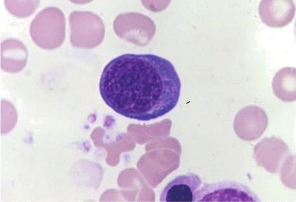
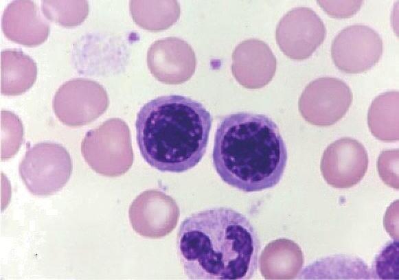
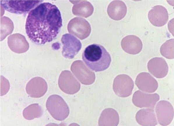
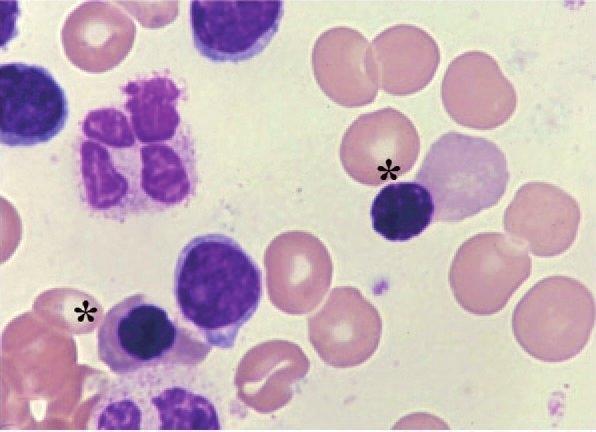
Figure1–2.Humanerythrocyteprecursors.Lightmicroscopicappearance.MarrowfilmsstainedwithWrightstain.Therearefivestagesoferythroblastdevelopmentrecognizablebylightmicroscopy.A.Proerythroblasts.Twoarepresentinthisfield.Theyarethelargestredcellprecursor,withafine nuclearchromatinpattern,nucleoli,basophiliccytoplasm,andoftenaclearareaatthesiteoftheGolgiapparatus.B.Basophilicerythroblast.Thecell issmallerthantheproerythroblast,thenuclearchromatinisslightlymorecondensed,andcytoplasmisbasophilic.C.Polychromatophilicerythroblasts.Thecellissmalleronaveragethanitsprecursors.Thenuclearchromatinismorecondensed,withacheckerboardpatternthatdevelops.Nucleoli areusuallynotapparent.Thecytoplasmisgray,reflectingthestainingmodulationinducedbyhemoglobinsynthesis,whichaddscytoplasmiccontentthattakesaneosinophilicstain,admixedwiththeresidualbasophiliaofthefadingproteinsyntheticapparatus.D.Orthochromicnormoblast. Smalleronaveragethanitsprecursor,increasedcondensationofnuclearchromatin,withhomogeneouscytoplasmiccolorationapproachingthatof aredcell.E.Lateorthochromaticerythroblasts(asterisks).Theorthochromaticerythroblasttotherightisundergoingapparentenucleation.Theother threemononuclearcellsarelymphocytes.Adegeneratingfour-lobedneutrophilisalsopresent.(ReproducedwithpermissionfromLichtmanMA,ShaferMS, FelgarRE,etal:Lichtman’sAtlasofHematology2016.NewYork,NY:McGrawHill;2017.)
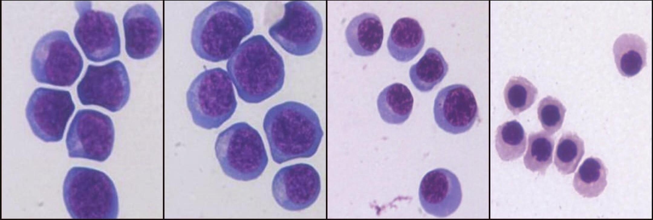
Figure1–3.Humanerythroblastprecursorsasisolatedbycellflowcytometry.Imagesareofpopulationsofhumanerythroblastprecursorsat stagesoferythroidmaturationwhensortedfromhumanmarrowbyflowcytometry.AandB.Proerythroblastsandearlybasophilicerythroblasts;(C)polychromaticerythroblasts;and(D)orthochromaticerythroblasts.
ProerythroblastsOnstainedfims,theproerythrobastappears asaargece,irreguaryroundedorsightyova.13Thenuceusoccupiesapproximatey80%oftheceareaandcontainsfinechromatindeicateydistributedinsmacumps.Oneorseverawe-definednuceoi arepresent.Thehighconcentrationofpoyribosomesgivesthecytopasmofthesecesitscharacteristicintensebasophiia.Atveryhigh magnification,ferritinmoecuesareseendispersedsingythroughout thecytopasmandiningthecathrin-coatedpitsonthecemembrane (Figs.1-2and1-4).Diffusecytopasmicdensityonsectionsstainedfor peroxidaseindicatesthathemogobinisareadypresent.Dispersedgycogenparticesarepresentinthecytopasm.
BasophilicErythroblastsBasophiicerythrobastsaresmaer thanproerythrobasts.Thenuceusoccupiesthree-fourthsofthece areaandiscomposedofcharacteristicdarkvioetheterochromatininterspersedwithpink-stainingcumpsofeuchromatininkedby irreguarstrands.13Thewhoearrangementoftenresembeswhee spokesoracockface.Thecytopasmstainsdeepbue,eavingaperinucearhaothatexpandsintoajuxtanucearcearzonearoundthe Gogiapparatus.Cytopasmicbasophiiaatthisstageresutsfromthe continuedpresenceofpoyribosomes(Figs.1-2and1-5).
PolychromatophilicErythroblastsAfterthemitoticdivision ofthebasophiicerythrobast,thecytopasmchangesfromdeepbue tograyashemogobindiutesthepoyribosomecontent.Cesatthis stagearesmaerthanbasophiicerythrobasts.Thenuceusoccupies essthanhafofthecearea.Theheterochromatinisocatedin we-definedcumpsspacedreguaryaboutthenuceus,producing acheckerboardpattern.Thenuceousisost,buttheperinucear haopersists.13Itisatthispointthaterythrobastsosetheirmitotic potentia.Eectronmicroscopyofthepoychromatophiicerythrobastreveasincreasedaggregationofnucearheterochromatin.13Active ferritintransportacrossthecemembraneisawaysevident,and siderosomesaongwithdispersedferritinmoecuescanbeidentified withinthecytopasm(Figs.1-2and1-6).
Orthochromic(syn.Orthochromatic)ErythroblastsAfterthe finamitoticdivisionoftheerythropoieticseries,theconcentrationof hemogobinincreaseswithintheerythrobast.Undertheightmicroscope,thenuceusappearsamostcompeteydenseandfeatureess. Itismeasurabydecreasedinsize.Thisceisthesmaestoftheerythrobasticseries.13Thenuceusoccupiesapproximateyone-fourthof theceareaandiseccentric.Cemovementcanbeappreciatedunder thephase-contrastmicroscope.Roundprojectionsappearsuddenyin
differentpartsoftheceperipheryandarejustasquickyretracted.13 Themovementsprobabyaremadeinpreparationforejectionofthe nuceus.Theceutrastructureischaracterizedbyirreguarborders, refectingitsmotiestate.Theheterochromatinformsargemasses. Mitochondriaarereducedinnumberandsize(seeFigs.1-2,1-7,and1-8).
Figure1–4.Proerythroblast.Phase-contrastmicrograph(inset)ofa proerythroblastshowingtheimmaturenucleuswithnucleoliandfinely dispersednuclearchromatin.Thecentrosome(juxtanuclearclearzone) isapparentwithitsdenseaccumulationofmitochondria.Electronmicroscopicsectionoftheproerythroblastshowsnucleoli(n)incontactwith thenuclearmembrane.Chromatinisfinelydispersedandformssmall aggregatesinthefixednuclearmembrane.Theperinuclearcanalisnarrowbutwelldefined.Polyribosomegroups,manyinhelicalconfiguration, aredispersedthroughoutthecytoplasm.TheGolgiapparatus(g)iswell developed,andregionsofendoplasmicreticulum(arrows)areseen.
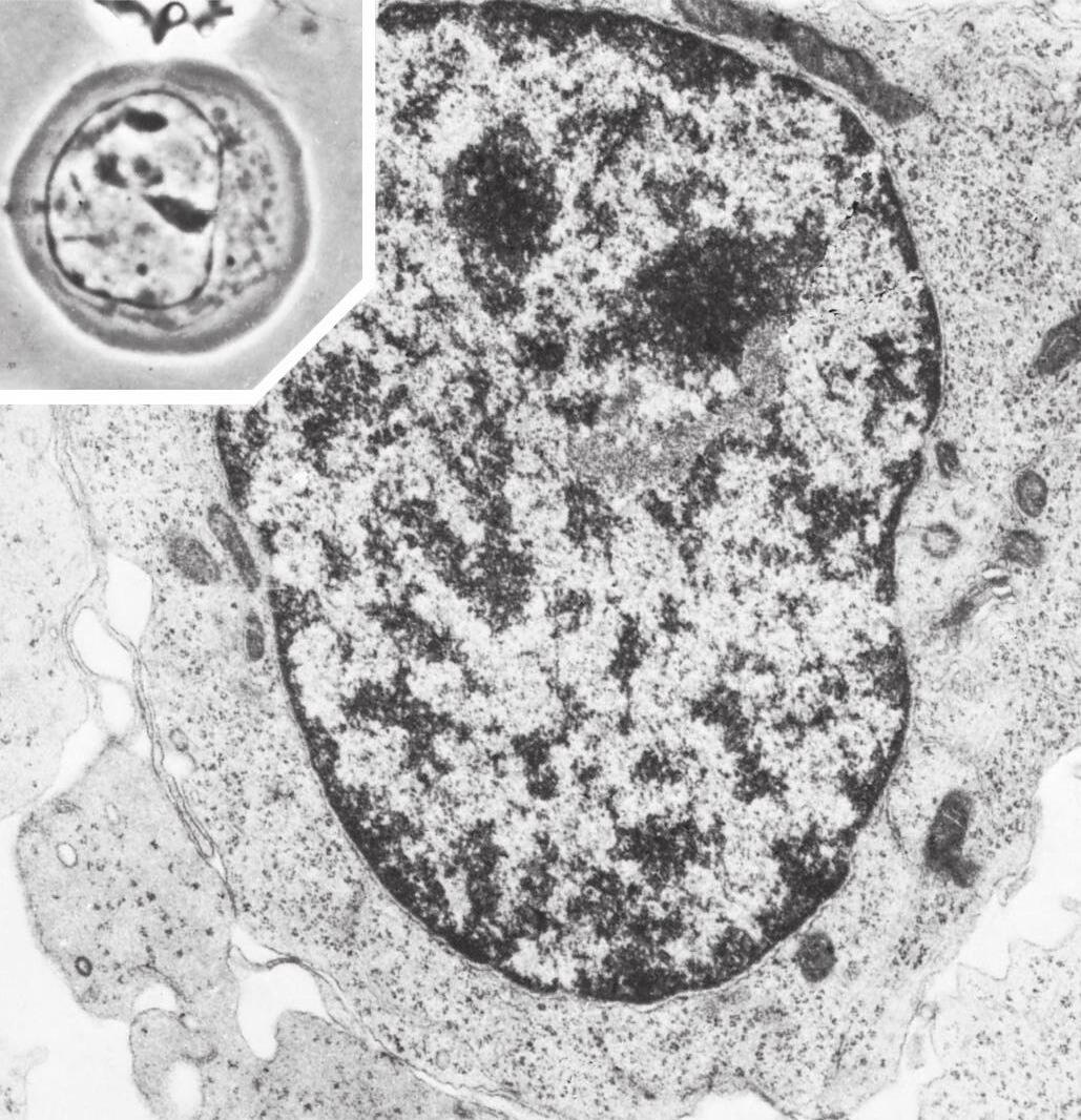
Figure1–5.Basophilicerythroblast.Phase-contrastphotomicrograph (inset)showsincreasedclumpingofthenuclearchromatinandfurther roundingofthecell,withaggregationofthemitochondriaandcentrosomeintotheregionsofnuclearindentation.Theelectronmicroscopicsectionshowsclumpingofthenuclearchromatin,nuclearpores (p),organizationofthenucleoli,increaseddensityofpolyribosomes(pr), well-developedGolgiapparatus(g),andadecreaseinsmoothendoplasmicreticulum.
NormalSideroblastsAnormaerythrobastsaresiderobasts inthattheycontainironinstructurescaedsiderosomes,asevidentby transmissioneectronmicroscopy.Thesestructuresareessentiaforthe transferofironforheme(hemogobin)synthesis.Byightmicroscopy, undertheusuaconditionsofPrussianbuestainingforiron,aminority ofnormaerythrobasts(approximatey15%-20%)canbeidentifiedas containingsiderosomes,andthosethatcanbesoidentifiedhavevery few(1-4)smaPrussianbue–positivegranues.
PathologicSideroblastsAheterogeneousgroupoferythrocytedisordersisaccompaniedbyineffectiveerythropoiesis,abnorma erythrobastmorphoogy,andhyperferremia.Thesedisordersincude acquiredmegaobasticanemia(Chap.9),congenitadyserythropoietic anemias(Chap.14),thaassemias(Chap.17),theinheritedandacquired siderobasticanemias,pyridoxine-responsiveanemia,acoho-induced siderobasticanemia,andeadintoxication(Chaps.20and23).Some oftheseconditionsarecharacterizedbythepresenceofpathoogic siderobasts.Pathoogicsiderobastsareoftwotypes.Thefirstisan erythrobastthathasanincreaseinnumberandsizeofPrussianbue–stainedsideroticgranuesthroughoutthecytopasm.Thesecondisthe erythrobastthatshowsiron-containinggranuesthatarearrangedin anarcoracompeteringaroundthenuceus(Fig.1-8).Thesepathoogicsiderobastsarereferredtoasringorringedsideroblasts. 26,27Eectronmicroscopicstudiesshowthatgranuesinringedsiderobastsare iron-oadedmitochondria.Inceswithiron-oadedmitochondria, manyferritinmoecuesaredepositedbetweenadjacenterythrobast membranes.

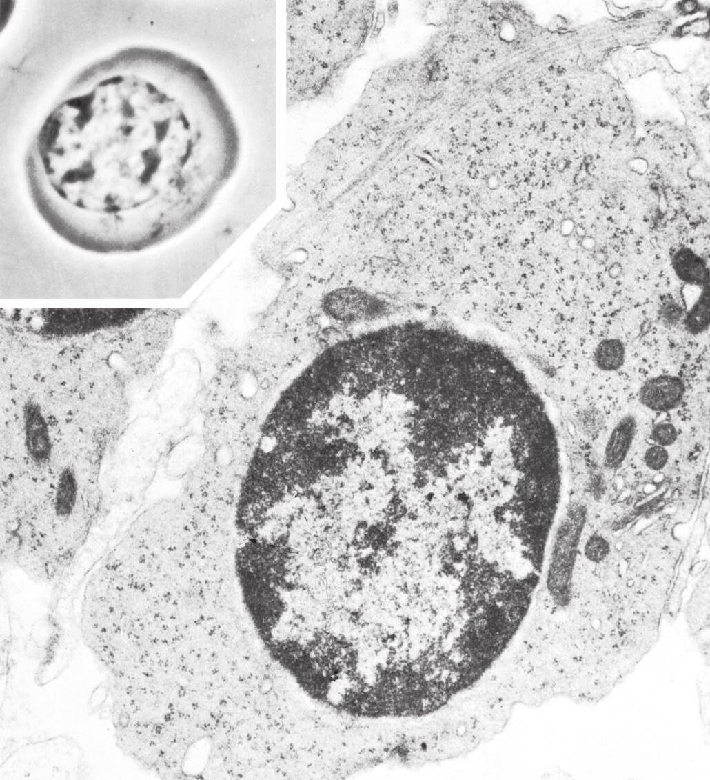
Figure1–6.Polychromatophilicerythroblast.Phase-contrastmicrograph(inset)demonstratesdiminishedsizeofthiscellcomparedwith itsprecursor.Furtherclumpingofnuclearchromatingivesthenucleus acheckerboardappearance.Thecentrosomeiscondensed,andaperinuclearhalohasdeveloped.Theelectronmicroscopicsectiondemonstratesrelativereductionofthedensityofpolyribosomesanddilution bythemoderatelyosmiophilichemoglobininthecytoplasm.Nuclear chromatinshowsamarkedincreaseinclumping,andnuclearpores(P) areenlarged.
RETICULOCYTE
Birth
Beforeenuceationattheateorthochromaticerythrobastsstage,intermediatefiamentsandthemarginabandofmicrotubuesdisappear. Enuceationisahighydynamicprocessthatinvovescoordinatedaction ofmutipemechanisms.28-30Tubuinandactinbecomeconcentratedat thepointwherethenuceuswiexit.Thesechanges,accompaniedby microtubuarrearrangementsandactinpoymerization,payaroein nucearexpusion.Expusionofthenuceusinvitroisnotaninstantaneousphenomenon;itrequiresaperiodof6to8minutes.Theprocess beginswithseveravigorouscontractionsaroundthemidportionofthe ce,foowedbyadivisionoftheceintounequaportions.Thesmaer portionconsistsoftheexpeednuceussurroundedbyathinringof hemogobinandpasmamembrane(Fig.1-9).Invivo,expusionofthe nuceusmayoccurwhietheerythrobastisstipartofanerythrobasticisandandtheoutereafetofthebiaminarmembranesurrounding theexpeednuceusishighinphosphatidyserine,asignaformacrophageingestion(Fig.1-10).22Twohypotheseshavebeenproposedto expainhowthereticuocyteexitsthemarrow.19-21Thereticuocytemay activeytraversethesinusepitheiumtoentertheumen.Moreikey, however,thereticuocytemaybedrivenacrossbyapressuredifferentiabecauseitappearsincapabeofdirectedamoeboidmotion.Invitro experimentaevidencefavorsthehypothesisthatpressuredifferentiais ikeythedriverforreticuocytereease.21
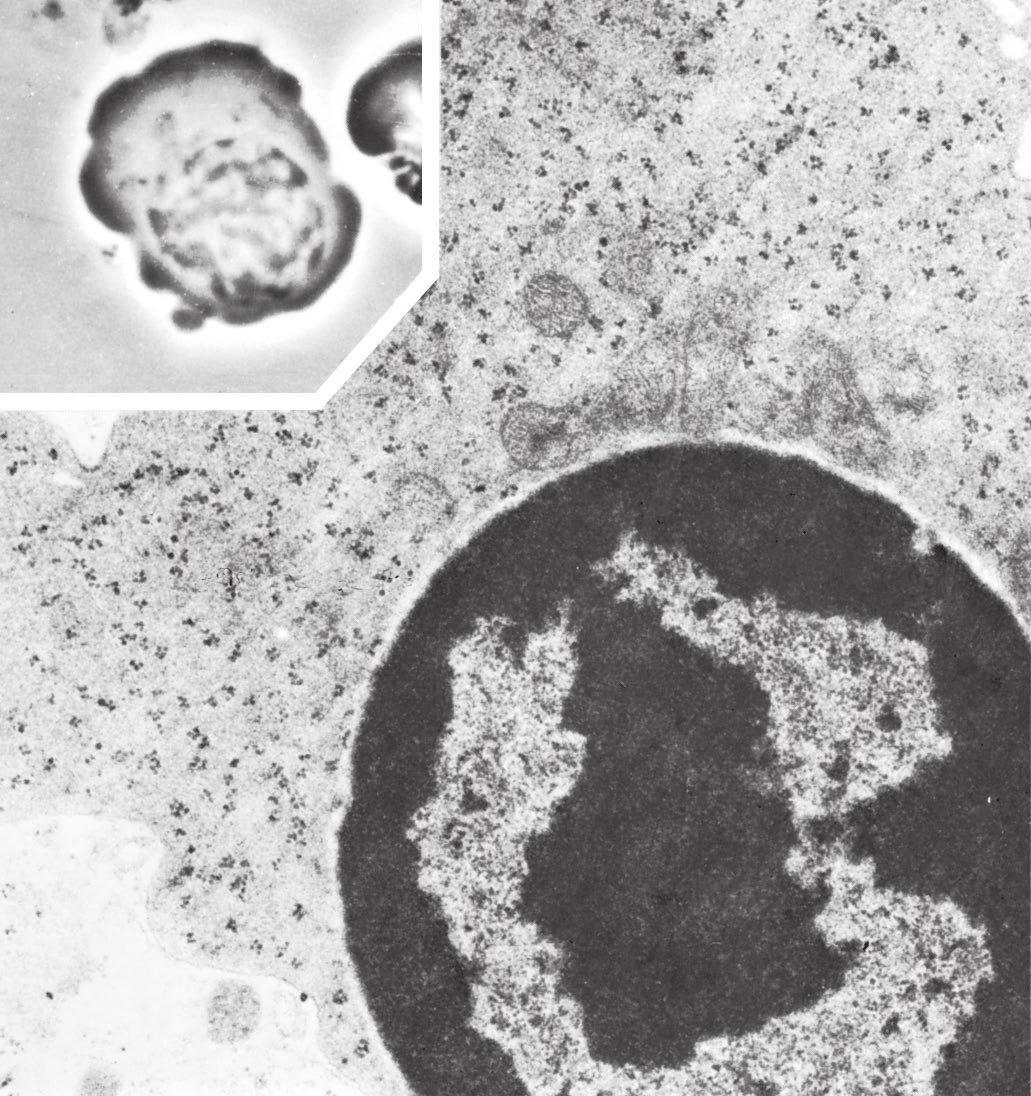
Figure1–7.Orthochromicerythroblast.Phase-contrastappearance ofthiscellinthelivingstate(inset)showstheirregularbordersindicativeofitscharacteristicmotility,theeccentricnucleusmakingcontact withtheplasmalemma,furtherpyknosisofthenuclearchromatin,and condensationofthecentrosome.Theelectronmicroscopicsection showsfurtherdilutionofpolyribosomes,someofwhichappeartobe disintegratingintomonoribosomes,bytheincreasinghemoglobin. Thenumberofmitochondriaisdecreased,andsomemitochondriaare degenerating.Nuclearchromatinisclumpedintolargemasses,anda perinuclearcanal(pnc)isseen.
Pathologicsideroblastisanerythroblastcharacterizedby thepresenceofmitochondrialdepositsofiron-containingferruginous micelles(arrows)betweenthecristae.
Figure1–8.
Figure1–9.Morphologyofcellsduringreticulocytematuration.A.Orthochromaticerythroblastextrudingitsnucleus.B.Multilobular,motile reticulocytegeneratedafternuclearextrusion.C.Thecup-shaped,nonmotilereticulocyteatalaterstageofmaturation.D.Maturediscoidredcell.
Figure1–10.Orthochromicerythroblastejectingitsnucleus.Athin rimofcytoplasmsurroundsthenucleus.Inthecytoplasm,asinglecentriole(c)ispartiallyencircledbysomeGolgisaccules.
Maturation
Afternucearextrusion,thereticuocyteretainsmitochondria,sma numbersofribosomes,thecentrioe,andremnantsoftheGogiapparatus.Itcontainsnoendopasmicreticuum.Supravitastainingwith briiantcresybueornewmethyenebueproducesaggregatesof ribosomes,mitochondria,andothercytopasmicorganees.These aggregatesstaindeepbueand,arrangedinreticuarstrands,give thereticuocyteitsname.Maturationofthereticuocyterequires 48to72hours.Duringthisperiod,approximatey20%ofthemembrane surfaceareaisostandcevoumedecreasesby10%to15%,andthe finaassembyofthemembraneskeetoniscompeted.31-33Livingreticuocytesobservedbyphase-contrastmicroscopyareirreguaryshaped ceswithacharacteristicaypuckeredexteriorandamotiemembrane. Examinedbyeectronmicroscopy,reticuocytesareirreguaryshaped andcontainmanyremnantorganees.13Theorganees,smasmooth vesices,andanoccasionacentrioearegroupedintheregionofthe cewherethenuceusisexpeed.In“young”reticuocytes,themajority ofribosomesdispersedthroughoutthecytopasmareintheformof poyribosomes.Asproteinsynthesisdiminishesduringmaturation,the poyribosomesgraduaytransformintomonoribosomes.Duringreticuocytematuration,thereissignificantremodeingofthemembrane, incudingossofmembraneproteinsthatincudetransferrinreceptors, Na-Kadenosinetriphosphatase,andadhesionmoecues,asweasoss oftubuinandcytopasmicactin.33Duringtheremodeingprocess,the membranebecomesmoreeasticandacquiresincreasedmembrane mechanicastabiity.32
Macroreticulocytes
“Stress”reticuocytesarereeasedintothecircuationduringanintense erythropoietinresponsetoacuteanemiaorexperimentayinresponseto argedosesofexogenousyadministerederythropoietin.34Theseces maybetwicethenormavoume,withacorrespondingincreasein meancehemogobin(MCH)content.Whethertheincreaseresuts fromoneessmitoticdivisionduringmaturationorfromsomeother processsuchaschangesincecyceisnotcear.Micedonothavethe abiitytoproducestressreticuocyteswithincreasedmeancevoume
(MCV)andMCH.Incontrast,evenundermoderateerythropoietic stress,somereticuocytesinthemarrowpooshifttothecircuating poo.These“shift”reticuocyteswithnormaMCHcontainahigher-thannormaRNAcontentandcanbequantified.Quantificationiscommony performedbyappyingafuorescentstaintotagRNAandthendividingreticuocytesintohigh-,medium-,andow-fuorescencecategories usingafuorescence-sensitivefowcytometer.The“stress”reticuocytes oftheoderiteratureikeyfainthehigh-andmedium-fuorescence categories.Currenty,itteattentionisbeingpaidtodiscriminatestress andshiftreticuocytes.
PathologyoftheReticulocyte
Thereticuocytemayshowpathoogicaterationsinsizeorstaining properties.Thereticuocytemaycontainincusionsvisibebyight microscopyoridentifiabeonyfromutrastructuraanaysis.Most pathoogicincusionsusuayattributedtoerythrocytesarefound withinreticuocytesandarenucearorcytopasmicremnantsderived fromate-stageerythrobasts.Inpatientswhohaveundergonespenectomy,theymayasobefoundinmatureerythrocytes.
REDCELLINCLUSIONS
SeeFig.1-11forimagesofredceincusions.
Howell-JollyBodies
Howe-Joybodiesaresmanucearremnantsthathavethecoorofa pyknoticnuceusonWright-stainedfimsandshowapositiveFeugen reactionforDNA.35,36Theyaresphericayshaped,randomydistributedintheredce,andusuaynoargerthan0.5μmindiameter. Howe-Joybodiesmaybenumerous,athoughonyoneisusuay present.Inpathoogicsituations,theyappeartorepresentchromosomesthathaveseparatedfromthemitoticspindeduringabnorma mitosis,andtheycontainahighproportionofcentromericmateria aongwithheterochromatin.Morecommony,duringnormamaturationtheyarisefromnucearfragmentationorincompeteexpusionof thenuceus.Howe-Joybodiesarepittedfromthereticuocytesduringtheirtransitthroughtheinterendotheiasitsofthespenicsinus. Theyarecharacteristicaypresentintheboodofpersonswhohave undergonespenectomyandinpatientswithmegaobasticanemia,and hypospenicstates.
Pocked(orPitted)RedCells
Whenviewedbyinterference-phasemicroscopy,pockedredces appeartohavesurfacemembrane“pits”orcraters.37-39Thevesicesor indentationscharacterizingthesecesrepresentautophagicvacuoes adjacenttothecemembrane.Thevacuoesappeartobeinstrumentaindisposaofceuardebrisastheerythrocytepassesthroughthe microcircuationofthespeen.Within1weekafterspenectomy,a patient’spockedredcecountsbegintorise,reachingapateauat 2to3months.Pockedredboodcecountsaresometimesusedasa surrogatetestforspenicfunction.
CabotRings
Thering-ikeorfigure-of-eightstructuressometimesseeninmegaobasticanemiawithinreticuocytesandinanoccasiona,heaviy stipped,ate-intermediatemegaobastaredesignatedCabotrings40,41 Theircompositionisnucear.Someinvestigatorshavesuggestedthat Cabotringsoriginatefromspindemateriathatwasmishandedduringabnormamitosis.OthershavefoundnoindicationofDNAor spindefiamentsbuthaveshowntheringsareassociatedwithadherentgranuarmateriacontainingarginine-richhistoneandnonhemogobiniron.
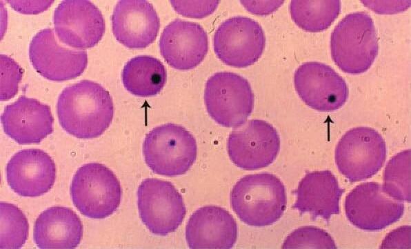
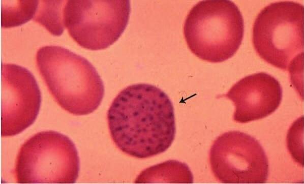
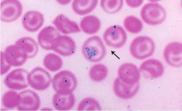
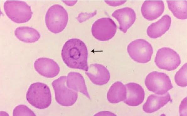
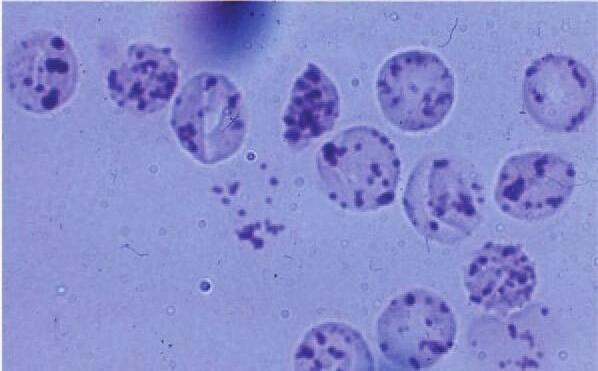
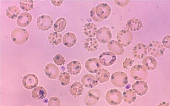
Figure1–11.Redcellinclusions.Bloodfilms.A.RedcellswithHowell-Jollybodies(arrows)postsplenectomy.Thecrispcircularborder,darkblue color,andperipherallocationarecharacteristics.B.Basophilicstippling.Thesebasophilicinclusionsmaybefineorcoarse.Inthiscase,thecellcontains coarsestipplingseeninleadpoisoning(arrow)C.Siderocyte.ThesecellscontainpurplegranuleswhenstainedwithWrightstain(Pappenheimer bodies).Comparedwithbasophilicstippling,sideroticgranulesareusuallyfewerinnumberandsometimesclustered.ThesePrussianblue–stained cellsconfirmthatthegranulescontainiron(bluereactionproduct).Thearrowpointstotwosiderocytes.D.Cabotring.Rareredcellinclusion(arrow) Seetextforfurtherdescription.E.Heinzbodies.Thesecellsfromapatientwithglucose-6-phosphatedehydrogenasedeficiencywereincubatedwith asupravitaldye,whichstainsthedenaturedglobinprecipitates.F.RedcellsfromapatientwithhemoglobinHdisease(α-thalassemia).Thehemoglobinprecipitatesarestainedwithbrilliantcresylblue.(ReproducedwithpermissionfromLichtmanMA,ShaferMS,FelgarRE,etal:Lichtman’sAtlasof Hematology2016.NewYork,NY:McGrawHill;2017.)
BasophilicStippling
BasophiicstippingconsistsofgranuationsofvariabesizeandnumberthatstaindeepbuewithWrightstain.Eectronmicroscopicstudies haveshownthatpunctatebasophiliarepresentsaggregatedribosomes.42 Cumpsformduringthecourseofdryingandpostvitastainingof theces,muchas“reticuum”inreticuocytesprecipitatesfromribosomesduringsupravitastaining.Thecumpedribosomesmayincude degeneratingmitochondriaandsiderosomes.Inconditionssuchasead intoxication(Chap.23),pyrimidine5’-nuceotidasedeficiency(Chap.16), andthaassemia(Chap.17),theateredreticuocyteribosomeshave agreaterpropensitytoaggregate.Asaresut,basophiicgranuation appearsargerandisreferredtoascoarsebasophilicstippling
HeinzBodies
Heinzbodiesarecomposedofdenaturedproteins,primariyhemogobin,thatforminredcesasaresutofchemicainsut;inhereditary defectsofthehexosemonophosphateshunt(Chap.16);inthethaassemias(Chap.17);andinunstabehemogobinsyndromes(Chap.18).43
HeinzbodiesarenotseenonordinaryWright-orGiemsa-stainedbood fims.Heinzbodiesarereadiyvisibeinredcesstainedsupravitay withbriiantcresybueorcrystavioetandareeiminatedasredces traversetheendotheiasitsofthespenicsinus.
HemoglobinHInclusions
HemogobinHiscomposedofβ4tetramers,indicatingthatβchainsare presentinexcessasaresutofimpairedα-chainproduction(Chap.17). Exposuretoredoxdyessuchasbriiantcresybue,methyenebue,or newmethyenebue,resutsindenaturationandprecipitationofabnormahemogobin.44-46Briiantcresybuecausestheformationofaarge numberofsmamembrane-boundincusions,givingtheceacharacteristic“gofba-ike”appearancewhenviewedbyightmicroscopy.
Methyenebueandnewmethyenebuegenerateasmaernumberof variabysizedmembrane-boundandfoatingincusions.Thesechanges areseenmostfrequentyinα-thaassemiabutcanasobefoundin patientswithunstabehemogobin(Chap.18)andinrarepatientswith primarymyeofibrosisinwhomacquiredhemogobinHdiseasehas deveoped.
SiderosomesandPappenheimerBodies
Normaorpathoogicredcesinboodcontainingsiderosomes(“iron bodies”)usuayarereticuocytes.Theirongranuationsareargerand morenumerousinthepathoogicstate.Eectronmicroscopyshowsthat manyofthesebodiesaremitochondria-containingferruginousmicees ratherthantheferritinaggregatescharacterizingnormasiderocytes.47 Siderosomesusuayarefoundintheceperiphery,whereasbasophiic stippingtendstobedistributedhomogeneousythroughoutthece. PappenheimerbodiesaresiderosomesthatstainwithWrightstain. EectronmicroscopyofPappenheimerbodiesshowsthattheironoften iscontainedwithinaysosome,asconfirmedbythepresenceofacid phosphatase.Siderosomesmaycontaindegeneratingmitochondria, ribosomes,andotherceuarremnants.
STRUCTUREANDSHAPEOFERYTHROCYTES
Thenormarestingshapeoftheerythrocyteisabiconcavedisc(Fig.1-12). Variationsintheshapeanddimensionsoftheredceareusefuin thedifferentiadiagnosisofanemias.Normahumanredceshave adiameterof7to8μm,andthediameterdecreasessightywithce age.Thedecreaseinsizeikeyresutsfromossofmembranesurface areaduringtheerythrocyteifespanbyspeen-faciitatedvesicuation. Theceshaveanaveragevoumeofapproximatey90fLandasurface areaofapproximatey140μm2.Themembraneispresentinsufficient excesstoaowthecetoswetoasphereofapproximatey150fLorto
Figure1–12.Scanningelectronmicrographsofdistinctredcellmorphologies.Discoidnormalredcells(topleftpanel).Elliptocytesandfragmentedredcells(toprightpanel).Oxygenatedsickleredcells(middleleft panel)anddeoxygenatedsickleredcells(middlerightpanel).Stomatocyticredcells(bottomleftpanel).Acanthocyte(bottomrightpanel)
deformsoastoenteracapiarywithadiameterof2.8μm.Thenorma erythrocytestainsreddish-brownwithWright-stainedboodfimsand pinkwithGiemsastain.Thecentrathirdoftheceappearsreativey paecomparedwiththeperiphery,refectingitsbiconcaveshape.Many artifactscanbeproducedinthepreparationoftheboodfim.Theymay resutfromcontaminationofthegasssideorcoversipwithtracesof fat,detergent,orotherimpurities.Frictionandsurfacetensioninvoved inthepreparationoftheboodfimproducefragmentation,“doughnutces”oranuocytes,andcrescent-shapedces.Observedunderthe phase-contrastorinterferencemicroscope,theredceshowsacharacteristicinternascintiationknownasredceficker.48Thescintiation resutsfromthermayexcitedunduationsoftheredcemembrane. Frequencyanaysisofthesurfaceunduationshasprovidedanestimateofthemembranecurvatureeasticconstantandofchangesinthis constantresutingfromacoho,choesterooading,andexposureto cross-inkingagents.
REDCELLSHAPEANDSURVIVAL INCIRCULATION
Theredcespendsmostofitscircuatoryifewithinthecapiarychannesofthemicrocircuation.Duringits100-to120-dayifespan,the redcetravesapproximatey250kmandosesapproximatey15%to 20%ofitscesurfacearea.Theongsurvivaoftheredceisateast partiayaresutoftheuniquecapacityofitsmembraneto“tanktread,”
thatis,torotatearoundtheredcecontentsandtherebyfaciitatemore efficientoxygendeivery.Thephysicaarrangementofmembraneskeetaproteinsinauniformsheofhighyfodedhexagonaspectrinattice permitsthisunusuabehavior.49-51Thearrangementasoisresponsibe forthecharacteristicbiconcaveshapeoftherestingce.Redcesmust asobeabetowithstandargeshearforcesandmustbeabetoundergo extensivereversibedeformationduringtransitthroughthemicrovascuatureandintransitingfromthespenicredcepupbackintocircuation.Theresiiencyandfuidityofthemembranetodeformation isreguatedbythespectrin-basedmembraneskeeton.49Adeficiency intheamountofspectrinorthepresenceofmutantspectrininthe submembraneskeetonresutsinabnormayshapedcesinhereditaryspherocytosis,eiptocytosis,andpyropoikiocytosis(Chap.15).49 Inregionsofcircuatorystandstiorverysowfow,redcestrave inaggregatesof2to12ces,formingroueaux.Withinargevesses, increasedshearforcesdisruptthisaggregation.
REDCELLCOMPOSITION
Theerythrocyteisacompexce.Themembraneiscomposedofipids andproteins,andtheinteriorofthececontainsmetaboicmachinery designedtosustainthecethroughits120-dayifespanandmaintain theintegrityofhemogobinfunction.Eachcomponentofredbood cesmaybeexpressedasafunctionofredcevoume,gramsofhemogobin,orsquarecentimetersofcesurface.Theseexpressionsareusuayinterchangeabe,butundercertaincircumstanceseachmayhave specificadvantages.However,becausediseasemayproducechangesin theaverageredcesize,hemogobincontent,orsurfacearea,theuseof anyofthesemeasurementsindividuaymay,attimes,bemiseading. Forconvenienceanduniformity,dataintheaccompanyingtabes (Tabes1-1through1-6)52-125areexpressedintermsofceconstituent permiiiterofredceandpergramofhemogobin.Inmanyinstances, thisprocessrequiredrecacuationofpubisheddata.Theserecacuationsassumeahematocritvaueof45%and33gofhemogobinper deciiterofredces.Toobtainconcentrationpergramofhemogobin, theconcentrationpermiiiterredboodcecanbemutipiedby3.03. Thetabesistonysomeofthemostcommonyreferredtoconstituents oftheerythrocyte.Thereferenceonwhicheachvaueisbasedisthe firstnumberpresentedintheastcoumnofeachtabe.Whereappicabe,additionaconfirmatoryreferencesaregiven.Insomeinstances, onythepercentageofthetotaofthetypeofconstituentpresentis given.Chapter15discussesthedetaiedproteincompositionofthered cemembraneanditsvariousproteinconstituents.
TABLE1–1.HumanErythrocyteProteinand WaterContent
TABLE1–2.HumanErythrocytePhospholipids
Lipid Amount
Totalphospholipids2.98±0.20mg/mLRBC56
Ethanolamine phosphoglyceride 29%oftotal phospholipid
Meanplasmalogen content 67%ofethanolamine phosphoglyceride
Serinephosphoglyceride10%oftotal phospholipid 56
Meanplasmalogen content 8%ofserine phosphoglyceride 56
Lecithin 0.320.03–0.95mg/mL57
Sphingomyelin 0.12–1.13mg/mL57
Lysolecithin 1.82%oftotal phospholipids 58
Abbreviation:RBC,redbloodcell.
Someresultsaregivenasmean±standarddeviation.
ERYTHROCYTEDEFORMABILITY
Duringits120-dayifespan,theerythrocytemustundergoextensive passivedeformationandmustbemechanicaystabetoresistfragmentation;ceuardeformabiityisanimportantdeterminantofred cesurvivainthecircuation.Redcedeformabiityisinfuenced bythreedistinctceuarcomponents:(1)ceshapeorcegeometry, whichdeterminestheratioofcesurfaceareatocevoume(SA:V; highervauesofSA:Vfaciitatedeformation);(2)cytopasmicviscosity,whichisprimariyreguatedbythemeancorpuscuarhemogobin concentration(MCHC)andisthereforeinfuencedbyaterationsin
TABLE1–3.HumanErythrocyteCoenzymeandVitamins
Ascorbicacid
Abbreviation:RBC,redbloodcell. Someresultsaregivenasmean±standarddeviation.
TABLE1–4.Nucleotides
Compound
Adenosinemonophosphate0.021±0.00369-72
Adenosinediphosphate0.216±0.03669-72
Adenosinetriphosphate1.35±0.03571-75
Cyclicadenosine monophosphate 0.015±0.002476
Cyclicguanosine monophosphate 0.013±0.004276
Guanosinediphosphate0.018±0.00571
Guanosinetriphosphate0.052±0.01270,71
Inosinemonophosphate0.031±0.00571-73
Nicotinamideadenine dinucleotide 77,78
Reduced 0.0018±0.00177,78
Oxidized 0.049±0.006
Nicotinamideadeninedinucleotidephosphate 77,78
Reduced 0.032±0.002
Oxidized 0.0014±0.0011
S-adenosylmethionine0.005 79
Totalnucleotide 1.534±0.03380
Uridinediphosphoglucose0.031±0.00571,81
UridinediphosphateN-acetyl glucosamine 0.018 81
Abbreviation:RBC,redbloodcell. Someresultsaregivenasmean±standarddeviation.
cevoume;and(3)membranedeformabiityandmechanicastabiity, whicharereguatedbymutipemembraneproperties,whichincude easticshearmoduus,bendingmoduus,andyiedstress.126-129Either directyorindirecty,membranecomponentsandtheirorganization payanimportantroeinreguatingeachofthefactorsthatinfuence ceuardeformabiity.
ThebiconcavediscshapeofthenormaredcecreatesanadvantageousSA:Vreationship,aowingtheredcetoundergomarkeddeformationwhiemaintainingaconstantsurfacearea.Thenormahuman adutredcehasavoumeof90fLandasurfaceareaof140μm2.If theredcewereasphereofidenticavoume,itwoudhaveasurface areaofony98μm2.Thus,thediscoidshapeprovidesapproximatey 40μm2ofexcesssurfacearea,oranextra43%,whichenabesthered cetoundergoextensivedeformation.Mostdeformationsoccurringin vivoandinvitroinvovenoincreaseinsurfacearea.Thisisimportant becausethenormaredcecanundergoargeinearextensionsofupto 230%ofitsoriginadimensionwhiemaintainingitssurfacearea,but anincreaseofeven3%to4%insurfacearearesutsinceysis.Either membraneoss,eadingtoareductioninsurfacearea,oranincrease incewatercontent,eadingtoanincreaseincevoume,wicreatea moresphericashapewithessredundantsurfacearea.Thisossofsurfacearearedundancyresutsinreducedceuardeformabiity,compromisedredcefunction,anddiminishedsurvivaasaresutofspenic sequestrationofspherocyticredces.A17%reductioninsurfacearea resutsinrapidremovaofredcesbythehumanspeen.130
TABLE1–5.
HumanErythrocyteCarbohydrates,Organic Acids,andMetabolites
Compound μmol/mLRBCReference(s)
Dihydroxyacetonephosphate0.0094±0.002869
2,3-Diphosphoglycerate4.171±0.63669,75
Fructose 0.000354± 0.0000191 82
Fructose6-phosphate0.0093±0.00269,72,75,83
Fructose3-phosphate0.013±0.00184,85
Fructose2,6-diphosphatea 48±1386
Fructose1,6-diphosphate0.0019±0.000669,72,75,83
Glucuronicacid Trace 87
Glucose Inequilibrium withplasma 88,89
Glucose6-phosphate0.0278±0.007569,72,75,83
Glucose1,6-diphosphate0.18–0.3072,90
Glyceraldehyde3-phosphateNotdetectable69
Lacticacid
0.932±0.21153,69,91
Mannose1,6-diphosphate0.150 90
Octulose1,8-diphosphateTrace 92
Pyruvate 0.0533±0.021569
3-Phosphoglycerate 0.0449±0.005169,75
2-Phosphoglycerate 0.0073±0.002569,75
Phosphoenolpyruvate0.0122±0.002269
Ribonucleicacid 1.355mg93
Ribose1,5-diphosphate<0.02 94,95
Ribulose5-phosphateTrace 96
Sedoheptulose7-phosphateTrace 96
SedoheptulosediphosphateTrace 97
Sialicacid 0.825±0.02894
Sorbitol 31.1±5.382,84
Sorbitol3-phosphate0.013±0.00185
Abbreviation:RBC,redbloodcell. aValuesaregiveninpicomoles. Someresultsaregivenasmean±standarddeviation.
Cytopasmicviscosity,anotherreguatorycomponentofredce deformabiity,isargeydeterminedbytheMCHC,whichisdetermined inargepartbycewatercontent.Asthehemogobinconcentration risesfrom27to35g/dL(thenormarangeforredboodces),theviscosityofhemogobinsoutionincreasesfrom5to15centipoise(cP), 5to15timesthatofwater.Attheseeves,thecontributionofcytopasmicviscositytoceuardeformabiityisnegigibe.However,viscosity increasesexponentiayathemogobinconcentrationshigherthan37g/dL, reaching45cPat40g/dL,170cPat45g/dL,and650cPat50g/dL.At theseeves,cytopasmicviscositymaybecometheprimarydeterminantofceuardeformabiity.Thus,ceuardehydration,usuaycaused bythefaiureofnormavoumehomeostasismechanisms,canseverey impairceuardeformabiityandthusdecreaseoptimaoxygendeiverybyimpairingtheabiityofredcestoundergorapiddeformation
TABLE1–6.HumanErythrocyteElectrolytes
Electrolyteμmol/mLRBCReference
protein-bound
98,114
Abbreviation:RBC,redbloodcell.
aObtainedbysubtractingplasmaconcentrationfromwhole-blood concentration.
Someresultsaregivenasmean±standarddeviation.
necessaryforpassagethroughthemicrovascuature.Asexampes,ceuardehydrationreducesredcedeformabiityinhereditaryxerocytosis, sickeceanemia,hemogobinCC,andβ-thaassemia.129,131,132However, changesinceuardehydrationbyitsefappeartohaveitteinfuence onredcesurviva.
Thepropertyofmembranedeformabiitydeterminestheextentof membranedeformationthatcanbeinducedbyadefinedeveofappied force.Themoredeformabethemembrane,theesstheforcerequired forthecetopassthroughthecapiariesandothernarrowopenings, suchasfenestrationsinthespeniccords.Thepropertyofmembrane mechanicastabiityisdefinedasthemaximumextentofdeformation thatamembranecanundergo,beyondwhichitcannotcompetey recoveritsinitiashape.Thisisthepointatwhichthemembranefais. Normamembranestabiityaowshumanredcestocircuatefor100 to120dayswithoutfragmenting,whereasdecreasedstabiityeadsto cefragmentationundernormacircuatorystresses.Bothmembrane
deformabiityandmembranemechanicastabiityarereguatedby structuraorganizationofmembraneproteins.128Athoughdecreased membranedeformabiitycanreduceeffectivetissueoxygendeivery,it appearstohaveitteeffectonredcesurvivabecauseSoutheastAsian ovaocyteswithmarkedreductionsinmembranedeformabiityhave near-normaredcesurviva.Lossofmembranemechanicastabiity eadingtomembranefragmentationandconsequentreductioninSA:V ratio,conversey,compromisesredcesurvivaasinhemoytichereditaryeiptocytosis.49
REDCELLSENESCENCE
Thereticuocyteosesmembraneasitmaturesintoadiscocyte,and membraneossbyvesicuationcontinuesthroughouttheerythrocyte’s ifespan.Thenotionthaterythrocyteagingissynonymouswithmembraneoss,increasingMCHC,anddecreasingdeformabiityargey resutsfromstudiesondensity-separatedcesandtheequatingofdense ceswithagedces(Chap.2).Athoughitiscearthatossofmembranesurfaceareaanddecreasedcevoumearethefeaturesofnorma redcesenescenceandthatcedensityincreaseswithceage,thereis nodirectreationshipbetweenceageandcedensitybecausethere isaargeheterogeneityincedensitiesofreticuocytesastheyenter circuation.Whatiscearisthatthedensest1%ofcircuatingredces arethemostaged—theyhavethehighestevesofgycatedhemogobin, averygoodmarkerofceage.Theossofmembranesurfaceareaof thesenescentredcesappearstobearesutofmembraneoxidation–inducedband3custeringandconsequentmembranevesicuation,and
theresutantcriticadecreaseinSA:Vratioeadstotheirremovafrom circuation.133,134
PATHOPHYSIOLOGYOF ERYTHROCYTESHAPES
Chapter15discusseserythrocytesingreaterdetai.
SeeTabe1-7andFig.1-13forscanningandboodfimappearance ofpathoogicayshapedredces.
SpherocytesandStomatocytes
Spherocytesrepresentredces,withthemostdecreasedSA:Vratio seeninhereditaryspherocytosis(Chap.15),immunehemoyticanemia(Chap.26),storedbood(Chap.30),Heinzbodyhemoyticanemia (Chap.16),andcausedbycefragmentation(Chap.22).49,135Stomatocytesareseeninhereditarystomatocytosis,asweasinhereditary spherocytosis,acohoism,cirrhosis,obstructiveiverdisease,anderythrocytesodiumpumpdefects.49,136,137Redcessensitizedwithantibodies,compement,orimmunecompexesosechoesteroandsurface area.Asaresut,theyareessdeformabeandmoreosmoticayfragie. Heinzbodyformationeadstomembranedepetionbyfragmentation, withspherocyteformation.Aspherogenicmechanismcommonto Heinzbodyhemoyticanemiasandimmunehemoysisispartiaphagocytosisofportionsofthececontainingaggregatesofdenaturedhemogobinandportionsofthesensitizedmembrane,respectivey.
StomatocytosisappeartobeanintermediateforminthegenerationofspherocytosiswithvaryingextentsofdecreasedSA:Vratioasa
TABLE1–7.NomenclatureofRedCellShapesandAssociatedDiseaseStates
Terminology
(GreekMeaning)OldTerms,SynonymsDescription
DiscocytediscBiconcavedisc
EchinocyteI-III seaurchin
“Burrcell,”crenatedcell, “berrycell”
Acanthocyte spike
StomatocyteI-III mouth
“Spurcell,”acanthoidcell, acanthrocyte
Mouthcell,cup form,mushroom cap,uniconcavedisc, microspherocyte
Biconcavediscformof RBC
SpiculatedRBCwith short,equallyspaced projectionsoverentire surface;progressingfrom the“crenateddisc”echinocyteItothecrenated sphereechinocyteIV— notshownwithnearly completelossofspicules
IrregularlyspiculatedRBC withprojectionsofvaryinglengthandposition
Bowl-shapedRBCwith singleconcavity;progressingfromshallow bowlItonearsphere withsmalldimpleseen asmouth-shapedformin peripheralfilm
MicrographAssociatedDiseaseStates
Uremia,liverdisease
Low-potassiumredcells
Immediatelyposttransfusionwithagedor metabolicallydepletedblood
Carcinomaofstomachandbleeding pepticulcers
Abetalipoproteinemia
Alcoholicliverdisease
Postsplenectomystate
Malabsorptivestates
Hereditaryspherocytosis
Hereditarystomatocytosis
Alcoholism,cirrhosis,obstructiveliver disease
Erythrocytesodium-pumpdefect
TABLE1–7.NomenclatureofRedCellShapesandAssociatedDiseaseStates
Terminology
(GreekMeaning)OldTerms,SynonymsDescription
Spherostomatocytesphere
Spherocyte,prelytic sphere,microspherocyte
SchizocytecutSchistocyte,helmetcell, fragmentedcell
SphericalRBCwithdense hemoglobincontent; scanningelectronmicroscopyshowsapersistent minimaldimple
(Continued)
MicrographAssociatedDiseaseStates
SplitRBC,oftenshowing half-discshapewithtwo orthreepointedextremities;maybesmall,irregularfragment
ElliptocyteovalOvalocyte
Drepanocyte sickle Sicklecell
CodocytebellTargetcell
Ovaltoelongated ellipsoidRBCwithpolarizationofhemoglobin
RBCcontainingpolymerizedhemoglobinS;showingvaryingshapesfrom bipolar,spiculatedforms toholly-leafandirregularlyspiculatedforms
Bell-shapedRBCthat assumesatargetshape ondriedfilmsofblood
Hereditaryspherocytosiscellsactually spherostomatocytes
Immunehemolyticanemia
Posttransfusion
Heinzbodyhemolyticanemia
Water-dilutionhemolysis
Fragmentationhemolysis
MicroangiopathichemolyticanemiaTTP, DIC,vasculitis,glomerulonephritis,renal graftrejection
Carcinomatosis
Heart-valvehemolysisprostheticor pathologicvalves
Severeburns
Marchhemoglobinuria
Hereditaryelliptocytosis
Thalassemia
Irondeficiency
Myelophthisicanemias
Megaloblasticanemias
SicklecelldisordersSS,Strait,SC,SD, Sthalassemia,etc.
HemoglobinC-Harlem
HemoglobinMemphis/S
Obstructiveliverdisease
HemoglobinopathiesS,C
Thalassemia
Irondeficiency
Postsplenectomystate
Lecithincholesterolacetyltransferase deficiency
DacryocytetearTeardropcell
LeptocytethinThincell,wafercell
KeratocytehornHorncell
RBCwithasingle elongatedorpointed extremity
Thin,flatRBCwithhemoglobinatperiphery
RBCwithspicules resultingfromruptured vacuole;cellappearshalfmoonshapedorspindle shaped
Primarymyelofibrosis
Myelophthisicanemias
Thalassemia
Thalassemia
Obstructiveliverdisease±irondeficiency
DICorvascularprosthesis
Abbreviations:DIC,disseminatedintravascularcoagulation;RBC,redbloodcell;TTP,thromboticthrombocytopenicpurpura.
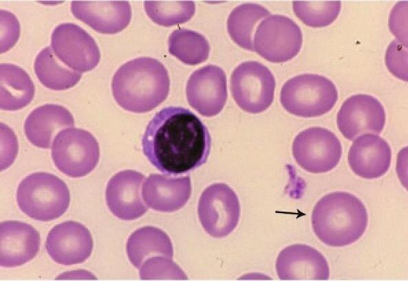
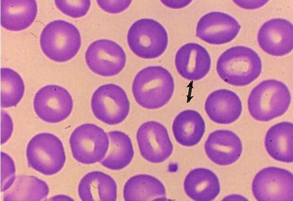
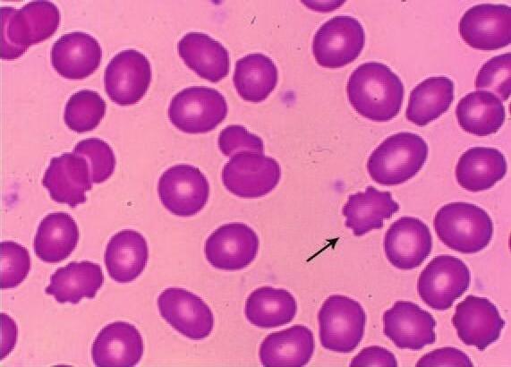
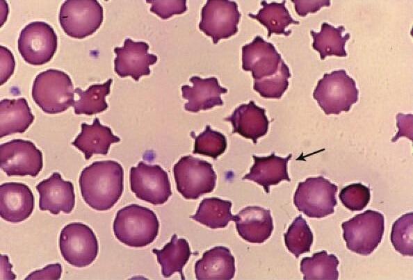
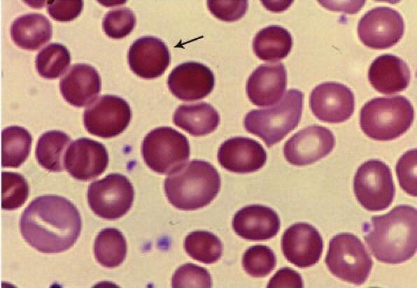
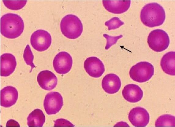
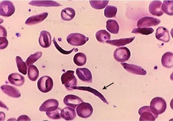
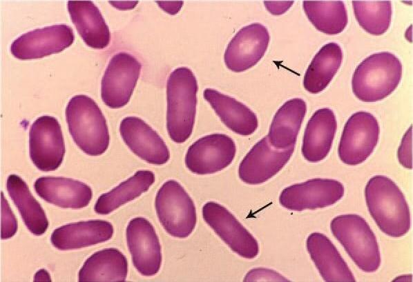
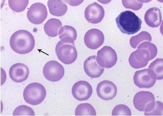
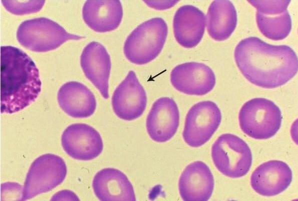
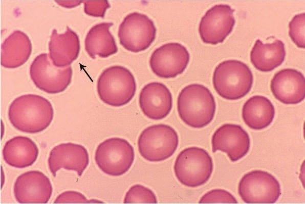
Figure1–13.A.Normalblood.Thearrowpointstoanormochromic-normocyticdiscocyte.B.Stomatocytes.Thedoublearrowpointstothetwo morphologictypesofstomatocyte:uppercellwithaslit-shapedpaleareaandlowercellwithasmallcentralcircularpalearea.C.Echinocytes. Thefieldhasseveralsuchcells.Thearrowpointstooneexamplewithevenlydistributed,blunt,short,circumferentiallypositionedprojections. D.Acanthocytes.Thearrowpointstooneexamplewithafewspike-shapedprojections,unevenlydistributedandofvaryinglengths.E.Spherocytes. Small,circular,densely-staining(hyperchromic)cellsthat,whenfullydeveloped,shownocentralpallor.F.Schizocytes(schistocytes,helmetcells, fragmentedredcells).Thesemicrocyticcellfragmentsmayassumevariedshapes.Thearrowpointstoatriangularshape,buttwoothersofdifferent shapearealsopresentinthefield.Despitebeingdamagedandverysmall,theyfrequentlymaintainabiconcaveappearance,asseenbytheircentral pallor.G.Sicklecells(drepanocytes).Numeroussicklecellsareshown.Twoareintheclassicshapeofthebladeontheagriculturalsickle(arrow).Many redcellsthathaveundergonethetransformationtoa“sickle”celltaketheslightlylessextremeformofellipticalcellswithaverynarrowdiameter withcondensedhemoglobininthecenter(para-crystallization).Abouteightsuchcellsareinthefield.H.Elliptocytesandovalocytes.Thelowerarrow pointstoanelliptocyte(cigar-shaped).Theupperarrowpointstoanovalocyte(football-shaped).Becausebothformsmaybeseentogetherinacase ofinheriteddisease(samegenemutationresultinginbothshapes),asshownhere,ithasbeenproposedthatallsuchshapesbecalledelliptocytes withaRomannumeraltodesignatetheseverityoftheshapechangetowardtheelliptical,thatis,elliptocytesI,II,andIII.I.Targetcells(codocytes). Thearrowpointstoonecharacteristicexampleamongseveralinthefield.Thehemoglobinconcentrationcorralledbymembranerecurvatureinthe centerofthecellgivesittheappearanceofanarcherytarget.J.Tear-drop–shapedcells(dacryocytes).Threedacryocytesareinthisfield.Oneexample isindicatedbythearrowK.Horncell(keratocyte).Severalexamplesareinthefield.Thearrowpointstoatypicalsuchcellwithtwosharpprojections. (ReproducedwithpermissionfromLichtmanMA,ShaferMS,FelgarRE,etal:Lichtman’sAtlasofHematology2016.NewYork,NY:McGrawHill;2017.)