List of Contributors
Departments of Dermatology, Whipps Cross University Hospital, and The Royal London Hospital, London, UK
The Dermatology Research Centre and Manchester Centre for Health Psychology, The University of Manchester, Manchester Academic Health Science Centre, Manchester, UK
Medical and Neuropsychology Unit, Faculty of Social and Behavioral Science, Leiden University, Institute of Psychology, Health, Leiden, Department of Medical Psychology, Radboud University Medical Centre, Nijmegen, The Netherlands
Psykiatri Nordväst, Karolinska University Hospital, Solna, Sweden
Department of Dermatology, Hadassah-Hebrew University Medical Center, Jerusalem, Israel
Department of Dermatology, Mälarsjukhuset, Eskilstuna, and Dermatology and Venereology Unit, Department of Medicine, Solna, Karolinska Institutet, Stockholm, Sweden
Palo Alto Foundation Medical Group, Department of Dermatology, Mountain View, CA, USA
Alcañiz Hospital, University of Zaragoza, Aragon Health Sciences Institute (IACS), Zaragoza, Spain
Department of Dermatology, Royal Free NHS Foundation Trust, London, UK
Department of Dermatology, Baylor University Medical Center, Dallas, USA
Department of Dermatology, University Hospital of Brest, France
Department of Dermatology, Royal Free NHS Foundation Trust, London, UK
Departments of Dermatology, Whipps Cross University Hospital, UK
Department of Dermatology, Karolinska University Hospital, Solna, Sweden
Psykiatri Sydväst, M46, Karolinska University Hospital, Huddinge, Sweden
Health, Medical and Neuropsychology Unit, Faculty of Social and Behavioral Science, Leiden University, Institute of Psychology, Leiden Department of Medical Psychology, Radboud University Medical Centre, Nijmegen, The Netherlands
University of Zaragoza, Aragon Health Sciences Institute (IACS), Zaragoza, Spain
J.C. Ulnik
S. Van Beugen
Pathophysiology and Psychosomatic diseases, Psychology School, Buenos Aires University, Buenos Aires, Argentina Argentine Psychoanalytical Association / Mental Health Department, Medicine School, Buenos Aires University, Buenos Aires, Argentina
Institute of Psychology, Health, Medical and Neuropsychology Unit, Faculty of Social and Behavioral Science, Leiden University, Leiden, The Netherlands Department of Medical Psychology, Radboud University Medical Centre, Nijmegen, The Netherlands
A. Zalewska-Janowska Medical University of Lodz, Psychodermatology Department, Poland
and Psyche, 2016, 3-24
Skin and Psyche: Biological Aspects
Laurent Misery*
CHAPTER 1
Department of Dermatology, University Hospital of Brest, 2 avenue Foch, 29200 Brest, France
Abstract: The skin has a dense innervation with synapses between nerve endings and many cells. These cells communicate via neurotransmitters and their receptors. Thus, the nervous system may influence different skin functions, including immunity. In skin diseases, the equilibrium of these neurotransmitters is disturbed. There are numerous disorders of this neuro-immuno-cutaneous system (NEICS). The present chapter aims at understanding the impact of psyche in inflammatory skin disorders.
Keywords: Itch, Nerve, Neurotransmitters, Pruritus, Psyche, Skin, Stress.
INTRODUCTION
Frequent interactions exist between the skin and the psyche. These interactions are understood through the organization of the neuro-immuno-cutaneous system (NEICS) [1, 2], and its interactions [3, 4].
INNERVATION OF THE SKIN
The skin is the organ of touch, this being necessary for human homeostasis. The absence of touch may be followed by death, such as reported in congenital pain insensitivity [5]. The skin is densely innervated, with nerve fibers up to its outermost layer [6]. This chapter aims to provide some data to illustrate that nerve endings are not only cellular endings in the skin in order to obtain information and transmit them to the central nervous system (CNS). But sensory and autonomic nerve endings are also involved in numerous interactions within the skin.
* Corresponding author Laurent Misery: Department of Dermatology, University Hospital of Brest, 2 avenue Foch, 29200 Brest, France; Tel: +3329822315; Fax: +33298223382; E-mail: laurent.misery@chu-best.fr
Klas Nordlind & Anna Zalewska-Janowska (Eds.) All rights reserved-© 2016 Bentham Science Publishers
4 Skin and Psyche Laurent Misery
Anatomical Connections
Skin neurons have contacts with cutaneous cell endings, which contain neurosecretory vesicles. These contacts may be viewed upon as synapses since the intercellular distance is less than 300 nm and being highly functional [7].
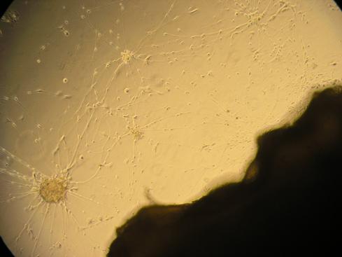
These contacts may be spontaneously produced in vitro (Fig. 1).
The first contacts between neurons and epidermal cells were described by Merkel [8], Merkel cells (or epidermal neuro-endocrine cells) being in contact with nerve endings [9]. Langerhans [10] also suspected the existence of such connections with Langerhans cells. These cells have then been shown to be in contact with axons via their cellular bodies [11, 12] and their dendrites [12]. In the epidermis, there are also contacts between nerve fibers and keratinocytes [13] and, more recently, connections with melanocytes have been reported [14].
In the dermis, there are contacts between nerve fibers and mast cells [15]. Recently dermal dendritic cells have been observed in contact with axons [16]. In contrast, perivascular nerves are found at the interface between the adventitia and
Fig. (1). In vitro co-culture of skin and neurons showing spontaneous growth of nerve endings from neurons (left) to the skin (right).
6 Skin and Psyche Laurent Misery
The quantity of skin neuromediators varies according to the individual, the disease, and their location. Neuropeptide concentrations range from 0.1 to 5.5 pmol/g of tissue [19].
Skin neuromediators are synthesized by nerve endings and Merkel cells but also by keratinocytes, Langerhans cells, melanocytes and immune cells (granulocytes, lymphocytes, monocytes-macrophages and especially mast cells). Neuro-secretory granules have been described only in nerve endings and Merkel cells, the mechanisms of neurotransmitter synthesis by other cells remaining unknown. The majority of the cutaneous cells have also been shown to possess receptors for the neuromediators [1].
Skin Cells Have Neuronal Capacities
Hence, like nerve cells, skin cells are able to express neuronal markers, receptors for neurotransmitters and produce neuromediators [4].
An epithelial or a neural origin of Merkel cells has been a matter of controversy [21 - 23]. However, it is now admitted that Merkel cells originate from epidermal stem cells [24]. These cells have certain nerve cell characteristics ((protein S-100, specific neuronal enolase, neurofilament protein, protein gene product (PGP) 9.5, chromogranin, synaptophysin)) and also show epithelial markers [22, 23]. They, furthermore, produce nerve growth factor (NGF) and its receptor. Merkel cells possess electric properties [25] and are mechanoreceptors [26]. They, as stated above, possess granules and neuro-secretory vesicles, containing neuropeptides. These granules are present in the entire cytoplasm, being more dense in juxtaaxonal position [27].
They express chromogranin and their contents are intended for nerve and dermal cells, while the vesicles expressing synaptophysin, are located on the epidermal cell side [28]. Thus, human Merkel cells probably synthesize neuromediators, which are secreted by fusing granules in neurosecretory vesicles with the plasmic membrane. Through these neurotransmitters Merkel cells are able to modulate skin inflammation [29].
Langerhans cells and their precursors express proteins usually encountered in cells
Skin and Psyche: Biological Aspects Skin and Psyche 7 of the nervous system, i.e. protein S-100 and specific neuronal enolase [30, 31]. When being denervated Langerhans cells are able to express PGP 9.5 [32, 33]. These cells produce pro-opiomelanocortin (precursor to ACTH, endorphins and MSH) [34]. Through receptors on the cell surface surface, neuromediators, CGRP [11], substance P [35], GRP [36] and -MSH [37], may modulate Langerhans cell functions. Thus, Langerhans cells are linking skin, immunity and nervous system [38].
Keratinocytes are able to produce neuromediators: pro-opiomelanocortin [34], ACh [39] dopamine, epinephrine and norepinephrine [40], and substance P [41]. These cells express receptors to substance P [42 - 46], VIP [47], CGRP [47], NPY [47], GRP [36], ACh (muscarinic or nicotinic) [48, 49] and MSH [50].
Melanocytes express protein S-100 and enzymes involved in catecholamine synthesis [51]. These cells express MSH receptors and possibly also to melatonin [52]. Catecholamines may induce the expression of 1 adreno-receptors [53].
In the dermis, fibroblasts have receptors to bombesin, SOM and substance P [54], while in the hypodermis catecholamine receptors of type 1, 2 and 3 are present on adipocytes [55].
FUNCTIONAL LINKS BETWEEN SKIN AND THE NERVOUS SYSTEM
An interdependence has been described between Merkel cells and cutaneous nerves [56], even though being facultative [57]. Merkel cells might offer metabolic support for axons. Eliminating the Merkel cells causes a higher excitability threshold for the nerve ending [58]. In addition, these cells might be regulating the nerve ending positions.
Neuromediators have effects on eccrine or apocrine glandular cells, endothelial cells, epidermal cells, fibroblasts, immune cells and muscle cells, most often via protein G coupled receptors.
Hence, substance P may inhibit antigen presentation, through NK-1-type receptors on both Langerhans cells and T lymphocytes [35]. In a similar murine model, CGRP inhibits antigen presentation [11]. Furthermore, such an incubation inhibits the induction of B7-2 in mice [59] and seems to limit the interleukin (IL)-1 and
Skin and Psyche: Biological Aspects
Skin and Psyche 9
Other chemical actors of the NEICS are neurotrophins: NGF, brain-derived nerve factor (BDNF), neurotrophins 3 and 4 (NT-3 and NT-4). The neurotrophins are growth factors for neurons but also keratinocytes [78, 79]. They have antiapoptotic effects, are involved in the release of neurotransmitters, control of inflammation [80], and hair growth and morphogenesis [81].
Such an organization is not skin specific. Thus, a NEICS analogue is also described in the gastro-intestinal (GI) tractus [82]. Contacts exist between nerve fibers and muscle cells, glandular cells, mast cells or lymphocytes. The nerve endings, but also lymphocytes, plasma cells and neuro-endocrine cells, secrete neuromediators. GI tractus cells, neuro-endocrine, muscle and glandular (or immune) cells possess receptors for neuromediators. In addition, neuromediators play a significant role in numerous symptoms/disorders of the GI tractus, i.e. inflammatory bowel disorders, etc.
WHEN THERE IS A SKIN DISORDER....
The nervous system plays a significant role in many skin disorders, especially inflammatory and/or auto-immune ones, e.g. psoriasis [83 - 88], atopic dermatitis [89 - 94], prurigo [95, 96], and vitiligo [97 - 99].
Psoriasis is a good example. One of its pathophysiological mechanisms is suggested to be psychological. Stress preceding psoriasis symptoms has been reported in varying degrees (from 32% to 90% of all cases) [83], and this by less than one month, and in two thirds of cases, less than two weeks. In addition, patients with psoriasis were reported to be more stress sensitive compared with a control group. In addition, the beneficial effects of psychotherapy or psychotropic drugs for psoriasis have been suggested. A psychiatric distress is presumably found in at least two-thirds of psoriasis patients [83]. However, psoriasis so far does not seem to occur due to a particular personality trait or a specific psychiatric disorder.
Psoriasis lesions often regress after surgical or traumatic denervation [84]. During the course of psoriasis, innervation increases, involving mainly intra-epidermal substance P+ nerve fibers [100]. The affinity, distribution and amount of epidermal receptors to substance P are strongly modified [42]. Moreover, NGF levels
10 Skin and Psyche Laurent Misery increase in psoriatic skin [88]. In addition, blood levels of neuromediators, such as b-endorphin [86] are altered during flare-ups of psoriasis.
Atopic dermatitis is another good example of the effects of neuro-cutaneous alterations in skin disorders [94]. SP+ CGRP+ nerve fiber density increases, while adrenergic innervation decreases [89] and SOM-immunoreactive (IR) fibers disappear [90]. Distribution of epidermal and dermal SOM+cells is highly disrupted [91], whereas NPY-IR dendritic cells appear in the epidermis [101]. The concentration of substance P decreases while the level of VIP increases [92].
Prolactin blood levels are enhanced during atopic dermatitis outbreaks [93].
During the course of prurigo nodularis, the nerve fiber density increases and there is an excessive release of substance P, CGRP and VIP [95]. These neuropeptide modifications do not seem to be secondary to mechanical trauma from scratching, since not being found in lichenified skin.
During the course of vitiligo, the Koebner phenomenon is important [98]. Nerve fibers are damaged, showing both deterioration and regeneration [99]. The nerve fiber NPY immunoreactivity increases [99]. Contacts are observed between melanocytes and axons [102] and the synthesis of catecholamines by keratinocytes and melanocytes is impaired.
Thus, the list of skin diseases being possibly modulated by the activation of nerve endings is not limited. The nervous system is also important in the control of the effects of UV light in the skin [3], wound healing [103] as well as in cosmetic disorders [104], and sensitive skin [105].
SKIN AND STRESS
The course of many skin disorders may be influenced by psychological factors and stress events [106 - 108]. The links between psyche and skin appear to be intimate, especially when inflammation being substantially involved.
There are some psychogenic concepts to explain why the onset of cutaneous lesions can be triggered by psychological factors and/or life stress events [109111]. Probably chemical factors, neurotransmitters and hormones, mediate an emotion or a stress into a cutaneous lesion [2].
Skin and Psyche: Biological Aspects
Skin and Psyche 11
Alterations of the NEICS are frequently observed in the course of numerous skin disorders. The levels of neuromediators are altered as well as the expression of their receptors or enzymes.
During a stress episode, similar phenomena occur in the nervous system and in the blood [112 - 117]. Thus, the skin production of mediators, such as substance P, is modified in response to stress [118]. Stress triggers mast cell degranulation through effects of local substance P and neurotensin [119]. In addition, dendritic cell functions are impaired by psychological stress via the release of substance P [120]. Serotonin [121] is another neurotransmitter with a potential role in such interactions.
However, stress does not induce psoriasis or other dermatoses in all patients. Firstly, the occurrence of a cutaneous disorder after stress appears to be linked to particular personality profiles and maybe to altered neurotransmitter profiles in response to stress. As a second option, this occurrence is linked to genetic and immune backgrounds.
Stress induces both local tissue releases of neurotransmitters and blood release of cortisol and catecholamines [122]. The response by the hypothalamic-pituitar-adrenal (HPA) axis is important in this respect [123]. This axis is characterized by a hierarchy in which the hypothalamus produces CRH, which induces the release of ACTH by the pituitary gland, in turn inducing the release of cortisol by adrenocortical glands [124]. Interestingly human skin itself also expresses molecules of the HPA axis, including pro-opiomelanocortin, CRH, the CRH receptor-1 (CRH-R1), and key enzymes related to corticosteroid synthesis. Also skin cells are able to synthesize glucocorticoids. Expression of these elements is organized into functional, cell type-specific regulatory loops, imitating the HPA axis, where differential CRH-driven responses of defined cutaneous cell populations are activated during stress.
ITCH
Itch (or pruritus) has been defined as an unpleasant sensation leading to the need of scratching [125]. The sensation of itch emanates from nerve fibres especially in the dermo-epidermal junction area [126]. In contrast inflammation of the reticular
Skin and Psyche: Biological Aspects Skin and Psyche 13 [139].
The itch centers in the brain may allow us to understand how itch can be modulated, and sometimes initiated locally, and how stress and other psychological factors are able to modulate its perception. This relationship between itch and stress has been especially studied in atopic dermatitis patients [140]. Such patients often report a close relationship between emotional distress, pruritus, and scratching [140, 141].
CONCLUSION
All these discoveries may increase our understanding of the interactions between skin and the psyche in skin disorders, being aggravated by psychological factors (Table 2).
Table 2. Classification of psychocutaneous disorders [from ref.142].
1. Psychological disorders secondary to skin disorders
2. Psychological disorders responsible for skin disorders
2.1. Psychological disorders responsible for psychogenic skin sensations
2.1.1.Delusional syndromes (e.g. delusional parasitosis, hypochondria)
2.1.2. Phobias (e.g. dysmorphophobia, bromidrosophobia)
2.1.3. Psychogenic pruritus
2.1.4. Psychogenic dysesthesia and paresthesia
2.1.5. Pain syndromes (e.g. glossodynia, vulvodynia, anodynia)
2.2. Psychological disorders responsible for skin lesions
2.2.1. Severe (e.g. dermatitis artefacta, psychogenic excoriations)
2.2.2. Not severe (e.g. trichotillomania)
3. Skin disorders influenced by mental disorders
3.1. Dermatological diseases (e.g. atopic dermatitis, psoriasis, urticaria)
3.2. Cosmetological disorders (e.g. telogen effluvium, skin aging)
4. Skin and mental disorders without any obvious relationship
CONFLICT OF INTEREST
The author is medical advisor to Almirall, Astellas, BASF, Bioderma, Clarins,
14 Skin and Psyche Laurent Misery Galderma, GSK, Johnson & Johnson, Maruho, Novartis, Pierre Fabre, Solabia, and Uriage.
Part of this chapter has been previously published in G Ital Dermatol Venereol 2005;140:677-84.
ACKNOWLEDGEMENTS
Declared none.
REFERENCES
[1]Misery L. Skin, immunity and the nervous system. Br J Dermatol 1997; 137(6): 843-50. [http://dx.doi.org/10.1111/j.1365-2133.1997.tb01542.x] [PMID: 9470898]
[2] Misery L. Are biochemical mediators the missing link between psychosomatics and dermatology? Dermatol Psychosom 2001; 2: 178-83. [http://dx.doi.org/10.1159/000049668]
[3] Misery L. The neuro-immuno-cutaneous system and ultraviolet radiation. Photodermatol Photoimmunol Photomed 2000; 16(2): 78-81. [http://dx.doi.org/10.1034/j.1600-0781.2000.d01-8.x] [PMID: 10823317]
[4] Misery L. The interactions between skin and nervous system. G Ital Dermatol Venereol 2005; 140: 677-84.
[5] Misery L, Hermier M, Staniek V, et al. Congenital insensitivity to pain with anhidrosis: absence of substance P receptors in the skin. Br J Dermatol 1999; 140(1): 190-1. [http://dx.doi.org/10.1046/j.1365-2133.1999.02694.x] [PMID: 10215808]
[6] Wang L, Hilliges M, Jernberg T, Wiegleb-Edström D, Johansson O. Protein gene product 9.5immunoreactive nerve fibres and cells in human skin. Cell Tissue Res 1990; 261(1): 25-33.
[http://dx.doi.org/10.1007/BF00329435] [PMID: 2143435]
[7] Chateau Y, Misery L. Connections between nerve endings and epidermal cells: are they synapses? Exp Dermatol 2004; 13(1): 2-4.
[http://dx.doi.org/10.1111/j.0906-6705.2004.00158.x] [PMID: 15009109]
[8] Merkel F. Tastzellen und Tastkorperchen bei den Haustieren und beim Menschen. Arch Mikrosk Anat 1875; 11: 636-52.
[http://dx.doi.org/10.1007/BF02933819]
[9] Hartschuh W, Weihe E. Fine structural analysis of the synaptic junction of Merkel cell-axo-complexes. J Invest Dermatol 1980; 75(2): 159-65.
[http://dx.doi.org/10.1111/1523-1747.ep12522555] [PMID: 6774030]
[10] Langerhans P. Uber die Nerven der menschlichen Haut. Virchows Arch Pathol Anat Physiol 1868; 44: 325-37.
[http://dx.doi.org/10.1007/BF01959006]
Skin and Psyche: Biological Aspects Skin and Psyche 15
[11] Hosoi J, Murphy GF, Egan CL, et al. Regulation of Langerhans cell function by nerves containing calcitonin gene-related peptide. Nature 1993; 363(6425): 159-63. [http://dx.doi.org/10.1038/363159a0] [PMID: 8483499]
[12] Gaudillere A, Misery L, Souchier C, Claudy A, Schmitt D. Intimate associations between PGP9.5positive nerve fibres and Langerhans cells. Br J Dermatol 1996; 135(2): 343-4. [http://dx.doi.org/10.1111/j.1365-2133.1996.tb01191.x] [PMID: 8881702]
[13] Hilliges M, Wang L, Johansson O. Ultrastructural evidence for nerve fibers within all vital layers of the human epidermis. J Invest Dermatol 1995; 104(1): 134-7. [http://dx.doi.org/10.1111/1523-1747.ep12613631] [PMID: 7798631]
[14] Hara M, Toyoda M, Yaar M, et al. Innervation of melanocytes in human skin. J Exp Med 1996; 184(4): 1385-95. [http://dx.doi.org/10.1084/jem.184.4.1385] [PMID: 8879211]
[15] Wiesner-Menzel L, Schulz B, Vakilzadeh F, Czarnetzki BM. Electron microscopical evidence for a direct contact between nerve fibres and mast cells. Acta Derm Venereol 1981; 61(6): 465-9. [PMID: 6177155]
[16] Sueki H, Telegan B, Murphy GF. Computer-assisted three-dimensional reconstruction of human dermal dendrocytes. J Invest Dermatol 1995; 105(5): 704-8. [http://dx.doi.org/10.1111/1523-1747.ep12324502] [PMID: 7594648]
[17] Chédotal A, Hamel E. L'innervation cholinergique de la paroi vasculaire. Med Sci (Paris) 1993; 9: 1035-42. [http://dx.doi.org/10.4267/10608/2806]
[18]Lotti T, Hautmann G, Panconesi E. Neuropeptides in skin. J Am Acad Dermatol 1995; 33(3): 482-96. [http://dx.doi.org/10.1016/0190-9622(95)91395-5] [PMID: 7657872]
[19] Eedy DJ, Shaw C, Johnston CF, Buchanan KD. The regional distribution of neuropeptides in human skin as assessed by radioimmunoassay and high-performance liquid chromatography. Clin Exp Dermatol 1994; 19(6): 463-72. [http://dx.doi.org/10.1111/j.1365-2230.1994.tb01248.x] [PMID: 7534221]
[20] Boissel JP, Ohly D, Bros M, Gödtel-Armbrust U, Förstermann U, Frank S. The neuronal nitric oxide synthase is upregulated in mouse skin repair and in response to epidermal growth factor in human HaCaT keratinocytes. J Invest Dermatol 2004; 123(1): 132-9. [http://dx.doi.org/10.1111/j.0022-202X.2004.22731.x] [PMID: 15191553]
[21]Boulais N, Misery L. Merkel cells. J Am Acad Dermatol 2007; 57(1): 147-65. [http://dx.doi.org/10.1016/j.jaad.2007.02.009] [PMID: 17412453]
[22]Gaudillère A, Misery L. [Merkel cell]. Ann Dermatol Venereol 1994; 121(12): 909-17. [PMID: 7632011]
[23] Moll I, Hartscuh W, Moll R. First International Merkel Symposium, Heidelberg, Germany. J Invest Dermatol 1995; 105: 851-3.
[http://dx.doi.org/10.1111/1523-1747.ep12326653]
[24] Woo SH, Stumpfova M, Jensen UB, Lumpkin EA, Owens DM. Identification of epidermal progenitors for the Merkel cell lineage. Development 2010; 137(23): 3965-71.
16 Skin and Psyche Laurent Misery [http://dx.doi.org/10.1242/dev.055970] [PMID: 21041368]
[25] Yamashita Y, Akaike N, Wakamori M, Ikeda I, Ogawa H. Voltage-dependent currents in isolated single Merkel cells of rats. J Physiol 1992; 450: 143-62. [http://dx.doi.org/10.1113/jphysiol.1992.sp019120] [PMID: 1331421]
[26] Boulais N, Pennec JP, Lebonvallet N, et al. Rat Merkel cells are mechanoreceptors and osmoreceptors. PLoS One 2009; 4(11): e7759.
[http://dx.doi.org/10.1371/journal.pone.0007759] [PMID: 19898622]
[27] Hartschuh W, Weihe E. Multiple messenger candidates and marker substance in the mammalian Merkel cell-axon complex: a light and electron microscopic immunohistochemical study. Prog Brain Res 1988; 74: 181-7.
[http://dx.doi.org/10.1016/S0079-6123(08)63012-5] [PMID: 3187030]
[28] Ortonne JP, Petchot-Bacque JP, Verrando P, Pisani A, Pautrat G, Bernerd F. Normal Merkel cells express a synaptophysin-like immunoreactivity. Dermatologica 1988; 177(1): 1-10. [http://dx.doi.org/10.1159/000248491] [PMID: 3141225]
[29] Boulais N, Pereira U, Lebonvallet N, et al. Merkel cells as putative regulatory cells in skin disorders: an in vitro study. PLoS One 2009; 4(8): e6528. [http://dx.doi.org/10.1371/journal.pone.0006528] [PMID: 19668696]
[30] Cocchia D, Michetti F, Donato R. Immunochemical and immuno-cytochemical localization of S-100 antigen in normal human skin. Nature 1981; 294(5836): 85-7. [http://dx.doi.org/10.1038/294085a0] [PMID: 7290214]
[31] Misery L, Campos L, Sabido O, et al. S100 protein and neuron-specific enolase on monocytic leukemic CD1+ cells, probable precursors of Langerhans cells. Eur J Haematol 1993; 51(3): 132-5. [http://dx.doi.org/10.1111/j.1600-0609.1993.tb00612.x] [PMID: 7691652]
[32] Hsieh ST, Choi S, Lin WM, Chang YC, Mcarthur JC, Griffin JW. Epidermal denervation and its effects on keratinocytes and Langerhans cells. J Neurocytol 1996; 25(9): 513-24. [http://dx.doi.org/10.1007/BF02284819] [PMID: 8910797]
[33] Hamzeh H, Gaudillère A, Sabido O, et al. Expression of PGP9.5 on Langerhans cells and their precursors. Acta Derm Venereol 2000; 80(1): 14-6. [http://dx.doi.org/10.1080/000155500750012423] [PMID: 10721824]
[34] Bhardwaj RS, Luger TA. Proopiomelanocortin production by epidermal cells: evidence for an immune neuroendocrine network in the epidermis. Arch Dermatol Res 1994; 287(1): 85-90. [http://dx.doi.org/10.1007/BF00370724] [PMID: 7726641]
[35] Staniek V, Misery L, Péguet-Navarro J, et al. Binding and in vitro modulation of human epidermal Langerhans cell functions by substance P. Arch Dermatol Res 1997; 289(5): 285-91. [http://dx.doi.org/10.1007/s004030050194] [PMID: 9164639]
[36] Staniek V, Misery L, Peguet-Navarro J, et al. Expression of gastrin-releasing peptide receptor in human skin. Acta Derm Venereol 1996; 76(4): 282-6. [PMID: 8869685]
[37] Shimizu T, Streilein JW. Influence of alpha-melanocyte stimulating hormone on induction of contact hypersensitivity and tolerance. J Dermatol Sci 1994; 8(3): 187-93.
Skin and Psyche: Biological Aspects Skin and Psyche 17
[http://dx.doi.org/10.1016/0923-1811(94)90053-1] [PMID: 7865476]
[38] Misery L. Langerhans cells in the neuro-immuno-cutaneous system. J Neuroimmunol 1998; 89(1-2): 83-7.
[http://dx.doi.org/10.1016/S0165-5728(98)00117-9] [PMID: 9726829]
[39] Grando SA, Kist DA, Qi M, Dahl MV. Human keratinocytes synthesize, secrete, and degrade acetylcholine. J Invest Dermatol 1993; 101(1): 32-6.
[http://dx.doi.org/10.1111/1523-1747.ep12358588] [PMID: 8331294]
[40] Schallreuter KU, Wood JM, Lemke R, et al. Production of catecholamines in the human epidermis. Biochem Biophys Res Commun 1992; 189(1): 72-8.
[http://dx.doi.org/10.1016/0006-291X(92)91527-W] [PMID: 1360208]
[41] Bae S, Matsunaga Y, Tanaka Y, Katayama I. Autocrine induction of substance P mRNA and peptide in cultured normal human keratinocytes. Biochem Biophys Res Commun 1999; 263(2): 327-33. [http://dx.doi.org/10.1006/bbrc.1999.1285] [PMID: 10491292]
[42] Staniek V, Doutremepuich J, Schmitt D, Claudy A, Misery L. Expression of substance P receptors in normal and psoriatic skin. Pathobiology 1999; 67(1): 51-4. [http://dx.doi.org/10.1159/000028051] [PMID: 9873229]
[43] Arenberger P, Leder RO, Abraham A, Chang JK, Farber EM. Substance P binding in normal neonatal foreskin. Br J Dermatol 1995; 132(1): 54-8. [http://dx.doi.org/10.1111/j.1365-2133.1995.tb08624.x] [PMID: 7538777]
[44] Kemény L, von Restorff B, Michel G, Ruzicka T. Specific binding and lack of growth-promoting activity of substance P in cultured human keratinocytes. J Invest Dermatol 1994; 103(4): 605-6. [http://dx.doi.org/10.1111/1523-1747.ep12396976] [PMID: 7523535]
[45] Pincelli C, Fantini F, Giardino L, et al. Autoradiographic detection of substance P receptors in normal and psoriatic skin. J Invest Dermatol 1993; 101(3): 301-4.
[http://dx.doi.org/10.1111/1523-1747.ep12365423] [PMID: 7690377]
[46] Song IS, Bunnett NW, Olerud JE, et al. Substance P induction of murine keratinocyte PAM 212 interleukin 1 production is mediated by the neurokinin 2 receptor (NK-2R). Exp Dermatol 2000; 9(1): 42-52.
[http://dx.doi.org/10.1034/j.1600-0625.2000.009001042.x] [PMID: 10688374]
[47] Takahashi K, Nakanishi S, Imamura S. Direct effects of cutaneous neuropeptides on adenylyl cyclase activity and proliferation in a keratinocyte cell line: stimulation of cyclic AMP formation by CGRP and VIP/PHM, and inhibition by NPY through G protein-coupled receptors. J Invest Dermatol 1993; 101(5): 646-51.
[http://dx.doi.org/10.1111/1523-1747.ep12371670] [PMID: 8228323]
[48] Grando SA, Horton RM, Pereira EF, et al. A nicotinic acetylcholine receptor regulating cell adhesion and motility is expressed in human keratinocytes. J Invest Dermatol 1995; 105(6): 774-81.
[http://dx.doi.org/10.1111/1523-1747.ep12325606] [PMID: 7490471]
[49] Grando SA, Zelickson BD, Kist DA, et al. Keratinocyte muscarinic acetylcholine receptors: immunolocalization and partial characterization. J Invest Dermatol 1995; 104(1): 95-100.
[http://dx.doi.org/10.1111/1523-1747.ep12613582] [PMID: 7528248]
18 Skin and Psyche Laurent Misery
[50] Chakraborty A, Slominski A, Ermak G, Hwang J, Pawelek J. Ultraviolet B and melanocytestimulating hormone (MSH) stimulate mRNA production for alpha MSH receptors and proopiomelanocortin-derived peptides in mouse melanoma cells and transformed keratinocytes. J Invest Dermatol 1995; 105(5): 655-9.
[http://dx.doi.org/10.1111/1523-1747.ep12324134] [PMID: 7594638]
[51] Schallreuter KU, Wood JM, Pittelkow MR, et al. Increased monoamine oxidase A activity in the epidermis of patients with vitiligo. Arch Dermatol Res 1996; 288(1): 14-8.
[http://dx.doi.org/10.1007/BF02505037] [PMID: 8750929]
[52] Almeida AL, Markus RP, Visconti MA, Castrucci AM. Presence of melatonin binding sites in S-91 murine melanoma cells. Pigment Cell Res 1995; 10: 31.
[53] Schallreuter KU, Körner C, Pittelkow MR, Swanson NN, Gardner ML. The induction of the alpha--adrenoceptor signal transduction system on human melanocytes. Exp Dermatol 1996; 5(1): 20-3.
[http://dx.doi.org/10.1111/j.1600-0625.1996.tb00088.x] [PMID: 8624607]
[54] Gaudillère A, Bernard C, Abello J, Schmitt D, Claudy A, Misery L. Human normal dermal fibroblasts express somatostatin receptors. Exp Dermatol 1999; 8(4): 267-73.
[http://dx.doi.org/10.1111/j.1600-0625.1999.tb00381.x] [PMID: 10439224]
[55] Arner P. The beta 3-adrenergic receptor--a cause and cure of obesity? N Engl J Med 1995; 333(6): 382-3.
[http://dx.doi.org/10.1056/NEJM199508103330612] [PMID: 7609759]
[56] Morohunfola KA, Jones TE, Munger BL. The differentiation of the skin and its appendages. I. Normal development of papillary ridges. Anat Rec 1992; 232(4): 587-98. [http://dx.doi.org/10.1002/ar.1092320414] [PMID: 1554108]
[57] Narisawa Y, Kohda H. Merkel cells do not require trophic maintenance from the nerves in adult human skin. Br J Dermatol 1995; 133(4): 553-6.
[http://dx.doi.org/10.1111/j.1365-2133.1995.tb02703.x] [PMID: 7577582]
[58] Diamond J, Holmes M, Nurse CA. Are Merkel cell-neurite reciprocal synapses involved in the initiation of tactile responses in salamander skin? J Physiol 1986; 376: 101-20. [http://dx.doi.org/10.1113/jphysiol.1986.sp016144] [PMID: 3795072]
[59] Gillardon F, Moll I, Michel S, Benrath J, Weihe E, Zimmerman M. CGRP and nitric oxide are involved in ultraviolet radiation-induced immunosuppression. Eur J Pharmacol 1995; 296: 395-400. [http://dx.doi.org/10.1016/0926-6917(95)90060-8] [PMID: 8748693]
[60] Hosoi J, Torii H, Fox F, Zan Z, Rook AH, Granstein RD. Alteration of cytokine expression by calcitonin gene-related peptide (CGRP). J Invest Dermatol 1995; 105: 859.
[61] Asahina A, Hosoi J, Beissert S, Stratigos A, Granstein RD. Inhibition of the induction of delayed-type and contact hyeprsensitivity by CGRP. J Immunol 1995; 154: 3056-61. [PMID: 7897198]
[62] Clausen OP, Thorud E, Iversen OH. Adrenalin has differential effects on epidermal cell cycle progression in mice. J Invest Dermatol 1982; 78(6): 472-6. [http://dx.doi.org/10.1111/1523-1747.ep12510172] [PMID: 7086167]