Other documents randomly have different content
and difficult in all, cases to cover only one spinous process when the head is resting on its side.
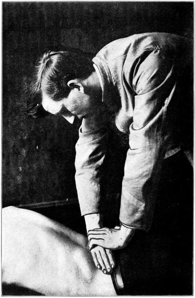
27. The edge contact in Lumbar region.
Palpation
Same as for Recoil or Heel Contact, q. v.
Fig.
Contact
Using the same adjusting hand as for the Heel Contact, place the middle of the ulnar edge of the fifth metacarpal bone in contact with the spinous process. If the vertebra be superior, place the edge of hand above, if inferior, place the hand below. This contact is especially good for S or I vertebrae.
Position of Hands and Arms
The fingers of adjusting hand cross the spine at a right angle to its long axis. The back of hand will be toward patient’s head except in adjusting the last two Lumbars, with which a change of hands is made necessary by the upward slant of the lower half of the Lumbar curve.
The palpating hand now grips the adjusting hand so that the fingers of the upper hand, held close together, press against and reinforce the lower on its dorsum and just above the contact point. The thumbs are hooked together as shown in Fig. 27, so that the hands may be stiffened and their tendency to roll avoided.
The elbows are outrotated and locked as in the Pisiform Double Transverse Move and both shoulders are loosened.
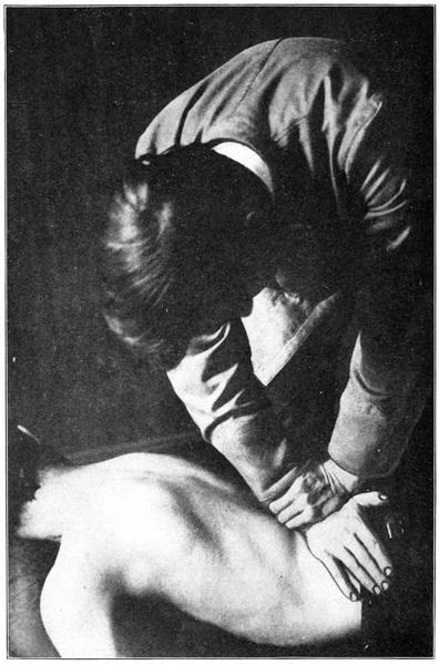
Movement
This is chiefly delivered with the upper arm, using upper hand to drive the lower. Force should be quickly delivered when patient is relaxed. The direction of force should be determined by the direction of subluxation and by the slant of the spinous process. Thus, when patient lies prone upon a bifid bench and sways downward against a lax abdominal support, the spinous processes of the lower dorsal make an acute angle with the plane of the floor. If one be superior, contact above it and force driven straight toward the floor will tend to correct the subluxation. There is a slightly different force angle for every subluxation correctable by this move.
This move is less painful than the pisiform contact and may often be used to advantage, especially in the Lumbar region.
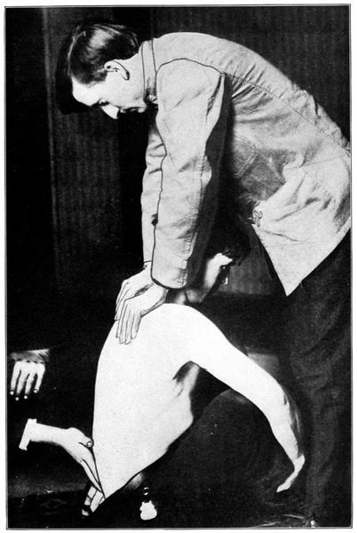
LUMBAR SINGLE TRANSVERSE
For the correction of a rotated Lumbar. Best used on second and third. This movement should never be attempted unless the transverse process can be palpated. Lumbar transverses are sometimes short or fragile, and unless they can be distinctly felt no force should be applied where they are believedto lie.
Contact
Pisiform bone with posterior transverse.
Palpation and Placing of Hands
Palpating as if for other movements, pause with the second finger of palpating hand indicating the spinous process of the vertebra to be moved. Note that if the spinous process be to the right of the median line the left transverse will be posterior, if to the left, the right transverse.
Fig. 28. Lumbar single transverse move.
The transverse may then be found as in the Dorsals; it should lie even with the interspace above the spinous process, deeply overlaid with strong muscles. When the transverse has been located by a deep, probing movement of the fingers, place adjusting hand, pisiform on transverse, close to the spinous process for greater solidity and fingers extending downward and outward from the midspinal line parallel with the lower rib curve.
If the adjuster stands on the side of the patient opposite to the transverse to be moved the hand opposite the palpating hand becomes the contact hand, as in other moves. But if the posterior transverse is on the same side with the adjuster, a change of hands is made and the palpating hand becomes contact hand. To accomplish this the adjuster must turn and face away from the patient with arm extended straight downward to the contact. After contact is made the remaining hand reinforces the adjusting hand by gripping the wrist.
Movement
In making the contact press downward, deeply and firmly, so as to crowd the muscles aside and place the pisiform directly upon the transverse. Movement is given after the patient’s body has been swung downward for a considerable distance, and is sharp and decisive, directed straight toward the floor.
LUMBAR DOUBLE TRANSVERSE MOVE
A movement sometimes applied to posterior or postero-rotary Lumbars.
Palpation and Contact
From the spinous, find first the more posterior transverse and make contact with it, since most force must be directed there. Stand
facing patient’s head and place right hand on right transverse and left hand on left.
Contact point in this move is the tuberosity of the scaphoid with the posterior surface of the transverse. Fingers curve away from median line so as to avoid the rib curve.
Movement
After heavy, steady pressure downward, force is delivered with a quick, throwing movement, most force on the posterior side.
THE “SPREAD” MOVE
Upon the theory that when two forces are simultaneously applied, the one to drive some vertebra cephalad (by its spinous process) and the other to drive some lower vertebra caudad, the intervening vertebrae tend, if anterior, to be drawn outward or toward a more posterior position, this move is predicated.
The author does not believe that it accomplishes its purpose, but will briefly describe it for the benefit of those who do.
Position
Patient is placed over a roll which rests under the thighs so as to flex thighs and pelvis on the Lumbar spine, or an adjustable table is so tilted, both sections sloping downward from the middle, as to accomplish the same result.
Contact
The usual method, if only a single vertebra is anterior, is to make contact with the vertebrae immediately adjacent, crossing the hands and having fingers of upper hand pointing toward head and of lower hand toward Sacrum. But some adjusters use this move differently,
making contact with Sacrum and with the mid-dorsal region in general and applying a slow force with both hands. Contact is with heel of hand upon spinous process.
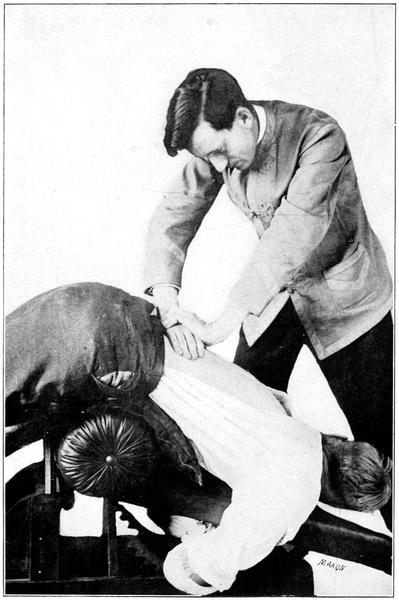
COCCYGEAL ADJUSTMENTS
Examination
Place patient on an angle table, i. e., one which rises in the center and slopes away toward either end. Separate the thighs slightly, patient lying face down, and insert the rubber-covered second finger, palmar surface upward, very carefully into the rectum. The tip of the coccyx may then be felt and its movability and position determined. Unless it is immovably fixed in an abnormal position it should not be molested; the movable coccyx responds to mere muscle tension by changes of position and cannot act as a primary cause of nerve impingement.
Usually this examination will be rendered unnecessary by the external palpation which may disclose the movability of the coccyx and at once render further exploration superfluous.
When the coccyx is anteriorly subluxated and ankylosed in that position it may be a factor in producing constipation, hemorrhoids, etc., but its influence in other diseases, especially of the nervous system, has been greatly overrated by those who have not yet fully accepted the doctrine that nerve impingement is the primary cause of all disease.
Fig. 30. Edge contact with “Roll,” q. v. Attitude of patient for coccygeal adjustment.
Movement
When it has been decided that the coccyx must be moved, the position and use of hand is the same as for the palpation. The finger hooks under the tip of the coccyx, draws upon it until a tight contact is secured and then jerks sharply backward upon it with a view to its
abrupt fracture. No mitigation of the jerk in the hope of previously loosening or gradually replacing the bone is of value for osseous tissuemust be broken before any movement may take place.
This movement is painful and the region of the newly fractured coccyx may remain sore for a period ranging from a few days to several weeks. It is wise to warn the patient of the facts before proceeding.
The fractured coccyx may be absorbed, or may be reankylosed in a proper position or in a new abnormal position, or may remain loose and movable.
ADJUSTMENT OF CURVATURES
We have previously discussed in detail the nature and discovery of curvatures. A few words should be said here about their correction.
If the sole object of the adjustment is to correct the curvature it is best to select for adjustment those vertebrae which are most subluxated in the direction of the curvature. According to the length of the curvature a series of from two to six, separated by some distance, are chosen. These are adjusted until they cease to be the most prominent ones in the curvature and then others, then most prominent, are chosen and adjusted until they in turn cease to be most prominent. In this way the curvature may eventually be straightened, or nearly so. It is doubtful if any curvature can be absolutely eradicated, although it may be straightened until unnoticeable except by the expert.
To overcome a curvature it may be necessary to break every rule which governs ordinary adjusting and to invent new ways of placing the hands or of delivering force. No two require exactly the same measures and he is most successful with curvatures who is most adaptable to changing conditions.
One rule may be safely laid down. Do not alternate from day to day, loosening at the same time many vertebrae, but choose the ones most in need of adjustment and followyourchoiceas long as it is indicated. The chief vertebra is nearly always the one at the angle or pointof the curvature.
The sharp, angular curve of Potts’ Disease, involving two or three vertebrae, should warn against adjustment, usually, since in this disease the vertebrae are fragile and easily fractured. If a case has not progressed too far a cure may be effected, but great caution in taking such cases must be exercised. Every Chiropractor should be well informed on the diagnosis of Potts’ Disease, or spinal caries.
Many months are usually required for the straightening of a curvature—how many can scarcely be estimated in advance of the experiment with any case. Often the case which seems simplest requires the longer time, while a very pronounced curvature, as in some cases of rachitis, may yield in a few months.
PREFERABLE ADJUSTMENTS
The selection of the move with which to correct each subluxation depends upon the adjuster’s concept of the kindand directionof the subluxation and of the mechanics of the different corrective moves in his repertoire. The move used should be one in which the application of force is exactly along opposite lines to the lines of force which originally produced the subluxation.
Omitting involved explanations as to the elements of each displacement and the manner of change in bone, muscle, ligament, cartilage, etc., and presupposing a comprehension of the principles of each adjustment named, there follows here a list of possible subluxations of each vertebra in turn, from Atlas down, with a simple statement of the RIGHT MOVE for that subluxation.
In each instance there are other moves than the one listed which would move the vertebra and some which would partially correct it, but none which would quite so definitely tend to correct thedisplacement. Unfortunately it is not a fact that every movement of a vertebra is an adjustment. If this were true subluxations would not exist, because they could never have been produced. Too often the adjuster uses a move because it is easy, because its use has become habitual with him, rather than because it is indicated by the conditions of the case—then blames Chiropractic because his results are negative or bad.
The move which is suited to a certain kind of subluxation of one vertebra may be quite out of place with another, in a different part of the spine. Thus the Recoil is quite proper for a posterior Lumbar and is contraindicated with a posterior middle Dorsal.
If all vertebrae were shaped exactly alike, if all were equal in size, if subluxation were possible only in one direction, then one method of adjustment would be quite sufficient. Diversity of technic is demanded, but a discriminating diversity, with a good reason for every move used.
First Cervical
Subluxation. Adjustment.
Right—R. Break, or straight lateral.
Right, posterior—R. P. Rotary lateral.
Right, anterior—R. A. Morikubo.
Right, superior—R. S. Break.
Right, inferior—R. I. Break.
Right, posterior, superior—R. P. S. Rotary lateral.
Right, posterior, inferior—R. P. I. Rotary lateral.
Right, anterior, superior—R. A. S. Morikubo.
Right, anterior, inferior—R. A. I. Morikubo.
Left—L. Break.
Left, posterior—L. P. Rotary lateral.
Left, anterior—L. A. Morikubo.
Left, superior—L. S. Break.
Left, inferior—L. I. Break.
Left, posterior, superior—L. P. S. Rotary lateral.
Left, posterior, inferior—L. P. I. Rotary lateral.
Left, anterior, superior—L. A. S. Morikubo.
Left, anterior, inferior—L. A. I. Morikubo.
Anterior (entire Atlas)—A. Morikubo (both sides).
Posterior (entire Atlas)—P. Rotary lateral (both sides).
NOTE.—All right subluxations adjusted from right side, all left from left side.
Second Cervical
Posterior—P.
Posterior Cervical move.
Posterior, right—P. R. Double contact on right side.
Posterior, left—P. L.
Double contact on left side.
Posterior, right, inferior—P. R. L. Double contact on right.
Posterior, right, superior—P. R. S. Double contact on right.
Posterior, left, inferior—P. L. I. Double contact on left side.
Posterior, left, superior—P. L. S. Double contact on left side.
Right (lateral)—R.
Right (rotary)—R.
Left (lateral)—L.
Left (rotary)—L.
Break (Same if R. I. or R. S.)
Rotary (Same if R. I. or R. S.)
Break (Same if L. I. or L. S.)
Rotary (Same if L. I. or L. S.)
Superior—S. Posterior Cervical move.
Inferior—I. Posterior Cervical move.
Anterior (entire Vertebra)—A. Ventral transverse contact on most anterior side.
Anterior, right (lateral)—A. R. Second metacarpal contact from right.
Anterior, right (rotary)—A. R. Pisiform Ant. Cerv. contact on right.
Anterior, left (lateral)—A. L. Second metacarpal contact from left.
Anterior, left (rotary)—A. L. Pisiform Ant. Cerv. contact on left.
Third Cervical
Same as second.
Fourth Cervical
Same as second.
Fifth Cervical
Same as second.
Sixth Cervical
Posterior—P. The Recoil, hands reversed.
Posterior, right—P. R. Recoil, hands reversed.
Posterior, left—P. L. Recoil, hands reversed.
Posterior, right, superior—P. R. S. Recoil, hands reversed.
Posterior, right, inferior—P. R. I. Recoil, hands reversed.
Posterior, left, superior—P. L. S. Recoil, hands reversed.
Posterior, left, inferior—P. L. I. Recoil, hands reversed.
Right (lateral)—R.
Right (rotary)—R.
Left (lateral)—L.
Left (rotary)—L.
Break (Same if R. I. or R. S.)
Rotary (Same if R. I. or R. S.)
Break, from left (Same if L. I. or L. S.)
Rotary (Same if L. I. or L. S.)
Superior S. Edge contact move.
Inferior—I. Edge contact move.
Anterior (entire vertebra)—A. Pisiform Ant. Cerv. contact on most anterior side.
Anterior, right (lateral)—A. R. Second metacarpal contact from right.
Anterior, right (rotary)—A. R. Pisiform Ant. Cerv. contact on right.
Anterior, left (lateral)—A. L. Second metacarpal contact from left.
Anterior, left (rotary)—A. L. Pisiform Ant. Cerv. contact on left.
Seventh Cervical
Same as sixth Cervical, except that T. M. may be used on right or left rotary subluxations.
First Dorsal
Posterior—P. Recoil, hands reversed.
Posterior, right—P. R. Recoil, hands reversed.
Posterior, right, superior—P. R. S. Recoil, hands reversed.
Posterior, right, inferior—P. R. I. Recoil, hands reversed.
Posterior, left—P. L. Recoil, hands reversed.
Posterior, left, superior—P. L. S. Recoil, hands reversed.
Posterior, left, inferior—P. L. I. Recoil, hands reversed.
Posterior, superior—P. S. Heel contact.
Posterior, inferior—P. I. Edge contact.
Superior S. Heel contact.
Inferior I. Edge contact.
Right—R.
Left—L.
T. M. (Same if R. S. or R. I.)
T. M. (Same if L. S. or L. I.)
Anterior—A. No correction.
Second Dorsal
Posterior—P. Heel contact.
Posterior, superior—P. S. Heel contact.
Posterior, inferior—P. I. Edge contact.
Posterior, right—P. R. Recoil.
Posterior, right, superior—P. R. S. Recoil.
Posterior, right, inferior—P. R. I. Recoil.
Posterior, left—P. L. Recoil.
Posterior, left, superior—P. L. S. Recoil.
Posterior, left, inferior—P. L. I. Recoil.
Left—L. T. M. (Same if L. S. or L. I.)
Right—R. T. M. (Same if R. S. or R. I.)
Anterior—A. No correction.
Third Dorsal
Posterior—P. Heel contact.
Posterior, superior—P. S. Heel contact.
Posterior, inferior—P. I. Edge contact.
Posterior, right—P. R. Recoil.
Posterior, right, superior—P. R. S. Recoil.
Posterior, right, inferior—P. R. I. Recoil.
Posterior, left—P. L. Recoil.
Posterior, left, superior—P. L. S. Recoil.
Posterior, left, inferior—P. L. I. Recoil.
Right—R. Pisiform single transverse (on left) (Same if R. S. or R. I.)
Left—L. Pisiform single transverse (on right) (Same if L. S. or L. I.)
Anterior—A. No correction.
Fourth Dorsal
Same as third Dorsal.
NOTE.—While the Recoil is here, the preferred move for posterior and postero-lateral subluxations, the pisiform double transverse or the two finger double transverse may be used if both transverses are palpable.
Fifth Dorsal
Posterior—P. Double transverse move.
Posterior, superior—P. S. Heel contact.
Posterior, inferior—P. I. Double transverse.
Posterior, right—P. R. Double transverse.
Posterior, right, superior—P. R. S. Double transverse.
Posterior, right, inferior—P. R. I. Double transverse.
Posterior, left—P. L. Double transverse.
NOTE.—The pisiform double transverse and the two-finger double transverse, apply force in exactly similar directions and may
therefore be used interchangeably. The latter is preferable for children.
Posterior, left, superior—P. L. S. Double transverse.
Posterior, left, inferior—P. L. I. Double transverse.
Right—R. Pisiform single transverse (Same if R. S. or R. I.)
Left—L. Pisiform single transverse. (Same if L. S. or L. I.)
Anterior—A. No correction.
Sixth Dorsal
Same as Fifth Dorsal.
Seventh Dorsal
Same as Fifth Dorsal.
Same as Fifth Dorsal.
Eighth Dorsal
Ninth Dorsal
Same as Fifth Dorsal.
Tenth Dorsal
Posterior—P. Heel contact.
Posterior, superior—P. S. Edge contact.
Posterior, inferior—P. I. Edge contact.
Posterior, right—P. R. Recoil.
Posterior, right, superior—P. R. S. Recoil.
Posterior, right, inferior—P. R. I. Recoil.
Posterior, left—P. L. Recoil.
Posterior, left, superior—P. L. S. Recoil.
Posterior, left, inferior—P. L. I. Recoil.
Right—R.
Left—L.
Recoil (Same if R. S. or R. I.) A
Recoil (Same if L. S. or L. I.) A
Anterior—A. No correction.
A Note.—The use of this move is not quite mechanically correct, but it is advised because of the possible danger of using the transverse processes as levers.
Eleventh Dorsal
Same as Tenth Dorsal.
Twelfth Dorsal
Same as Tenth Dorsal.
First Lumbar
Posterior—P. Heel contact.
Posterior, superior—P. S. Heel contact.
Posterior, inferior—P. I. Heel contact.
Posterior, right, superior—P. R. S. Recoil.
Posterior, right, inferior—P. R. I. Recoil.
Posterior, left—P. L. Recoil.
Posterior, left, superior—P. L. S. Recoil.
Posterior, left, inferior—P. L. I. Recoil.
Right—R.
Left—L.
Lumbar single transverse move, if transverse is palpable, otherwise Recoil. (Same if R. S. or R. I.)
Lumbar single transverse move, if transverse is palpable, otherwise Recoil. (Same if L. S. or L. I.)
Anterior—A. No correction.
Second Lumbar
Same as First Lumbar.
Third Lumbar
Same as First Lumbar.
Fourth Lumbar
Posterior—P. Heel contact.
Posterior, superior—P. S. Heel contact.
Posterior, inferior—P. I. Heel contact.
Posterior, right—P. R. Recoil, hands reversed.
Posterior, right, superior—P. R. S. Recoil, hands reversed.
NOTE.—The Heel contact may be substituted for the Recoil above if force be carefully directed in the proper direction in delivery.
Posterior, right, inferior—P. R. I. Recoil, hands reversed.
Posterior, left—P. L. Recoil, hands reversed.
Posterior, left, superior—P. L. S. Recoil, hands reversed.
Posterior, left, inferior—P. L. I. Recoil, hands reversed.
Right—R.
Left—L.
Lumbar single transverse move, if transverse is palpable, otherwise Recoil. (Same if R. S. or R. I.)
Lumbar single transverse, if transverse is palpable, otherwise Recoil. (Same if L. S. or L. I.)
Anterior—A. No correction.
Fifth Lumbar
Posterior—P. Heel contact.
Posterior, superior—P. S. Edge contact.
Posterior, inferior—P. I. Edge contact.
Posterior, right—P. R. Recoil.
Posterior, right, superior—P. R. S. Recoil.
Posterior, right, inferior—P. R. I. Recoil.
Posterior, left—P. L. Recoil.
Posterior, left, superior—P. L. S. Recoil.
Posterior, left, inferior—P. L. I. Recoil.
Right—R. Recoil (Same if R. S. or R. I.)
Left—L. Recoil (Same if L. S. or L. I.)
Anterior A. “Bohemian” anterior fifth Lumbar move. (Not always advisable.)
Sacrum
Posterior base—B. of S.—P. Heel contact on base.
Posterior apex A. of S.—P. Heel contact on apex.
Entire Sacrum posterior Sac. P. Heel contact between sacroiliac articulations.
Coccyx
To be adjusted only when ankylosed in an abnormal position and then by leverage of finger through rectum.
A FINAL WORD
Some useful information pertaining to adjustment will be found in section entitled, “Practice,” q. v.
After a careful and painstaking study of the foregoing pages it will still be found that the student is not by any means equipped for the work. He must practice these things to learn them. We learn to do by doing. The chief use of this section will be as a reference and guide during the practice of adjusting.
Vital Energy
Irritability is the property of being susceptible to excitement or stimulation. Stimulation is the process of increasing the functional activity of any organ. Inhibition is the act of checking, restraining, or holding back the functional activity of any organ. These definitions, taken from Gould, are here introduced as a necessary preface to an attempt to set forth, without unnecessary reference to, or discussion of, any other theory as to the etiology of disease, the Chiropractic explanation of its presence.
Chiropractic maintains that all the chemical and physical activities of the human organism are controlled, directly or indirectly, through a third form of energy transmitted through the Nerve System; that while all three forms of energy are interdependent and closely related in their ultimate expression, one of the three is the primaryand most essential form, and especially indicative of life. We may call this third form VitalEnergy.
There are several good reasons for believing that this nerve force is the primary form in which energy is expressed in man and for believing that it controls and directs the others in greater degree than it is controlled and directed by them.
Of the four forms of tissue of which the body is composed— connective, epithelial, muscular, and nervous—the latter is the one damage to which is followed by the greatest and most permanent consequences.
It is a fact that there are several organs whose removal leads to certain death because of their importance in the general economy of the body, but it is also true that section of the nerves leading to these organs just as certainly causes death by the cessation of their functions. There is no organ in the body aside from the nerves themselves which does not immediately cease to act upon withdrawal of its nerve force and at once begin a process of degeneration or atrophy.
Pathologic changes in the Nerve System invariably are followed by pathologic changes in the organs controlled by the diseased segment but the converse is not true. Excitation or inhibition of nerve activity produces corresponding and responsive change in the activity of the organs innervated, but excitation of an organ does not necessarily produce similar changes in the Nerve System. That system possesses the power of inhibiting or permitting responsive action, in other words, the power of choice.
Research in Comparative Anatomy develops the fact that the differences in power of complex action possessed by different organisms are entirely measurable by differences in the structure and complexity of their nerve mechanisms.
Further, by studying the effects of removal or extirpation, or of pathologic changes in various parts of the nerve system it has been demonstrated that the Brain is the center for those higher forms of activity known as psychic, for the power of accelerating or inhibiting the responses of the lower centers of the nerve system to stimulation from without, and for the conveyance of authority to act to all the lower centers. The Nerve System is the morphologic, physiologic, and dynamic center of the organism and the Brain the center of the Nerve System. We may, then, logically expect to find in the Brain, or in the channels by which power is distributed from the Brain to lower centers or organs, the initial step in the disease process, which is our present quest.
One Nerve System
All nerve tissue in the body is organized and linked together in a complicated aggregation of individual units, communicating by contact, and forming one great Nerve System having its directing center in the Brain. It is said by some writers to consist of two distinct systems—cerebro-spinal and sympathetic—but would better be described as consisting of central organs—brain and spinal cord— and peripheral organs—cranial, spinal, and sympathetic peripheral axons connecting with cells in the central axis and linked together in
a net-work improperly separable into separate or distinct divisions, the fibres of different parts being bound together in such a way as to establish an intricate intercommunication, closest on the one hand between the cranial and sympathetic and on the other between the spinal and sympathetic. The sympathetic system may be regarded as nothing more than a medium for proper distribution of impulses originating in the cerebro-spinal system, and a series of reflex centers deriving their power to act from the central axis. The proper action of sympathetic ganglia has been demonstrated to depend upon the integrity of the spinal nerve fibres, or rami communicantes, which pass to and terminate in the ganglia with their telodendria (terminal arborizations) in contact with the dendrites (cellulipetal processes) of the ganglion cells.
It will appear that interference with one division or part of the nerve system may be followed by effects partly manifested through a distant part; that excitation or inhibition of a spinal nerve may correspondingly excite or inhibit sympathetic fibres.
Chiropractic Hypothesis
Chiropractic has accepted, as a convenient working hypothesis amply justified by years of clinical experiment and anatomical and physiological research, the proposition that all disease in the human body is primarily made possible by injury to (stimulation or inhibition of) some part of the nervous mechanism.
Injury to other tissues, unless the injury also involves nerve tissue, is quickly repaired and the body goes on without disease. Or the injury is sufficient at once to render the body untenable and death ensues. Few pathological changes follow trauma unless nerve tissue be injured.
This theory to be logical must and does include the entire nerve system. Also, since it is noted that each nerve cell presides over the nutrition of its own processes and possesses its own power of repair, it follows that unless an injury be of fatal nature or of permanent
duration, even injuries to nerves tend toward automatic cure. We must seek a permanently operating interference with nerve tissue.
The brain, enclosed within the comparatively solid cranium, is so well protected that nothing except fracture of the skull, violent concussion, or shutting off of its blood supply from without, will produce permanent change there. Also, unless there be pressure by foreign substance against the brain, an injury will be repaired in time and the body resume its normal functional activity. It has been demonstrated that comparatively few diseases occur in this way. Such as do are called traumatic; i. e., caused by wound or injury.
In the broadest sense all disease is caused by trauma, as we shall presently show.
The upper or cephalic peripheral nerves, called cranial, leave the skull by foramina in its base (except the auditory) and are so protected by the immobility of the bones of the skull as to be comparatively free from direct injury. Peripheral injuries occur to cranial nerves but are repairable; even section of the trigeminal for neuralgia is usually followed after an interval by a reunion of the severed parts. As will be shown later, the special end organs of the cranial nerves are not free from the effects of spinal subluxation and their nuclei (deep origins) often share in morbid changes in the brain tissue due to nutritional disturbances.
The sympathetic portion of the nervous system might be classed with the cranial as regards infrequency of permanent interference were it not for the proximity of the great gangliated cord to the transverse processes and bodies of the vertebrae. This proximity renders it liable to sustain permanent impingement in vertebral subluxation.
Trauma Affects Spinal Nerves
With the exception of the first pair of Cervical nerves and the Sacral and Coccygeal, all spinal nerves pass through foramina of exit which are composed each of two movable vertebrae. The
Chiropractic hypothesis is based upon the discovery that in addition to the part these vertebrae may take in general movements of the spine it is possible that their relation to each other may be changed by the application of force from without, and that this change once produced tends to remain permanently. These permanent vertebral subluxations occur with great frequency, a fact clinically demonstrable by palpation and by the X-Ray.
The discovery of this fact led to the ascertaining of two more, namely,
Nodiseaseiseverfoundwithoutaccompanyingsubluxation.
Since each organ or tissue is connected with some definite and special vertebra, subluxations accompanying disease bear a relation to disease which is controlled by a general law, operative alike on all human organisms.
The latter fact required one other for its complete demonstration; namely, that the removal of the subluxation is always followed by the complete disappearance of the disease. Given more perfect methods of correcting subluxations it would follow that proof of the Chiropractic theory would be so complete and overwhelming as to meet at once with general acceptance. The difficulty lies in the fact that with our present methods much time is often required for complete correction of the vertebral displacement and much skill is needed even for successful investigation of the results obtainable. The theory is too often judged by unskilled or imperfect applications of it.
Every school of Chiropractic accepts the presence of the subluxation and has spent much thought and time in the effort to deduce the law governing its connection with disease. Diverse conclusions have been reached owing to the difficulty experienced in completely eradicating the subluxation. When it isaccomplished the results are absolutely conclusive. When it is partially or relatively accomplished the results are so good in a great per cent of cases as to lead sometimes to the erroneous belief that the subluxation did
not cause the disease since mere partial correction of the subluxation suffices to bring about the apparent total removal of the disease. In every case of thorough experiment the results warrant the recommendation of the subluxation theory as at least a proper working hypothesis.
Without attempting here to review all the various conclusions reached or the methods by which they have been attained, we would simply state our own conclusion, which we believe is the only one compatible with demonstrable facts. It is briefly this: Since every portion of the body is connected through the nervous system with the spinal nerves and since it has been proven that this connection is reasonably constant and anatomically demonstrable; since the removal or correction of a subluxation leads in all cases to the complete disappearance of disease from the organs or tissues innervated from the subluxated portion of the spinal column, we conclude that the subluxation is the primarycauseof disease.
The final test of the correctness of any theory is the result of its application. Since Chiropractic secures a larger percentage of results than any other known system of healing it is safe to assume, at least, that it has discovered the way to remove the primary cause of disease.
That the Chiropractic theory, or more properly the subluxation theory, does not include all of the etiology of disease is evidenced by the facts of contagion and infection, by the effect upon the organism of the introduction of poison, by the consequences of worry, anger, and other abnormal mental states and conditions. These facts do not in the least invalidate the theory. They merely require explanation which will make clear their relation to the subluxation. That such explanation is abundantly at hand strengthens the position of Chiropractic more than would negation of all other causes save the one we concentrate upon.
The Mentalist who holds that all diseases exist in and are but figments of the mind is as far afield as the Physicist who holds that special nerve energy is nonexistent. The Chiropractor views Man as a