(Ebook) Cambridge IGCSE and O Level History Workbook 2C - Depth Study: the United States, 1919-41 2nd Edition by Benjamin Harrison ISBN 9781398375147, 9781398375048, 1398375144, 1398375047
(Ebook) Big Data Computing and Communications: Second International Conference, BigCom 2016, Shenyang, China, July 29-31, 2016. Proceedings by Yu Wang, Ge Yu, Yanyong Zhang, Zhu Han, Guoren Wang (eds.) ISBN 9783319425528, 9783319425535, 3319425528, 3319425536
Volume1417 Advances in Experimental Medicine and Biology
Series Editors
Wim E. Crusio, Institut de Neurosciences Cognitives et Intégratives
d’Aquitaine, CNRS and University of Bordeaux, Pessac Cedex, France
Haidong Dong, Departments of Urology and Immunology, Mayo Clinic, Rochester, MN, USA
Heinfried H. Radeke, Institute of Pharmacology and Toxicology, Clinic of the Goethe University Frankfurt Main, Frankfurt am Main, Hessen, Germany
Nima Rezaei , Research Center for Immunodeficiencies, Children's Medical Center, Tehran University of Medical Sciences, Tehran, Iran
Ortrud Steinlein, Institute of Human Genetics, LMU University Hospital, Munich, Germany
Junjie Xiao, Cardiac Regeneration and Ageing Lab, Institute of Cardiovascular Sciences, School of Life Science, Shanghai University, Shanghai, China
Advances in Experimental Medicine and Biology provides a platform for scientific contributions in the main disciplines of the biomedicine and the life sciences. This series publishes thematic volumes on contemporary research in the areas of microbiology, immunology, neurosciences, biochemistry, biomedical engineering, genetics, physiology, and cancer research. Covering emerging topics and techniques in basic and clinical science, it brings together clinicians and researchers from various fields.
Advances in Experimental Medicine and Biology has been publishing exceptional works in the field for over 40 years, and is indexed in SCOPUS, Medline (PubMed), EMBASE, BIOSIS, Reaxys, EMBiology, the Chemical Abstracts Service (CAS), and Pathway Studio.
2021 Impact Factor: 3.650 (no longer indexed in SCIE as of 2022)
YouchunWang Editor
HepatitisEVirus
SecondEdition
Editor
Youchun Wang
Institute of Medical Biology
Chinese Academy of Medical Science & Peking
Union Medical College
Kunming, China
ISSN 0065-2598ISSN 2214-8019 (electronic)
Advances in Experimental Medicine and Biology
ISBN 978-981-99-1303-9ISBN 978-981-99-1304-6 (eBook) https://doi.org/10.1007/978-981-99-1304-6
# The Editor(s) (if applicable) and The Author(s), under exclusive license to Springer Nature Singapore Pte Ltd. 2023
This work is subject to copyright. All rights are solely and exclusively licensed by the Publisher, whether the whole or part of the material is concerned, specifically the rights of translation, reprinting, reuse of illustrations, recitation, broadcasting, reproduction on microfilms or in any other physical way, and transmission or information storage and retrieval, electronic adaptation, computer software, or by similar or dissimilar methodology now known or hereafter developed. The use of general descriptive names, registered names, trademarks, service marks, etc. in this publication does not imply, even in the absence of a specific statement, that such names are exempt from the relevant protective laws and regulations and therefore free for general use. The publisher, the authors, and the editors are safe to assume that the advice and information in this book are believed to be true and accurate at the date of publication. Neither the publisher nor the authors or the editors give a warranty, expressed or implied, with respect to the material contained herein or for any errors or omissions that may have been made. The publisher remains neutral with regard to jurisdictional claims in published maps and institutional affiliations.
This Springer imprint is published by the registered company Springer Nature Singapore Pte Ltd. The registered company address is: 152 Beach Road, #21-01/04 Gateway East, Singapore 189721, Singapore
Preface to the Second Edition
The first edition of this book was published in 2016 and was welcomed by readers. As commented by Professor Sven Pischke in Lancet Infectious Disease (2018,18(1):36), “this book is the first to focus on hepatitis E virus (HEV) infections. Overall, Hepatitis E Virus (Advances in Experimental Medicine and Biology) is a well written book based on a detailed analysis of the recent science on HEV. This book is a useful and practical overview for specialists in hepatology or infectiology, and novices in this field. The volume enables readers to gain some insight into the fascinating world of HEV. Everyone who wants to know something about HEV should read this book ” By the end of July 2022, this book has been downloaded and viewed more than 18,000 times, surpassing our expectations and also encouraging us to develop new content.
It has been almost 6 years since the publication of the first edition, during which significant progress has been made in the field of hepatitis E and its related diseases. Thus, the content of the first edition required updating. The first edition contained 13 chapters, covering topics such as etiology, genetic evolution, transmission, zoonosis, epidemiology, cell culture, animal models, diagnosis, clinical manifestation and treatment, prophylaxis, and others.. All the 13 chapters has been updated with new developments and references, and four new chapters have been added to the second edition. With the advancement of novel technologies for developing different types of liver organoids, it has become possible to culture hepatitis E virus in organoids. The organoids become promising tools to culture hepatitis E virus and study the biological characteristics of hepatitis E virus. Thus, a new chapter on hepatitis E virus cultured in organoids has been added. With the progress of cell culture models in vitro and genetic tractable animal models in vivo, progress has been made in understanding the HEV life cycle and its interaction with host cells, and thus a second new chapter on the HEV life cycle has been added. Recently, a lipid membrane-associated form of HEV termed as “quasi-enveloped” HEV has been identified and changed this long-held notion about HEV. The biogenesis and mechanisms underlying the composition, biogenesis regulation, and functions of these novel quasi-enveloped virions have been reviewed in the third new chapter. Despite significant progress, many unexplained challenges and phenomena remain. Thus, a fourth new chapter on the puzzles surrounding hepatitis E virus has been added. In this chapter, we explore future challenges and prospects in HEV research and identify important
questions that need to be addressed. Through this provide readers with more comprehensive infor and its associated diseases.
updatededition,weaimto mationonhepatitisEvirus
Although some authors were very busy conducting research on SARSCoV-2 during the COVID-19 pandemic, they still wholeheartedly contributed to this edition and supported me. I would like to thank all the authors for their excellent contributions, and also like to thank Drs. Chenyan Zhao, Yansheng Geng, and Weijing Huang for their valuable assistance in reading and editing the manuscript. Without their support, the new edition would not be successfully published. Finally, I hope this edition will offer new and useful information to all readers.
Kunming, ChinaYouchun Wang
1
Youchun Wang, Chenyan Zhao, Ying Qi, and Yansheng Geng
2 Characteristics and Functions of HEV Proteins ..........
Yan Zhou, Chenyan Zhao, Yabin Tian, Nan Xu, and Youchun Wang
3 Epidemiology of Hepatitis
Yansheng Geng, Tengfei Shi, and Youchun Wang
4 Hepatitis E as a Zoonosis
Fusheng Si, Frederik Widén, Shijuan Dong, and Zhen Li
5 Genetic Evolution of Hepatitis E Virus
Qiyu He, Yulin Zhang, Wanyun Gong, Hang Zeng, and Ling Wang
Yansheng Geng, Tengfei Shi, and Youchun Wang
7 Immunobiology and Host Response to
Yi-Hua Zhou and Hong Zhao 8
Feng Zhang and Youchun Wang 9 Liver Organoid Potential Application for Hepatitis
Kuanhui Xiang and Hui Zhuang
Xiaohui Ju, Lin Dong, and Qiang Ding 11 Morphogenesis of Hepatitis E
Xing Liu, Shuhui Qi, and Xin Yin
12
Tianxu Liu, Lin Wang, and Ling Wang
Shaoli You, Bing Zhu, and Shaojie Xin
Chenyan Zhao and Youchun Wang
Wei Hui and Linlin Wei
16 Prophylactic Hepatitis E Vaccine
Jun Zhang, Zizheng Zheng, and Ningshao Xia
17 Puzzles for Hepatitis E Virus
Lin Wang, Youchun Wang, and Hui Zhuang
Editor and Contributors
Youchun Wang is no vaccines at the National co-director of WHO Coll of Biologicals, and direct and Evaluation of Biologi Epidemiology from Peki Virology from Universit in the quality control of a and Dohme Pty Ltd fro postdoctoral grant from t mutant analysis of the He Medicine in the UK from received a grant from the each year working at the College London, where Besides Hepatitis E viru such as HIV, HPV, and e quality control of viral va in 2003, he has been f emerging viruses and ot pseudotyped viruses to compounds. So far, more genotypes, and other varia The biological characteri comprehensively analyze several grants from Well Technology, National Fo has published more than Cell, Nature, Science, Cel and other SCI journals as t positions in academic ass Virology and Vice Chair
Jun Zhang M.Sc State Key Laboratory of M Molecular Diagnostics, National Institute of Diag opment in Infectious Diseases, School of Public Xiamen, China
Yulin Zhang PhD Department of Microbiolo Center, School of Basic Medical Sciences, Peking Center, Beijing, China
Chenyan Zhao PhD Division of HIV/AIDS Vaccines, National Institutes for Food and Drug
gyandInfectiousDisease UniversityHealthScience
andSex-transmittedVirus Control,Beijing,China
Zizheng Zheng State Key Laboratory of Molecul ular Diagnostics, National Institute of Diagnostics in Infectious Diseases, School of Public Health, X China
Yan Zhou MSc RegCMC, Great Regulatory Aff ment Co., Ltd, Beijing, China
Yi-Hua Zhou Departments of Experimental Diseases, Nanjing Drum Tower Hospital, Nanjing Nanjing, China
Hui Zhuang MD Department of Microbiology a ter, School of Basic Medical Science, Peking Center, Beijing, China
Since the sequence of hepatitis E virus (HEV) was determined from a patient with enterically transmitted non-A, non-B hepatitis in 1989, similar sequences have been isolated from many different animals, including pigs, wild boars, deer, rabbits, bats, rats, chicken, and trout. All of these sequences have the same genomic organization, which contains open reading frames (ORFs) 1, 2, and 3, although their genomic sequences are variable. Some have proposed that they be classified as new family, Hepeviridae, which would be further divided into different genera and species according to their sequence variability. The size of these virus particles generally ranged from 27 to 34 nm. However, HEV virions produced in cell culture differ in structure from the viruses found in feces. Those from
Y. Wang (✉)
Institute of Medical Biology, Chinese Academy of Medical Science & Peking Union Medical College, Kunming, China
Division of HIV/AIDS and Sex-Transmitted Virus Vaccines, National Institutes for Food and Drug Control, Beijing, China
e-mail: zhaochenyan@nifdc.org.cn
Y. Geng
Key Laboratory of Public Health Safety of Hebei Province, School of Public Health, Hebei University, Baoding, China
cell culture have a lipid envelope and either lack or have a little ORF3, whereas the viruses isolated from feces lack a lipid envelope but have ORF3 on their surfaces. Surprisingly, most of the secreted ORF2 proteins from both these sources are not associated with HEV RNA.
SISPA Sequence-independent single primer amplification
UTR Untranslated region
# The Author(s), under exclusive license to Springer Nature Singapore Pte Ltd. 2023
Y. Wang (ed.), Hepatitis E Virus, Advances in Experimental Medicine and Biology 1417, https://doi.org/10.1007/978-981-99-1304-6_1 1
1.1The Discovery of ET-NANBH
The enterically transmitted, non-A, non-B hepatitis virus is named ET-NANB hepatitis virus. The first documented outbreak of ET-NANBH was in New Delhi, India in 1957, when 29,000 cases of hepatitis were identified following the widespread fecal contamination of drinking water. This outbreak was originally thought to be caused by hepatitis A virus (HAV); however, a retrospective serological analysis of serum samples from documented cases revealed that neither HAV nor hepatitis B virus (HBV) could be implicated as the etiological agent [1, 2]. Similar outbreaks of ET-NANBH were reported in Nepal, Burma, Pakistan, Mexico, and China [3–6]. Both HAV and ET-NANBH are transmitted enterically and can cause outbreaks due to inadequate sanitary conditions. However, epidemiological investigation revealed that two notable features differ between HAV and ET-NANBH. Unusually high mortality rates of approximately 20% were observed in pregnant women infected with ET-NANBH, while significantly lower mortality rates were seen in HAV-infected pregnant women [7]. The other notable feature of ET-NANBH is the relatively low incidence of clinical disease observed in case contacts. Only 2.4% of household contacts of ET-NANBH patients developed clinical hepatitis, while about 10–20% of household contacts of HAV patients developed clinical hepatitis in the same region [4]. The ET-NANBH virus was originally designated as hepatitis E virus at the international meeting on non-A, non-B hepatitis that was held in Tokyo on September 27–30, 1989.
1.2Molecular Cloning of the ET-NANBH Virus Genome
The first cloning source was bile obtained from cynomolgus macaques (cyno#121) infected with a third-passage Burma ET-NANBH isolate. A cDNA library was made from the RNA extracted from the cloning source, constructed in the
lambda gt10 (λgt10) phage vector, and screened by hybridization to random-primed 32P-labeled cDNA probes derived from either infected (cyno#121) or uninfected (cyno#126) bile. Several clones were identified through screening. By testing the inserts of clones, only the 1.3-kb cDNA, derived from clone ET1.1, detected a uniquely hybridizing band in DNA prepared from the ET-NANBH library. To exclude ET1.1 as a potential source of exogenous sequence contamination, primers derived from the ET1.1 sequence were used in polymerase chain reaction (PCR) assays to investigate the various sources. All of the sources that were unrelated to ET-NANBH were negative. Meanwhile, sequence-independent single primer amplification (SISPA) was also used to amplify the cDNA from infected or uninfected cynomolgus macaque bile [8, 9]. The amplified cDNA was separated by electrophoresis and hybridized with probes from the ET1.1 clone. Only the amplified cDNA from infected cynomolgus macaque bile was positive for hybridization. This indicated that ET1.1 was derived from the infected bile source and was not a cloning artifact. The oligonucleotides based on the end sequence of ET1.1 were used as a hybridization probe to rescreen the original bile-derived cDNA library. Another clone, BET6-1, containing an insert of approximately 2.6 kb was found, and comparative analysis between ET1.1 and BET6-1 revealed that the insert of ET1.1 was contained within clone BET6.1. The oligonucleotides from BET6-1 were then used as probes to screen the oligo (dT)-primed cDNA libraries. Another overlapping clone, BET1, was identified. Further study showed that a long poly(A) tail was located at the 3′ end of BET1. This finding indicates that the 3′ end of the viral genome was present in the clone BET1. The 5′ end of the viral genome was isolated from the cDNA library by primer extension using HEV sequence-specific primers. The final clone, BET-SP1, was identified as being located in the 5′ end of the viral genome. The resulting composite cDNA map spanned approximately 7.5 kb from the 5′ end of BET-SP1 to the 3′ end of clone BET1. In summary, the
ET-NANBH virus has a polyadenylated, single-stranded RNA genome of approximately 7.5 kb [8, 10].
The second cloning approach was to use SISPA to construct cDNA libraries using stool samples from a case from a Mexican outbreak of ET-NANBH. Briefly, cDNA was synthesized using random primers. The blunt-ended cDNA was modi fied for SISPA by the ligation of oligonucleotide linkers, and the modified cDNA was then subjected to amplification with SISPA. The SIPSA PCR products were digested with the appropriate restriction enzymes for the restriction sites located in the linkers and were ligated into the λgt10 phage vector. The cDNA library was then immunoscreened with the convalescent serum from another well-documented case of ET-NANBH. Several overlapping, virus-specific clones were identified. The full-length sequence of the Mexican HEV isolate was reported, and it was variable as compared with the first reported ET-NANBH virus sequences [11]. This isolate was designated as HEV genotype 2.
The third cloning approach was to use an affinity capture method (anti-HEV immunoglobulin M [IgM]) in combination with reverse transcription (RT)-PCR [12]. Briefly, microcentrifuge tubes were coated with goat anti-human IgM. These tubes were incubated with anti-HEV-positive acute HEV patient sera to allow for capture of the anti-HEV IgM. The stool suspension was then incubated with these tubes to capture HEV particles. Next, the captured virus was disrupted, and the enclosed viral RNA was used as a template for RT-PCR. The sequence of each oligonucleotide primer was based on the Burmese HEV sequence. This method was found to be much more efficient than previously used approaches for cloning HEV RNA.
After that, many similar strains of HEV isolates from Pakistan, Nepali, India, China, and Africa were amplified using normal RT-PCR or long-range RT-PCR [13–19]. The partial genomes or full-length genomes of these HEV strains were sequenced and analyzed. The identity at the nucleic acid level among them was very high, over 90%.
In 1997, primers near the 5′-end of open reading frame (ORF) 1 of the Mexican strain were used in RT-PCR to detect HEV RNA from the serum of a 62-year-old white man (US-1). The resulting PCR product was cloned and sequenced, and the entire genome of US-1 HEV has been extended using a gene-walking method that is dependent on RT-PCR. These PCR reactions, which used standard amplification or touchdown amplification, utilized three kinds of PCR primer pairs: (1) two HEV consensus primers, each based on a Mexican or Burmese isolate within conserved regions; (2) one HEV consensus primer and one US-1HEV-specific primer; and (3) two US-1 HEV-specific primers [20]. The resulting full-length sequence has only ~80% identity to the reported genotypes 1 and 2, so it was designated as HEV genotype 3. Notably, US-1 HEV was isolated from an American patient who had never traveled abroad. Meng et al. was the first to discover that the majority of adult pigs in the USA are positive for anti-HEV immunoglobulin G (IgG). To identity the agent responsible for the anti-HEV IgG seropositivity in pigs, a novel virus was cloned and sequenced from piglets. The results confirmed that the novel virus has high identity to US-1 HEV [21], and this virus was designated as swine HEV.
Before 1999, when sera from patients with acute hepatitis in China were assessed as negative for hepatitis viruses A–E, most of this assessment was based on serology results, but HEV was excluded based only on the results from testing for anti-HEV IgG antibodies. One study later searched for HEV sequences in these patient samples by using RT-PCR based on degenerate primers designed within the conserved sequences of the HEV ORF1 and ORF2 regions and found that some HEV isolates were very similar to each other, but divergent from all other known HEV sequences (74–83% nucleotide identity in ORF1 or ORF2). These results indicate that the sequences may belong to a novel genotype of HEV [22]. Further, the complete genomic sequence of a representative isolate of this novel genotype HEV was amplified directly from the stool of an acutely infected patient. Analysis of
the entire sequence confirmed that these Chinese isolates belong to a novel genotype, designated as genotype 4 [23].
Payne et al. was the first to report HEV-related sequences isolated from chickens with big liver and spleen disease in Australia. These sequences shared approximately 62% nucleotide sequence identity with human HEV [24]. The first fulllength sequence of avian HEV was isolated from bile samples of chickens with hepatitissplenomegaly syndrome in the USA. Its genomic organization is very similar to human HEV, but it shares approximately 60% identity with human HEV at the nucleic acid level [25].
When the serum samples from the farmed rabbits in China were tested for anti-HEV antibody using enzyme immunoassays (EIAs) and for HEV RNA using nested RT-PCRs with ORF2 primers, 57.0% (191/335) and 7.5% (25/335) of them were positive for anti-HEV antibody and HEV RNA, respectively. The nucleotide sequences isolated from these rabbits had 84–99% identity to each other but less than 82% identity to other HEV genotypes. Two representative full-length sequences were also determined, and they had less than 80% identity to other HEV genotype full-length nucleotide sequences [26]. Koch’s postulates were fulfilled when speci fic-pathogen-free (SPF) rabbits were experimentally infected with rabbit HEV and the same viruses were also recovered from the experimentally infected rabbits [27]. This study further indicated that rabbits are more susceptible to rabbit HEV than to genotype 4 or genotype 1; only a small percentage of rabbits can be infected with genotype 4 HEV, and none can be infected by genotype 1 HEV. After that, more strains of rabbit HEV were isolated from the USA and France [28, 29].
The positivity of anti-HEV IgG was also detected in rats worldwide, with seroprevalence rates varying from 13 to 90% [30]. Additionally, a HEV-related virus was isolated from rats. Its nucleotide sequence shares approximately 55.9% identity with genotype 1–4 HEV [31]. Recently, novel HEV strains have been isolated not only from domestic animal species including goats, horses, cattle, and cows [32–35],
but also from many wild animal species such as wild boars in Japan [36–38], deer [39, 40], wild hares [41], as well as from cutthroat trout [42], bats [43], ferret [44], and camels in the Middle East [45].
To date, HEV genomes have been isolated not only from humans, but also from diverse animal species. In general, the genomic organization of these HEV isolates is similar, but their sequences are genetically variable.
1.3Classification of HEV
Before 2000, HEV was classified in the family Caliciviridae because the virion size, morphology, viral particle sedimentation coefficient, and buoyant density of HEV are all very similar to those of caliciviruses [10]. The Caliciviridae family of viruses is composed of non-enveloped viruses whose particles are round, 27–34 nm, and have a “ragged” outer edge that lacks a definite surface structure. The buoyant density of calicivirus particles is 1.33–1.411 g/cm3. Additionally, calicivirus genomes are composed of single-stranded, positive-sense RNA that is polyadenylated at its 3′-terminus, and they have three ORFs [46]. ORF1 encodes a nonstructural protein, ORF2 encodes a capsid protein, and ORF3 encodes a small protein, whose function is not clear. Between 2000 and 2004, HEV was removed from the Calicivirdae family into an “unassigned” classification status by the International Committee on Taxonomy of Viruses (ICTV) because HEV lacks a phylogenetic relatedness with other members of the Caliciviridae family and because both the types of putative replicative enzymes used by HEV and the cap structure at the 5′ end of the viral genome are much different from those of other Caliciviridae family members [47]
Through HEV cDNA cloning and sequencing, comparative sequence analyses mainly based on the larger nonstructural protein were made between HEV and “alpha-like” viruses, such as alphavirus, rubivirus (RubV), and beet necrotic yellow vein virus (BNYVV). Three shared domains, encoding a putative methyltransferase,
a putative RNA helicase, and a putative RNA polymerase, were found on the genomes of both HEV and “alpha-like” viruses. Four additional domains, including the Y domain, a putative papain-like protease, a proline-rich hinge domain, and the X domain were also found in the genomes of both HEV and Rubella virus, and a highly conserved motif was observed in the RNA-dependent RNA polymerase.
RNA helicase, a putative methyltransferase, and Y domain also showed significant similarity between HEV and “alpha-like” viruses. However, the virions of HEV are 27–34 nm, non-enveloped particles, whereas alphavirus, rubivirus, and BNYVV all have enveloped viral particles [48], indicating that HEV is clearly different from alphavirus, rubivirus, and BNRVV. The classification of HEV remained unsettled for several years, until 2004, when it was designated as the Hepeviridae family [49].
The HEV variants used to be classified into genotypes and subtypes within Hepeviridae, based on their degree of sequence relatedness to existing variants. As more sequences of HEV-related viruses were isolated from a wide range of mammalian species, as well as from chickens and trout, this classification system caused both controversy and confusion because it could not cover all of the new HEV strains isolated from difference species. A new proposal for reclassification of the family Hepeviridae was published in 2014 [50]. This revised system contains three classification levels, namely genus, species, and genotype. The family Hepeviridae is composed of Orthohepevirus and Piscihepevirus at the genus level, with Orthohepevirus into A–D at the species level and Piscihepevirus containing only one species, named Piscihepevirus A (Table 1.1; Fig. 1.1). These demarcation criteria are based on phylogenetic analyses of HEV nucleotide and amino acid sequences. Maximum-likelihood trees were produced by using the program models and phylogeny in MEGA 6.0. A pairwise ( p)-distance model was used to calculate the genetic distances of viral variants. Within species orthohepevirus A, the phylogenetic analysis of the amino acid sequences of concatenated ORF1 and ORF2
(excluding the HVR) revealed seven branches. Variants derived from genotypes 1, 3, 4, and rabbit HEV show ranges of amino acid sequence distances from each other with values of 0.004–0.041,0–0.053,0–0.053,and 0.012–0.081, respectively. Minimum distances between variants derived from rabbit and genotype 3 variants are lower (0.061) than those between them and other genotypes (0.108). Therefore, the rabbit HEV sequences were considered to belong to genotype 3. On this basis, an amino acid sequence p-distance of 0.088 could act as a threshold to demarcate intra-and intergenotype distances. Using this criterion, the two grouping variants isolated from wild boars would become genotype 5 and 6 by >0.10, and the variants derived from camels (differing from all other sequences by >0.095) would belong to genotype 7 [50].
Recently, ICTV has accepted the proposal for an extensive taxonomy revision and expansion for Hepeviridae. The proposal displays that the family Hepeviridae (currently 2 genera and 5 species) is expanded to include 2 subfamilies (Orthohepevirinae and Parahepevirinae), 5 genera (Paslahepevirus, Avihepevirus, Rocahepevirus,Chirohepevirus, and Piscihepevirus), and 10 species (balayani, alci, magniiecur, egretti, ratti, eothenomi, eptesici, rhinolophi, desmodi and heenan). This reorganization is based on the phylogenetic analysis of three genome regions including ORF1 amino acid positions 1–450 (methyltransferase), ORF1 amino acid positions 971–1692 (RNA-directed RNA polymerase), and ORF2 amino acid positions 121-473 (capsid protein not including the region encoded by the overlapping ORF3), and host associations. Trees were obtained using the JTT model with bootstrap replication. Consistent host-associated groups are obtained for these three regions supporting the proposed taxonomy [51] (Table 1.1).
To date, only species balayani and ratti can be subdivided into genotypes: balayani has eight genotypes and ratti has three genotypes. All genotypes that can infect humans are located within balayani and have been designated as genotypes 1–4. Genotypes 1 and 2, which are
Table 1.1 Proposed classification of the family Hepeviridae
Genus Species Predominant host species
Paslahepevirus balayani Human
Human
Human, pig, wild boar, rabbit, deer, mongoose
Human, pig, wild boar
Wild boar
Wild boar
Dromedary camel
Bactrian camel
Genotype Reference accession
HEV-1 M73218
HEV-2 M74506
HEV-3 AF082843
HEV-4 AJ272108
HEV-5 AB573435
HEV-6 AB602441
HEV-7 KJ496143
HEV-8 KX387865 alci Moose KF951328
Avihepevirus magniiecur Chicken, sparrow AY535004
egretti Little egret KX589065
Rocahepevirus ratti Rat
Ferret
Field mice
HEV-C1 GU345042
HEV-C2 JN998606
HEV-C3 MG020022
eothenomi Vole MG020024
Chirohepevirus eptesici Bats JQ001749 rhinolophi Horseshoe bat KJ562187 desmodi Bat MW249011
Piscihepevirus heenan Trout HQ731075
Modified from Smith and Purdy et al. [50, 51]
Fig. 1.1 HEV genome structure. A schematic of the genomic structure shared by all known isolates of HEV. UTR untranslated region, ORF open reading frame, MeT
mainly distributed in Asia, Africa, and North America, have been identified exclusively in humans and are mainly associated with large waterborne epidemics, although they can cause sporadic cases of hepatitis E. Genotypes 3 and 4, which seem to be mainly porcine strains but have also been found in other mammalian reservoirs (wild boar, deer, and mongooses), are mainly responsible for sporadic cases of hepatitis E in humans [52, 53]. Genotype 3 is predominantly distributed in western countries, while
genotype 4 is mainly found in the Asia region. Rabbit HEV strains have been identified from farm rabbits in China, the USA, and France [26, 28, 29, 54]. The overall nucleotide similarity between rabbit HEV and genotypes 1–4 is about 77–79% [26]. Although rabbit HEV is most closely related to genotype 3 HEV, with 79% identity of its full-length nucleotide sequence, and it was provisionally assigned as genotype 3, rabbit HEV has different biological characteristics than genotype 3. No rabbit HEV
has been found in swine populations, even in areas where pig farms are located in close proximity to rabbit farms. There has been no evidence of natural cross-species HEV transmission between rabbit and pigs [55]. It has been demonstrated that a quarter to half of pigs can be experimentally infected with rabbit HEV when inoculated intravenously; however, compared with pigs that have been experimentally infected with the genotype 3 swine HEV, the pigs infected with rabbit HEV have a delayed onset, a shorter duration of viremia and fecal virus shedding, and no detectable level of seroconversion to anti-HEV antibodies in the serum. These findings suggest that at least some rabbit HEV strains are able to infect pigs, but they do so less robustly than swine HEV strains [28]. The amino acid sequence of the ORF3 region is more highly variable than that of the other two ORFs. Although rabbit HEV strains are assigned to genotype 3, the speci fic sequence of rabbit HEV ORF3 indicates that it is different from that of genotype 3 [55]. Another unique feature of rabbit HEV is that all rabbit HEV ORF1s have an insertion of 93 nt in the X domain compared with HEV genotypes 1–4[29].
HEV genotypes 1 and 2 differ from genotypes 3 and 4 in their transmission patterns. Genotypes 1 and 2 have been identified exclusively in humans and are mainly associated with large waterborne epidemics; genotypes 3 and 4 are recognized as zoonotic pathogens and are mainly responsible for sporadic cases of hepatitis E [52–54]. Thus, genotypes 3 and 4 have circulated in different animals, which may result in genetic variability. Consistent with this hypothesis, genotypes 3 and 4 present a greater range of nucleotide sequence differences at the subtype level than genotypes 1 and 2 [56]. The differences among the complete nucleotide sequences for genotypes 3 and 4 range from12.1% to 18.0%, while those for genotypes 1 and 2 only range from 6.2% to 11.0%.
Species balayani also includes variants that infect wild boars and camels. According to amino acid sequences p-distances (>0.10), the wild boar isolates would comprise genotypes 5 and 6, while the variants from camels, with p-
distances greater than 0.095, would belong to genotype 7 and 8 [51, 57].
1.4Structure of the HEV Genome
HEV contains a single-stranded, positive-sense RNA genome of approximately 7.2 kb. Molecular analysis of the HEV genome indicates that it contains three separate ORFs, flanked by untranslated regions (UTR). The 5′ non-translating region (NTR) is about 26 nucleotides in length. However, a 5′ NTR was not reported for the Mexican strain. The HEV-US2 strain has the longest 5′NTR among all the known HEV isolates, at 35 nucleotides in length. The 3′NTR of HEV is about 65 nucleotides in length and is variable among different isolates. The putative nonstructural ORF is located at the 5′-end, and the structural ORF is at the 3′-end of the genome. ORF1 begins at the 5′-end of the viral genome and extends 5079 bp before termination at nucleotide position (nt) 5107. Before an alternative strategy for the translation of ORFs 2 and 3 of genotype 4 HEV was identified, the second major ORF (ORF2) was thought to begin 41 nucleotides downstream of ORF1 and the third ORF (ORF3), which contains 369 nucleotides, was believed to overlap ORF1 by one nucleotide and overlap ORF2 by 328 nucleotides [58, 59]. One single nucleotide (U) was found to be inserted at nt 5159 for genotype 4 HEV, and this difference in sequence for genotype 4 HEV changed the translation of ORF3 to start at nt 5174 and end at nt 5509 (total length: 336 nucleotides) [23]. Genotype 4 HEV ORF3 starts 28 bases downstream of ORF1, unlike the ORF1 and ORF3 in other isolates, which reportedly overlap by only one base. The ORF3 polypeptide length is 112 amino acids for genotype 4, whereas it is considered to be 123 amino acids for other genotypes.
To determine whether or not the initiation strategy of the HEV ORF3 for other genotypes is same as that for genotype 4 HEV, Graff et al. and Huang et al. examined the ORF3 translation for genotypes 1 and 3 [60, 61]. An additional T
residue was inserted after nt 5116 of an infectious cDNA clone of a genotype 1 strain, which mimicked the gene structure in this region of genotype 4 HEV strains. The ORF3 protein produced by the recombinant mutant contained only 114 amino acids, rather than the 123 amino acids predicted if initiation occurred at the first in-frame AUG of the genotype 1 wild-type parent. These results suggest that the first AUG codon previously assumed to serve as the initiation codon for ORF3 protein synthesis was not used for this purpose in the genotype 1 strain, but that the third in-frame AUG codon was used instead, in the same manner as in genotype 4. Huang et al. found that genotype 3 mutations with substitutions in the first in-frame AUG in the junction region or with the same T insertion at the corresponding position of HEV genotype 4 did not affect the virus infectivity or rescue [61]. However, a single mutation at the third in-frame AUG completely abolished virus infectivity in vivo. These results indicate that the third in-frame AUG in the junction region is required for virus infection and is likely the authentic initiation site for ORF3. Thus, the initiation strategy of HEVORF3 for all genotypes is same as that firstly reported for genotype 4.
Through bioinformatics-based protein homology analysis, several function domains were found within ORF1, which is about 1693 codons in length, including methyltransferase domain, Y domain, papain-like cysteine protease (PCP), hypervariable region containing previously assigned hypervariable domain and proline-rich domain, X domain, RNA helicase domain, and RNA-dependent RNA polymerase domain. The methyltransferase domain and Y domain together are thought to constitute the functional unit of RNA capping enzyme [62, 63]. The PCP domain might function to cleave the ORF1 product into smaller, functional proteins [63–65]. The RNA helicase and RNA-dependent RNA polymerase may be involved in the replication of the viral RNA. A hypervariable region was found to be located from nt 2002 to nt 2424 within ORF1, which exhibited a high degree of sequence diversity between all reported HEV sequences [48, 66, 67].
ORF2 encodes the putative capsid protein of HEV virion with approximately 660 amino acids residues. Previous attempts to characterize ORF2 glycosylation sites indicated that the deduced amino acid sequence of the protein contains three Asn-X-Ser/Thr (N-X-S/T) sequences in the Burmese strains and two such sequences in the Mexican isolates, they are potential N-linked glycosylation sites, suggesting that the protein product is likely to be a glycoprotein. By the rapid development of efficient HEV cell culture system, researchers demonstrated that there are various forms of the ORF2 proteins with multiple functions in cultured cells and patient samples, including infectious ORF2 (ORF2i), glycosylated ORF2 (ORF2g), and cleaved ORF2 (ORF2c) [68, 69]. The ORF2i protein is the structural component of infectious particles, likely stays in the cytosolic compartment. ORF2g and ORF2c proteins are secreted in large amounts in culture supernatant and infected patient sera, N-and O-glycosylated but are not associated with infectious virions [69]. Meanwhile, another study revealed the initiation codons for the secreted form of ORF2 product (ORF2s) and the actual capsid protein (ORF2c). ORF2s is glycosylated and lacks the binding site of the cellular receptor, but the protein inhibits antibody-mediated neutralization of HEV. Although the names are not uniform between these studies, the functional similarity of ORF2s and ORF2g, ORF2c, and ORF2i was showed [70].
A putative signal peptide sequence was identified at the N-terminal 112 amino acid residues of the ORF2 protein. This sequence probably directs the newly synthesized protein to the endoplasmic reticulum and may be important for capsid assembly and secretion. Moreover, the C-terminal 52 amino acid residues are involved in HEV genome encapsidation and stabilization of the capsid particles [71]. There is evidence that the form of capsid protein present on virions is C-terminally truncated [68, 70, 72].
ORF3 also encodes another protein, about 112 amino acids in length, which has a few immunoreactive epitopes [73, 74]. Analysis of its structure indicated that there are two hydrophobic domains at its N-terminal half and two
proline-rich domains at the C-terminal portion [75, 76]. This protein seems to be phosphorylated at a serine residue in the first proline-rich domain. Two hydrophobic domains are separated by a short hydrophilic segment. The hydrophobic segment nearest the amino end has eight cysteine residues. Some progress has also been made in the biological role of the ORF3-encoded protein. It interacts with microtubules and multiple host cellular proteins in modulating the host environment for HEV replication [77]. The primary amino acid sequence suggests that this protein may be membrane-associated. Recent studies demonstrated that HEV-ORF3 is indispensable for viral particle egress and biogenesis of lipid membrane-wrapped HEV particles, which is now recognized as quasi-enveloped particles [78–80].
ORF4 is a novel viral protein derived from ORF1 and located in a +1 reading frame of genotype 1 strains. ORF4 is induced under endoplasmic reticulum (ER) stress to promote genotype-1 HEV replication. The exact function of HEV-1 speci fic ORF4 protein is still unclear. It was demonstrated that ORF4 protein interacts with multiple ORF1 domains in vitro and enhances viral polymerase activity and HEV-pORF4 specific antibodies can be detected in HEV-infected patients [81].
All of the reported HEV isolates have been shown to possess the basic genomic features described above and illustrated in Fig. 1.1, even though they may belong to different genotypes of HEV.
1.5Morphological Appearance and Physiochemical Properties of ET-NANB
The virus-like particles (VLP) from stool samples from the acute phase of ET-NANB hepatitis from Nepal, Burma, Pakistan, Somalia, and Mexico were observed using immune electron microscopy [3, 4]. The size of these virus particles ranged from 27 to 34 nm. The variability in the HEV particle size reported by various laboratories may be related to the proteolytic digestion of HEV in its passage through the gut, to its
sensitivity to freeze–thawing, or to differences in the storage of stool preparations. The HEV particles have characteristic surface features, including indentations and projections.
When the virus particles ranging from 27 to 34 nm isolated from stools of sporadic ET-NANB hepatitis cases in former Soviet Central Asia were inoculated in a human volunteer [82], the virus particles were subsequently recovered from the volunteer ’s acute phase stool and had the same size and morphology as the inoculated particles. That indicated that the virus particles are infectious. In another study, the acute phase or serum pools from cases of ET-NANB hepatitis in the former Soviet Union, Pakistan, Burma, Nepal, Somalia, Sudan, and Mexico were all shown to aggregate VLPs from a case of ET-NANB hepatitis in Tashkent, the former Soviet Union. In contrast, neither normal human sera nor one serum positive for antibody to HAV could aggregate these 27–34 nm VLPs from Tashkent [83]. The same results were also observed for VLPs from cases of ET-NANB hepatitis in Pakistan, Burma, and Mexico. These results indicate that these particles might be etiologically related to ET-NANB hepatitis.
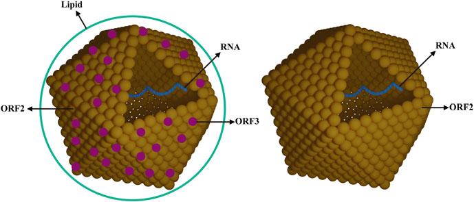
Although the virus particles were observed in stool samples of patients, the detailed information about structure of the virus particles was not clear. Thus, the study on structure of the virus particles is important, especially on comparing the structure difference for different sources of HEV virus particles. When HEV from cell culture systems or from the feces of infected patients was purified by ultracentrifugation, the ORF2 protein mainly stayed in the top fractions for both of these sample types. This finding indicates that most of the secreted ORF2 protein is not associated with HEV RNA. Infectious virions from these two sources, feces and cell culture supernatant, both formed single virion bands but their densities differed. The density of the virions from the cell culture supernatant changed to that of virions from feces when the lipid was removed with NP40. This indicates that, like HAV, HEV from cultured cells can hijack the host membrane to form an envelope. Several studies showed that the binding percentage of virions to anti-ORF3
antibody increased significantly when the lipids were removed from cell culture-derived virions. However, minimal binding to anti-ORF3 antibody was detected for virions from feces. This result indicates that infectious HEV virions produced in cell culture differ in structure from the virus found in feces. HEV infectious virions from cell culture have a lipid envelope containing little ORF3 and most of the ORF3 protein on these virions is protected by lipid, whereas the virions from feces have no lipid and more ORF3 (Fig. 1.2). In 2016, existing in both non-enveloped and enveloped (“eHEV ”) forms of HEV was declared to be a “quasi-enveloped” virus. The quasi-enveloped nature is a strategy used to protect the virus from neutralizing antibodies against protein ORF2 and ORF3 with the “eHEV” form in the blood stream [84]. However, the attachment and entry efficiency of eHEV particles is far less than that of non-enveloped HEV particles [85, 86].
HEV has been reported to be labile, which is extremely sensitive to freeze-thawing and spontaneously degrades when held at 4–8 °C for periods of time exceeding 3–5 days, and it will not tolerate exposure to high concentrations of salt. The computed sedimentation coefficient of HEV particles is approximately 183S, in contrast to157S for HAV. Additionally, the density of HEV particles from cell culture supernatant and sera is 1.10–1.15 g/cm3, while viruses derived from urine and feces have a density of
1.20–1.27 g/cm3. A detailed analysis of the quasi-enveloped particles revealed that they had a diameter of 39.6 ± 1.0 nm. After treatment with detergent and protease, the diameter of these VLPs shifted to 26.9 ± 0.9 nm [84].
1.6Conclusions and Perspective
Because the host-range of HEV is very wide and several genotypes or subtypes within each species have been reported, the virulent, pathogenic, and higher prevalence genotypes or subtypes of HEV still need to be defined. Studies on the HEV structure have indicated the ORF2 protein and the viral RNA band at different densities following ultracentrifugation. ORF3 protein and lipid are on the surface of HEV virus particles from the cell culture supernatant, but they are not present on HEV virus particles from feces and urine. The impact of the structure differences among viruses from different sources on the diagnosis and prevention of HEV infection should be investigated.
References
1. Khuroo MS (1980) Study of an epidemic of non-A, non-B hepatitis. Possibility of another human hepatitis virus distinct from post-transfusion non-A, non-B type. Am J Med 68(6):818–824
Fig. 1.2 Models of HEV from cell culture and feces. A model of HEV from cell culture is shown on the left and a model of HEV from feces is shown on the right
2. Wong DC, Purcell RH, Sreenivasan MA, Prasad SR, Pavri KM (1980) Epidemic and endemic hepatitis in India: evidence for a non-A, non-B hepatitis virus aetiology. Lancet 2(8200):876–879
3. De Cock KM, Bradley DW, Sandford NL, Govindarajan S, Maynard JE, Redeker AG (1987) Epidemic non-A, non-B hepatitis in patients from Pakistan. Ann Intern Med 106(2):227–230
4. Kane MA (1984) Epidemic non-A, non-B hepatitis in Nepal. Recovery of a possible etiologic agent and transmission studies in marmosets. JAMA 252(22): 3140–3145
5. Myint H, Soe MM, Khin T, Myint TM, Tin KM (1985) A clinical and epidemiological study of an epidemic of non-A non-B hepatitis in Rangoon. Am J Trop Med Hyg 34(6):1183–1189
6. Zhuang H (1991) [Advances in the research on non-A, non-B hepatitis in China]. Zhonghua Liu Xing Bing Xue Za Zhi 12(6):377–379
7. Hussaini SH, Skidmore SJ, Richardson P, Sherratt LM, Cooper BT, O’Grady JG (1997) Severehepatitis E infection during pregnancy. J Viral Hepat 4(1):51–54
8. Reyes GR, Purdy MA, Kim JP, Luk KC, Young LM, Fry KE et al (1990) Isolation of a cDNA from the virus responsible for enterically transmitted non-A, non-B hepatitis. Science 247(4948):1335–1339
9. Reyes GR, Yarbough PO, Tam AW, Purdy MA, Huang CC, Kim JS et al (1991) Hepatitis E virus (HEV): the novel agent responsible for enterically transmitted non-A, non-B hepatitis. Gastroenterol Jpn 26(suppl 3):142–147
10. Tam AW, Smith MM, Guerra ME, Huang CC, Bradley DW, Fry KE et al (1991) Hepatitis E virus (HEV): molecular cloning and sequencing of the full-length viral genome. Virology 185(1):120–131
11. Huang CC, Nguyen D, Fernandez J, Yun KY, Fry KE, Bradley DW et al (1992) Molecular cloning and sequencing of the Mexico isolate of hepatitis E virus (HEV). Virology 191(2):550–558
12. Bi SL, Purdy MA, McCaustland KA, Margolis HS, Bradley DW (1993) The sequence of hepatitis E virus isolated directly from a single source during an outbreak in China. Virus Res 28(3):233–247
13. Buisson Y, Grandadam M, Nicand E, Cheval P, van Cuyck-Gandre H, Innis B et al (2000) Identification of a novel hepatitis E virus in Nigeria. J Gen Virol 81 (pt 4):903–909
14. Chatterjee R, Tsarev S, Pillot J, Coursaget P, Emerson SU, Purcell RH (1997) African strains of hepatitis E virus that are distinct from Asian strains. J Med Virol 53(2):139–144
15. Gouvea V, Snellings N, Popek MJ, Longer CF, Innis BL (1998) Hepatitis E virus: complete genome sequence and phylogenetic analysis of a Nepali isolate. Virus Res 57(1):21–26
16. Meng J, Cong M, Dai X, Pillot J, Purdy MA, Fields HA et al (1999) Primary structure of open reading frame 2 and 3 of the hepatitis E virus isolated from Morocco. J Med Virol 57(2):126–133
17. Tsarev SA, Binn LN, Gomatos PJ, Arthur RR, Monier MK, van Cuyck-Gandre H et al (1999) Phylogenetic analysis of hepatitis E virus isolates from Egypt. J Med Virol 57(1):68–74
18. Tsarev SA, Emerson SU, Reyes GR, Tsareva TS, Legters LJ, Malik IA et al (1992) Characterization of a prototype strain of hepatitis E virus. Proc Natl Acad Sci U S A 89(2):559–563
19. van Cuyck-Gandre H, Zhang HY, Tsarev SA, Clements NJ, Cohen SJ, Caudill JD et al (1997) Characterization of hepatitis E virus (HEV) from Algeria and Chad by partial genome sequence. J Med Virol 53(4):340–347
20. Schlauder GG, Dawson GJ, Erker JC, Kwo PY, Knigge MF, Smalley DL et al (1998) The sequence and phylogenetic analysis of a novel hepatitis E virus isolated from a patient with acute hepatitis reported in the United States. J Gen Virol 79(pt 3):447–456
21. Meng XJ, Purcell RH, Halbur PG, Lehman JR, Webb DM, Tsareva TS et al (1997) A novel virus in swine is closely related to the human hepatitis E virus. Proc Natl Acad Sci U S A 94:9860
22. Wang Y, Ling R, Erker JC, Zhang H, Li H, Desai S et al (1999) A divergent genotype of hepatitis E virus in Chinese patients with acute hepatitis. J Gen Virol 80 (pt 1):169–177
23. Wang Y, Zhang H, Ling R, Li H, Harrison TJ (2000) The complete sequence of hepatitis E virus genotype 4 reveals an alternative strategy for translation of open reading frames 2 and 3. J Gen Virol 81(pt 7): 1675–1686
24. Payne CJ, Ellis TM, Plant SL, Gregory AR, Wilcox GE (1999) Sequence data suggests big liver and spleen disease virus (BLSV) is genetically related to hepatitis E virus. Vet Microbiol 68(1–2):119–125
25. Haqshenas G, Shivaprasad HL, Woolcock PR, Read DH, Meng XJ (2001) Genetic identification and characterization of a novel virus related to human hepatitis E virus from chickens with hepatitis-splenomegaly syndrome in the United States. J Gen Virol 82(pt 10): 2449–2462
26. Zhao C, Ma Z, Harrison TJ, Feng R, Zhang C, Qiao Z et al (2009) A novel genotype of hepatitis E virus prevalent among farmed rabbits in China. J Med Virol 81(8):1371–1379
27. Ma H, Zheng L, Liu Y, Zhao C, Harrison TJ, Ma Y et al (2010) Experimental infection of rabbits with rabbit and genotypes 1 and 4 hepatitis E viruses. PLoS One 5(2):e9160
28. Cossaboom CM, Cordoba L, Sanford BJ, Pineyro P, Kenney SP, Dryman BA et al (2012) Cross-species infection of pigs with a novel rabbit, but not rat, strain of hepatitis E virus isolated in the United States. J Gen Virol 93(pt 8):1687–1695
29. Izopet J, Dubois M, Bertagnoli S, Lhomme S, Marchandeau S, Boucher S et al (2012) Hepatitis E virus strains in rabbits and evidence of a closely related strain in humans, France. Emerg Infect Dis 18(8): 1274–1281
30. Yugo DM, Meng XJ (2013) Hepatitis E virus: foodborne, waterborne and zoonotic transmission. Int J Environ Res Public Health 10(10):4507–4533
31. Johne R, Plenge-Bonig A, Hess M, Ulrich RG, Reetz J, Schielke A (2010) Detection of a novel hepatitis E-like virus in faeces of wild rats using a nested broad-spectrum RT-PCR. J Gen Virol 91(pt 3): 750–758
32. Di Martino B, Di Profio F, Melegari I, Sarchese V, Robetto S, Marsilio F et al (2016) Detection of hepatitis E virus (HEV) in goats. Virus Res 225:69–72
33. Huang F, Li Y, Yu W, Jing S, Wang J, Long F et al (2016) Excretion of infectious hepatitis E virus into milk in cows imposes high risks of zoonosis. Hepatology 64(2):350–359
34. Li S, Liu M, Cong J, Zhou Y, Miao Z (2017) Detection and characterization of hepatitis E virus in goats at Slaughterhouse in Tai’an region, China. Biomed Res Int 2017:3723650
35. Yan B, Zhang L, Gong L, Lv J, Feng Y, Liu J et al (2016) Hepatitis E virus in yellow cattle, Shandong, Eastern China. Emerg Infect Dis 22(12):2211–2212
36. Sato Y, Sato H, Naka K, Furuya S, Tsukiji H, Kitagawa K et al (2011) A nationwide survey of hepatitis E virus (HEV) infection in wild boars in Japan: identification of boar HEV strains of genotypes 3 and 4 and unrecognized genotypes. Arch Virol 156(8): 1345–1358
37. Takahashi K, Kitajima N, Abe N, Mishiro S (2004) Complete or near-complete nucleotide sequences of hepatitis E virus genome recovered from a wild boar, a deer, and four patients who ate the deer. Virology 330(2):501–505
38. Takahashi M, Nishizawa T, Sato H, Sato Y, Jirintai, Nagashima S et al (2011) Analysis of the full-length genome of a hepatitis E virus isolate obtained from a wild boar in Japan that is classifiable into a novel genotype. J Gen Virol 92(pt 4):902–908
39. Neumann S, Hackl SS, Piepenschneider M, VinaRodriguez A, Dremsek P, Ulrich RG et al (2016) Serologic and molecular survey of hepatitis E virus in German deer populations. J Wildl Dis 52(1): 106–113
40. Thiry D, Mauroy A, Saegerman C, Licoppe A, Fett T, Thomas I et al (2017) Belgian wildlife as potential zoonotic reservoir of hepatitis E virus. Transbound Emerg Dis 64(3):764–773
41. Hammerschmidt F, Schwaiger K, Dahnert L, VinaRodriguez A, Hoper D, Gareis M et al (2017) Hepatitis E virus in wild rabbits and European brown hares in Germany. Zoonoses Public Health 64(8):612–622
42. Batts W, Yun S, Hedrick R, Winton J (2011) A novel member of the family Hepeviridae from cutthroat trout (Oncorhynchus clarkii). Virus Res 158(1-2):116–123
43. Drexler JF, Seelen A, Corman VM, Fumie Tateno A, Cottontail V, Melim Zerbinati R et al (2012) Bats worldwide carry hepatitis E virus-related viruses that form a putative novel genus within the family Hepeviridae. J Virol 86(17):9134–9147
44. Raj VS, Smits SL, Pas SD, Provacia LB, MoormanRoest H, Osterhaus AD et al (2012) Novel hepatitis E virus in ferrets, the Netherlands. Emerg Infect Dis 18(8):1369–1370
45. Woo PC, Lau SK, Teng JL, Tsang AK, Joseph M, Wong EY et al (2014) New hepatitis E virus genotype in camels, the Middle East. Emerg Infect Dis 20(6): 1044–1048
46. Xi JN, Graham DY, Wang KN, Estes MK (1990) Norwalk virus genome cloning and characterization. Science 250(4987):1580–1583
47. Green KY, Ando T, Balayan MS, Berke T, Clarke IN, Estes MK et al (2000) Taxonomy of the caliciviruses. J Infect Dis 181(suppl 2):S322–S330
48. Koonin EV, Gorbalenya AE, Purdy MA, Rozanov MN, Reyes GR, Bradley DW (1992) Computerassisted assignment of functional domains in the nonstructural polyprotein of hepatitis E virus: delineation of an additional group of positive-strand RNA plant and animal viruses. Proc Natl Acad Sci U S A 89(17): 8259–8263
49. Emerson SU, Nguyen H, Graff J, Stephany DA, Brockington A, Purcell RH (2004) In vitro replication of hepatitis E virus (HEV) genomes and of an HEV replicon expressing green fluorescent protein. J Virol 78(9):4838–4846
50. Smith DB, Simmonds P, Members of the International Committee on the Taxonomy of Viruses Study Group, Jameel S, Emerson SU, Harrison TJ et al (2014) Consensus proposals for classification of the family Hepeviridae. J Gen Virol 95(pt 10):2223–2232
51. Purdy MA (2022) ICTV virus taxonomy profile: Hepeviridae 2022. J Gen Virol (In press)
52. Meng XJ (2010) Hepatitis E virus: animal reservoirs and zoonotic risk. Vet Microbiol 140(3-4):256–265
53. Pavio N, Meng XJ, Renou C (2010) Zoonotic hepatitis E: animal reservoirs and emerging risks. Vet Res 41(6):46
54. Geng Y, Zhao C, Song A, Wang J, Zhang X, Harrison TJ et al (2011) The serological prevalence and genetic diversity of hepatitis E virus in farmed rabbits in China. Infect Genet Evol 11(2):476–482
55. Geng Y, Zhang H, Li J, Huang W, Harrison TJ, Zhao C et al (2013) Comparison of hepatitis E virus genotypes from rabbits and pigs in the same geographic area: no evidence of natural cross-species transmission between the two animals. Infect Genet Evol 13:304–309
56. Lu L, Li C, Hagedorn CH (2006) Phylogenetic analysis of global hepatitis E virus sequences: genetic diversity, subtypes and zoonosis. Rev Med Virol 16(1): 5–36
57. Smith JH, Schwedt TJ (2014) Consensus treatment of medication overuse headache in Latin America and Europe. Cephalalgia 34(9):643–644
58. Bradley DW, Purdy MA, Reyes GR (1991) Hepatitis E virus genome. Molecular features, expression of immunoreactive proteins and sequence divergence. J Hepatol 13 suppl 4:S152–S154
59. Reyes GR, Huang CC, Tam AW, Purdy MA (1993) Molecular organization and replication of hepatitis E virus (HEV). Arch Virol Suppl 7:15–25
60. Graff J, Torian U, Nguyen H, Emerson SU (2006) A bicistronic subgenomic mRNA encodes both the ORF2 and ORF3 proteins of hepatitis E virus. J Virol 80(12):5919–5926
61. Huang YW, Opriessnig T, Halbur PG, Meng XJ (2007) Initiation at the third in-frame AUG codon of open reading frame 3 of the hepatitis E virus is essential for viral infectivity in vivo. J Virol 81(6): 3018–3026
62. Ahola T, Karlin DG (2015) Sequence analysis reveals a conserved extension in the capping enzyme of the alphavirus supergroup, and a homologous domain in nodaviruses. Biol Direct 10:16
63. Nan Y, Zhang YJ (2016) Molecular biology and infection of hepatitis E virus. Front Microbiol 7:1419
64. Parvez MK (2013) Molecular characterization of hepatitis E virus ORF1 gene supports a papain-like cysteine protease (PCP)-domain activity. Virus Res 178(2): 553–556
65. Parvez MK (2017) The hepatitis E virus nonstructural polyprotein. Future Microbiol 12:915–924
66. Dosztanyi Z, Chen J, Dunker AK, Simon I, Tompa P (2006) Disorder and sequence repeats in hub proteins and their implications for network evolution. J Proteome Res 5(11):2985–2995
67. Kenney SP, Xiang-Jin M (2015) The lysine residues within the human ribosomal protein S17 sequence naturally inserted into the viral nonstructural protein of a unique strain of hepatitis E virus are important for enhanced virus replication. J Virol 89:3793
68. Ankavay M, Montpellier C, Sayed IM, Saliou JM, Wychowski C, Saas L et al (2019) New insights into the ORF2 capsid protein, a key player of the hepatitis E virus lifecycle. Sci Rep 9(1):6243
69. Montpellier C, Wychowski C, Sayed IM, Meunier JC, Saliou JM, Ankavay M et al (2018) Hepatitis E virus lifecycle and identification of 3 forms of the ORF2 capsid protein. Gastroenterology 154(1):211–223. e218
70. Yin X, Ying D, Lhomme S, Tang Z, Walker CM, Xia N et al (2018) Origin, antigenicity, and function of a secreted form of ORF2 in hepatitis E virus infection. Proc Natl Acad Sci U S A 115(18):4773–4778
71. ShiotaT,LiT-C,YoshizakiSetal(2013)ThehepatitisE virus capsid C-terminal region is essential for the viral life cycle: implication for viral genome encapsidation and particle stabilization. J Virol 87(10):6031
72. Nishiyama T, Umezawa K, Yamada K, Takahashi M, Kunita S, Mulyanto et al (2021) The capsid (ORF2) protein of hepatitis E virus in feces is C-terminally truncated. Pathogens 11(1):24
74. Yang Y, Lin S, Nan Y, Ma Z, Yang L, Zhang Y (2016) A linear surface epitope in a proline-rich region of ORF3 product of genotype 1 hepatitis E virus. Viruses 8(8):227
75. Ahmad I, Holla RP, Jameel S (2011) Molecular virology of hepatitis E virus. Virus Res 161(1):47–58
76. Holla RP, Ahmad I, Ahmad Z, Jameel S (2013) Molecular virology of hepatitis E virus. Semin Liver Dis 33(1):3–14
77. Kannan H, Fan S, Patel D, Bossis I, Zhang YJ (2009) The hepatitis E virus open reading frame 3 product interacts with microtubules and interferes with their dynamics. J Virol 83(13):6375–6382
78. Graff J, Nguyen H, Yu C, Elkins WR, St Claire M, Purcell RH et al (2005) The open reading frame 3 gene of hepatitis E virus contains a cis-reactive element and encodes a protein required for infection of macaques. J Virol 79(11):6680–6689
79. Nagashima S, Takahashi M, Jirintai TT, Yamada K, Nishizawa T et al (2011) A PSAP motif in the ORF3 protein of hepatitis E virus is necessary for virion release from infected cells. J Gen Virol 92(2):269–278
80. Yamada K, Takahashi M, Yu H, Takahashi H, Okamoto H (2009) ORF3 protein of hepatitis E virus is essential for virion release from infected cells. J Gen Virol 90(pt 8):1880–1891
81. Nair VP, Anang S, Subramani C, Madhvi A, Bakshi K, Srivastava A et al (2016) Endoplasmic reticulum stress induced synthesis of a novel viral factor mediates efficient replication of genotype-1 hepatitis E virus. PLoS Pathog 12(4):e1005521
82. Balayart MS, Andjaparidze AG, Savinskaya SS, Ketiladze ES, Braginsky DM, Savinov AP et al (1983) Evidence for a virus in non-A, non-B hepatitis transmitted via the fecal-oral route. Intervirology 20(1):23–31
83. Bradley DW, Krawczynski K, Cook EH Jr, McCaustland KA, Humphrey CD, Spelbring JE et al (1987) Enterically transmitted non-A, non-B hepatitis: serial passage of disease in cynomolgus macaques and tamarins and recovery of disease-associated 27-to 34-nm virus like particles. Proc Natl Acad Sci U S A 84(17):6277–6281
84. Kiyoshi H, Daniela B, Eberhard H (2018) Life cycle and morphogenesis of the hepatitis E virus. Emerg Microbes Infect 7:196
85. Nagashima S, Takahashi M, Kobayashi T, Tanggis NT, Nishiyama T et al (2017) The characterization of the quasi-enveloped hepatitis E virus particles released by the cellular exosomal pathway. J Virol 91:e0082217
86. Xin Y, Charuta A, Yurong L et al (2016) Distinct entry mechanisms for nonenveloped and quasi-enveloped hepatitis E viruses. J Virol 90:4232
73. Osterman A, Vizoso Pinto MG, Haase R, Nitschko H, Jager S, Sander M et al (2012) Systematic screening for novel, serologically reactive hepatitis E virus epitopes. Virol J 9:28
Hepatitis E virus (HEV) is a non-enveloped virus containing a single-stranded, positivesense RNA genome of 7.2 kb, which consists of a 5′ non-coding region, three open reading frames (ORFs), and a 3′ non-coding region. ORF1 is diverse between genotypes and encodes the nonstructural proteins, which include the enzymes needed for virus replication. In addition to its role in virus replication, the function of ORF1 is relevant to viral adaption in culture and may also relate to virus infection and HEV pathogenicity. ORF2 protein is the capsid protein, which is about 660 amino
Y. Zhou
RegCMC, Great Regulatory Affairs, Sanofi (China) Investment Co., Ltd, Beijing, China
e-mail: Yan5.Zhou@sanofi.com
C. Zhao · N. Xu
Division of HIV/AIDS and Sex-Transmitted Virus Vaccines, National Institutes for Food and Drug Control, Beijing, China
acids in length. It not only protects the integrity oftheviralgenome,butisalsoinvolvedinmany important physiological activities, such as virus assembly, infection, host interaction, and innate immune response. The main immune epitopes, especially neutralizing epitopes, are located on ORF2 protein, which is a candidate antigen for vaccine development. ORF3 protein is a phosphoprotein of 113 or 114 amino acids with a molecular weight of 13 kDa with multiple functions that can also induce strong immune reactivity. A novel ORF4 has been identified only in genotype 1 HEV and its translation promotes viral replication.
Keywords
Expression · Function · Protein · Pathogenicity · Replication · Adaption
# The Author(s), under exclusive license to Springer Nature Singapore Pte Ltd. 2023
Y. Wang (ed.), Hepatitis E Virus, Advances in Experimental Medicine and Biology 1417, https://doi.org/10.1007/978-981-99-1304-6_2
16Y.Zhouetal.
Hel RNA helicase
HVR Proline-rich hypervariable region
IRF3 IFN regulatory factor 3
ISG IFN-stimulated gene
MAPK Mitogen-activated protein kinases
MeT Methyltransferase
ORF Open reading frame
PCP Papain-like cysteine protease
PLG Plasminogen
RdRp RNA-dependent RNA polymerase
RIG-I Retinoic acid-inducible gene I
STAT Signal transducer and activator of transcription
VDAC Voltage-dependent anion channel
VLP Virus-like particle
Hepatitis E virus (HEV) contains three open reading frames (ORFs) and encodes three proteins, each of which has unique features and functions.
Although HEV can be divided into many genotypes within the genus and species of family Hepeviridae, all HEV genotypes that can infect humans are located in orthohepevirus A and have been designated as genotypes 1–4. Thus, the three HEV proteins described in this chapter focus on genotypes 1–4, and the positions of nucleic acids and amino acids referred to those of genotype 1. Besides the three ORFs, a noval ORF4 was found only in genotype 1, which overlaps within ORF1.
2.1ORF1 Protein
2.1.1
Structural Features of ORF1 Proteins
ORF1, which is 5082 bp long, is located on the 5′ terminus of the HEV genome and encodes a nonstructural polyprotein of 1693 amino acid residues. The functional domains of this polyprotein consist of methyltransferase (MeT), Y domain, papain-like cysteine protease (PCP), proline-rich hypervariable region (HVR or polyproline region; PPR), X domain (macro domain),RNAhelicase(Hel),and RNA-dependent RNA polymerase (RdRp) [1, 2] (Fig. 2.1).
2.1.2 Expression of ORF1
Two products, N-78 kDa and C-107 kDa, were obtained when expressing ORF1 in mammalian cells by using recombinant vaccinia virus [3]. Expressing ORF1 in an Escherichia coli plasmid expression system or in HapG2 carcinoma cells yielded only an unprocessed polyprotein of 186 kDa, but no processed functional unit products [4, 5]. In contrast, in vitro transfection of HepG2 cells with infectious clones containing the whole HEV genome yielded different ORF1 processed products. The bands 35 kDa (MeT), 38 kDa (Hel), and 36 kDa (RdRp) were identified from these expression products by using antiMeT, anti-Hel, and anti-RdRp antibodies [6]. Only a sole 191 kDa polyprotein was produced when the recombinant plasmid pTriExORF1 expressed in an in vitro transcription –translation system, but when this plasmid was transfected into S10-3 cells, an N-terminal product of 35 kDa and a C-terminal product of 78 kDa were detected by an immunoprecipitation assay [7]. When expressing ORF1 in a baculovirusinsect system in the form of fusion protein His6ORF1-Flag, a polyprotein of 192 kDa was produced, and the number of processed short fragments that reacted with anti-His and antiFlag antibodies increased overtime. This processing procedure could be inhibited by the cysteine proteinase inhibitor (E-64d) [8]. The 410–610 amino acid ORF1 fragment expressed in E. coli C43 showed disintegrating activity to nonstructural protein ORF1 and structural protein ORF2. The results of a mass spectrometry analysis indicate that ORF1 protein can be digested into N-terminal 35 kDa methyltransferase and C-terminal 35 kDa replicase by the expressed 410–610 amino acid ORF1 protein. The cleavage sites were G-15/I-16 and A-1364/V-1365, which confirmed the in vitro ORF1 protein disintegrating activity of this PCP-like proteinase [9].
Presently, the difference in function among the ORF1 proteins expressed by different systems and the extent of the involvement of host proteinase in ORF1 expression are not clear. Additionally, the expression of nonstructural ORF1 proteins after HEV infection has not been
Fig. 2.1 HEV ORF1 protein domains. A schematic of the ORF1 protein domains: methyltransferase (MeT), Y domain, papain-like cysteine protease (PCP), proline-rich
reported. Furthermore, it is not completely clear whether or not the functional domains of ORF1 proteins are processed to produce entities having biochemical function. Further studies are needed to address these questions.
2.1.3 Virus Infection and Pathogenicity Relevant to ORF1
An investigation into the heterogeneity of the HEV ORF1 gene and the outcome of infection in solid-organ transplant patients during the hepatitis E acute phase found that the entropy and genetic distance of HEV sequences in chronic hepatitis E patients were higher than those in patients who cleared the virus. Specifically, the PPR and macro domains of ORF1 were dramatically higher. The high genetic heterogeneity of the PPR and macro domains may be associated with persistent infection of HEV virus in the acute period due to regulation of the host immune response by mutation [10]. The macro domain protein was identified interacts with light chain subunit of human ferritin (FTL), the FTL is the acute phase protein elevated secretion in acute viral hepatitis. In the hepatoma culture cells, macro domain lowered the secretion of ferritin, but did not influence the FTL expression level and did not interfere the cellular iron homeostasis/metabolism either. The inhibition secretion of ferritin by macro domain possibly suppresses innate immune response [11].
The HVR domain may play a vital role in HEV pathogenicity as described in Chap. 5. Bu et al. [12] sequenced a strain of genotype 4 HEV that was collected from a patient with hepatic failure and compared it with other HEV genotype
hypervariable region (V), X domain (macro), RNA helicase (Hel), and RNA-dependent RNA polymerase (RdRp)
4 isolates; they found that 12 amino acid residues in ORF1 and three amino acid residues in ORF2 were substituted. Moreover, a comparative analysis of the mutations present in the nucleic acid/ amino acid sequences of ORF1 in genotypes 4 and 3 found mutations in 12 amino acid residues, with 11 mutations in the PCP domain and the remaining one in the RdRp domain [13]. Mishra et al. [14] compared amino acid sequences between strains of genotype 1 from patients with fulminant hepatic failure, as well as with the genotype 1 strains from acute virus hepatitis patients in the same subcontinent. Six identical substitutions in HEV strains of all fulminant patients occurred only in ORF1, namely F179S, A317T, T735I, L1110F, V1120I, and F1439Y. These mutations were significantly associated with the fulminant hepatic failure caused by genotype 1. It was reported that the nonsense mutation of U3148 in the Hel domain of ORF1 was associated with the severity of hepatitis E [15]. Billam et al. [16] aligned the complete sequences of a non-pathogenic and a pathogenic poultry HEV strain and found that the highest number of mutations was in ORF1 with 41 mutated sites, whereas there were only 10 mutated sites in the other ORFs. These discoveries indicate that ORF1 may have relationship with the pathogenicity of HEV.
2.1.4 ORF1 and Virus Replication
Capped RNA transcripts of HEV cDNA clones were able to be transfected into Huh-7 cells where they successfully replicated. These transcripts showed infectivity and were also able to produce virions when inoculated intravenously into swine. In contrast, uncapped RNA transcripts did not
show these abilities [17]. Notably, the activities of methyltransferase and guanyltransferase in the MET domain could be detected in the 110 kDa polyprotein expressed in baculovirus [18]. Additionally, the capping of genomic RNA could be confirmed by HEV 5′ RNA ligase-mediated rapid amplification of the cDNA ends, which selectively amplify capped RNAs [19]. The methyltransferase activities catalyze RNA capping, and the removal of the 5′ terminal γ-phosphorous group on the initial transcript by RNA triphosphatase is the key step of capping. Study showed that co-incubation of HEV helicase with 5′-[γ-32P] RNA and 5′-[α-32P] RNA released 32P from 5′-[γ-32P] RNA only, indicating the speci ficity of the helicase to a γ-β-triphosphate bond. These findings suggest that HEV RNA helicase might mediate the first step of 5′-terminal capping [20]. RNA helicase is necessary for the genomic replication of positive-sense RNA viruses. HEV RNA helicase displays nucleotide triphosphatase activity and has an RNA-binding domain [20]. When the Hel domain on HEV ORF1 amino acid position (aa)960–1204 was expressed in prokaryotic cells, the HEV RNA helicase was able to hydrolyze all rNTPs (ribonucleotide triphosphates) but showed lower hydrolysis activity against dNTPs (deoxyadenosine nucleoside triphosphates). This enzyme has only unwinding activity in the 5′→3′ direction to 5′-sticky double-stranded RNA [21].
A recombinant HEV RdRp expressed in E. coli was able to bind to the 3′-terminal non-coding region of the HEV genome and used 3′-polyadenylated HEV RNA as a template to synthesize complementary strands [22]. A study of HEV infection in A549 cells and suckling pigs found that RNA interference to RdRp could effectively inhibit the replication of HEV [23]. Karpe et al. [24] found that an active ubiquitin-proteasome system was necessary for HEV replication and that this could be inhibited by a proteasome inhibitor. Notably, the overexpression of ubiquitin in proteasome inhibitor-treated cells partially reversed the inhibition of HEV replication.
The protein expressed by the PCP domain has de-ubiquitin enzymatic activity, and PCP may be involved in the replication of HEV via this enzymatic activity. Notably, the mutations of G816V and G817V in G815-G816-G817 of the X domain prevented virus replication. Additionally, the mutation N806A did not preclude RNA replication, whereas the mutations N809A and H812L resulted in a lack of live virus, indicating the involvement of X domain amino acid residues at the post-translation stage of HEV replication [7, 25].
Besides, universally conserved residues C336, C337, and W413 in Y-domain was predicted for cytoplasmic membrane binding. The site-directed mutant replicons on the three residues (C336A, C337A, and W413A) showed no replication. Saturation mutants corresponded to the aa C336,C337, and W413 codons (nts 1031–1033, 1034–1036, and 1213–1215, respectively) showed very mild or insigni ficant effect on replication, which indicated C336, C337, and W413 were indispensable for the post-translational functional/structural of virus replication, probably through membrane binding in intracellular replication complexes.
Saturation mutants of three sequences (nts 788–856, nts 857–925, nts 926–994) predicted of stable RNA hairpin/stem-loop structures severely affected virus replication and virion infectivity. The mutations could completely unzip and destabilize the RNA secondary structures, which is critical for virus replication and infectivity. These indicated Y domain plays crucial role in the RNA replication of HEV virus [26].
A putative factor Xa cleavage site and two thrombin cleavage sites on HEV pORF1 were found, which highly conserved in HEV genotypes [27]. Mutations using a reverse genetics approach in the cleavage sites of either thrombin or factor Xa showed the reduction of HEV replication in cell culture. Thus, cleavage site sequences in pORF1 are essential for HEV replication in cell culture. Factor Xa and thrombin are serine protease, which may involve in the HEV pORF1 processing [27].
2.1.5 ORF1 and Viral Adaption
Aided by biological software, Purdy et al. [74] computerized and forecasted PPRs, informatics entropy, selective pressure, homoplastic density, intrinsically disordered regions (IDRs), linear motifs, electrostatic surfaces, secondary structures, structure-based functions, and protein-binding sites of ORF1 PPR sequences from four HEV genotypes, and found that the PPRs from four HEV genotypes were IDRs that all contained seven putative linear binding motifs for ligands. Structural analysis of the molecular functions of these motifs indicated that PPRs tended to bind to various ligands. The existence of nucleotide mutations in PPR was due to high frequencies of insertion and deletion. Although the mutation rate of PPR is the same as that of other ORF1 domains, PPR has a higher tolerance than the other ORF1 domains for substitution between its first and second codes. This high mixture led to more proline, glycine, serine, and threonine instead of histidine, phenylalanine, tryptophan, and tyrosine, indicating that these regions are typical proline-rich IDRs. Alignment analysis on PPR sequences from HEV strains of all genotypes found a common origin for animal strains and a higher tolerance to mutation in carboxyl moieties than in the remaining PPR domain amino acid residues. In contrast with other nonstructural polyproteins, the evolution of HEV PPR appears to have been shaped under selective pressure to use more proline and fewer aromatic amino acids, a ratio which favors the formation of an IDR structure. IDRs are able to bind to various ligands and have a regulatory effect on transcription and translation [73]. Therefore, PPR may play a key role in the ability of HEV adaption to different circumstances.
Izopet et al. [45] compared the sequence of rabbit HEV with that of human HEV, and they found that one human HEV strain was very close to rabbit HEV. There was an insertion of 93 nucleotides in the ORF1 X domain of the human HEV strain and rabbit HEV strains. This study suggested that the host range of HEV had
been expanding and that rabbit HEV may be transmitted among animals. By using recombination software RDP and SimPlot to analyze the intra-genotype and inter-genotype differences in the HEV genome, it was found that the recombinant fragments are non-randomly distributed in the HEV genome. The X domain, Hel, and RdRp were all hot spots with high recombination rates. These nonrandom distributions were due to the high adaption of recombination in this region as well as to the effects of natural selection [32].
2.2ORF2 Protein
ORF2 protein, which is approximately 660 amino acids in length, is translated from a 2.2 kb subgenome into the capsid protein. There is a conserved stem-loop structure in ORF2 that may be related to early-stage viral replication [37]. Moreover, it was reported that ORF2 could speci fically bind to the HEV genome RNA at the 5′ end and plays an essential part in the HEV assembly process [83]. As the capsid protein, ORF2 not only protects the integrity of the viral genome but is also involved in many important physiological activities, such as virus assembly, infection, and host immunity. It has an N-terminal signal sequence by which it is co-translated into the endoplasmic reticulum (ER) where it is sequentially N-linked glycosylated [28]. Since the C-terminal of the ER signal sequence of ORF2 includes an arginine-rich domain, it is expected to play a key role in genomic RNA-binding and HEV assembly. Moreover, the C-terminal 52 amino acid residues of ORF2 are also involved in HEV genome encapsidation and stabilization of the capsid particles [82].
2.2.1 Expression of HEV ORF2 Protein
Although growth of HEV in cell culture has been reported [43, 51, 97], the quantities of natural HEV proteins that are produced in this manner are not sufficient for further study. The structural
proteins of HEV have been expressed using various expression systems, including bacteria, mammalian cells, baculovirus, yeast, and vaccinia.
The full ORF2 [71], the carboxyl-terminal one-third of ORF2, and the carboxyl-terminal two-thirds of ORF2 were expressed in E. coli as fusion proteins with glutathione S-transferase (GST) or as trpE-HEV fusion proteins [56, 75]. Based on the primary structure of synthetic peptides possessing HEV-specific antigenic activity, mosaic proteins of HEV representing aa394–470, aa562–580, and aa631–660 of ORF2 and aa91–123 of ORF3 from the Burmese strain and the same regions from the Mexican strain were designed and similarly expressed in E. coli as GST-fusion proteins [53, 54]; for each strain, only one recombinant protein band of HEV ORF2 or ORF3 was observed in the E. coli expression system and this band had a molecular weight of the expected size of a protein lacking glycosylation and proteolytic posttranslation processing. Another group found that the ORF2 E239 peptide could be expressed in an E. coli system, and this expression successfully forms particles, which have been developed as an HEV vaccine [57].
Full ORF2 proteins have been expressed in COS-1, HepG2, and BHK-21 cells. Three forms of ORF2 protein were observed in normal transfected cells using both plasmid-based expression [46] and the Semliki Forest virus (SFV) vector [90] with molecular weights of 72–74 kDa, 79–82 kDa, and 84–88 kDa, even though the expected size for ORF2 protein is approximately 72 kDa. The glycosylation status of ORF2 was evaluated in experiments in which tunicamycin was used to inhibit glycosylation in transfected cells [46]. This experiment confirmed that the 74 kDa protein is the ORF2 protein without glycosylation. The two larger proteins were identified as glycosylated forms of ORF2 with different extents of glycosylation. Through pulse-chase analysis, tunicamycin inhibition, and endoglycosidase sensitivity, studies have found that ORF2 protein is likely co-translationally translocated via its N-terminal signal sequence into the ER. The protein may be glycosylated in the ER at asparagine residues at
one or more sites. When the signal peptide sequence was deleted, the modified ORF2 construct was found to express only one form of protein, and no glycosylated forms were observed, even though all three of the potential glycosylation sites were located in the construct. These results suggest that the signal peptide sequence can direct the protein into the ER for glycosylation. An expression analysis of ORF2 also found that ORF2 protein is expressed intracellularly, as well as on the cell surface [104]
The main advantage of using baculovirus for expression is that, in most cases, proteins expressed in insect cells are processed in the same way they would be in mammalian cells, resulting in authentic, functional proteins. Additionally, the protein expression levels in insect cells may also be much higher than those in mammalian cells. The full ORF3 and ORF2 from the Burmese strain were expressed in Sf9 cells using a baculovirus-expressing vector [41]. ORF2 protein, with a molecular weight of approximately 70.9 kDa, was observed in cell pellets, and no recombinant proteins were identified in cell supernatants. In another study, the major protein band had an apparent molecular weight of 75 kDa [92]. Minor bands were also observed, and some of these bands had molecular weights of more than 75 kDa, possibly due to varying extents of glycosylation, while others had lower molecular weights, which may reflect proteolytic processing post-translation. In a further study, three major bands were observed, including the complete structural protein at the earlier stages of recombinant baculovirus infection as well as two products of proteolytic cleavage (55 kDa and 63 kDa) at the later stages [91]. The 75 kDa protein is slightly larger than the predicted size for the entire ORF2, and this size difference could be due to glycosylation. An analysis of the other two proteins (55 kDa and 63 kDa) found that they were co-terminal at the amino end and shared the same N-terminal Ala-112. The 63 kDa protein had an intact C-terminus, while the 55 kDa protein was the product of an additional cleavage of 51 amino acids from the C-terminus [80]. The C-terminally truncated ORF2 was shown in
2CharacteristicsandFunctionsofHEVProteins21
fecal HEV, which molecular weight is 60 kDa [70].
The entire ORF2 was also expressed in two insect cell lines, Sf9 and Tn5. Three major proteins with molecular weights of 72, 58, and 50 kDa were found in the lysates of both cell types. All three of these proteins have reactivity to anti-HEV antibody-positive sera. They were tightly cell-associated and were not found in the culture supernatant. However, when the structural protein derived from ORF2 aa112–660 was expressed in the Tn5 cell line, a large amount of protein with a molecular weight of 50 kDa was produced and efficiently released into the culture medium [60]. Through electron microscopy, the 50 kDa protein was found to form empty viruslike particles (VLPs) in the culture medium. Li et al. later found that these HEV VLPs induced a strong immune response after their oral administration in mice and monkeys [58, 59]. They demonstrated that both ORF2 aa126–601 and aa112–608 can form T = 1 particles. When the RNA fragment was contained, ORF2 aa112–608 could also form T = 3 particle. The average size of T = 1 particles was 27 nm and that of T = 3 particles was 41 nm [40, 96, 99].
When the peptide sequence of aa126–621 in HEV genotype 4 was expressed in insect cells, two forms were obtained in the cells, VLP and non-VLP. The two forms have obvious differences in their granularity, which was reflected in their behavior during ultracentrifugation, dynamic light scattering, and chromatographic analysis, as well as by their appearance during electron microscopy [76]. VLPs have a more uniform granularity than non-VLPs. Although VLPs and non-VLPs behaved identically on SDS-PAGE, different peptides were produced when they were digested with the same enzymes under the same conditions. The peptide mapping detected by using LC-MS/MS showed that they have different post-translational modi fications. Additionally, VLPs induced stronger immune responses than non-VLP. Thus, increasing the yield of VLPs in insect expression system is important.
The translation of HEV ORF2 is predicted to yield a 72 kDa protein including a putative signal
sequence and potential sites of N-linked glycosylation [88]. When the full-length ORF2 of the Burmese, Pakistan, and Japanese strains was expressed in the baculovirus system, the predicted 72 kDa protein products were processed into a 50, 55, 58, or 62 kDa protein due to posttranslational processing [60, 108]. However, similar processing of HEV ORF2 protein was not observed in mammalian cells. When the fulllength ORF2 was expressed in mammalian cells [46, 90], multiple ORF2-specific proteins with molecular weights estimated as 72–74, 79–84, and 84–88 kDa were detected. The smallest of these proteins with a molecular weight of 72–74 kDa corresponds to the predicted size, and the larger proteins, 79–84 kDa and 84–88 kDa, reflect the super-glycosylation of the HEV ORF2 protein in mammalian cells [89]. Thus, different expression systems may result in different post-translation modifications of the same protein.
2.2.2 ORF2 Involved in HEV Infection and Intercellular Transduction
Interactions between ORF2 and extracellular matrix proteins were detected in host cells. It was reported that once heparan sulfate was removed from the cell surface by heparinase or sodium chlorate treatment, the binding of HEV VLP to the Huh-7 cell surface was significantly reduced. Syndecan-1 plays an important role in the ORF2 binding process because it is a ubiquitous proteoglycan on the cell [48]. However, the interaction between ORF2 and heparan sulfate proteoglycan is a nonspecific adsorption, and the identity of a HEV-specific receptor remains unknown. Although HEV is a non-enveloped virus, it may use heparan sulfate proteoglycan as its cell adhesion receptor, similarly to enveloped viruses.
Studies found that some chaperones and cytoskeleton proteins take part in HEV intercellular transportation through their interaction with HEV capsid protein ORF2. During the protein synthesis process, chaperones recognize partially folded polypeptides, and they participate in peptide
folding and help the protein assemble correctly. The heat-shock proteins are the main subset of chaperones. Some heat-shock proteins, such as heat-shock protein 90 (HSP90), were demonstrated to take part in the early stage of HEV infection. The p239 VLPs formed from recombinant ORF2 proteins were used to investigate the cellular interactions. HSP90, Grp78/Bip, and α-tubulin were identified as binding ORF2 by matrix-assisted laser desorption/ionization time of flight mass spectrometry. Inhibition of HSP90 blocked the p239 transportation in HepG2 cells, but it did not affect p239 cellular entry. The speci fic HSP90 inhibitor also significantly obstructed the transportation of HEV. Together, these findings indicate that HSP90 plays a key role in HEV intercellular transportation [109].
Asialoglycoprotein receptor 2 (ASGR2) is a transmembrane glycoprotein that is highly expressed in liver tissue. ASGR2 and ASGR1 can be composed of homologous or heterologous dimmers of ASGPR, which mediate the lysosomal-dependent degradation of various desialylated glycoproteins in hepatocytes. In recent years, ASGPR was found to play an important role in hepatitis A virus, hepatitis B virus, and hepatitis C virus infection, indicating that it may mediate hepatitis virus entry into cells [35, 81, 106]. Additionally, ASGPR is involved in and facilitates HEV infection by binding to ORF2, but the mechanism is still unclear [107].
2.2.3 ORF2 and the Endoplasmic Reticulum Stress Response (ERSR)
Numerous proteins are translated and modified on the ER. Some pathological changes can destroy the balance of intercellular protein translation and post-translational modi fication, leading to the accumulation of incorrectly folded peptides and inappropriately modi fied proteins. The unfolded or misfolded proteins form many aggregates, causing ER stress. John et al. found that overexpression of HEV ORF2 in cells induced ER stress by activating the amino acid response
elements of the pro-apoptotic gene C/EBP homologous protein (CHOP), which is a pro-apoptotic gene. The ORF2 proteins activate the transcription of CHOP by increasing the phosphorylation level of eukaryotic initiation factor 2 alpha (eIF2α). However, the ERSR caused by ORF2 overexpression did not cause the expected apoptosis of host cells. At the same time, it was reported that the expression of heat-shock proteins Hsp72, Hsp70B, and Hsp40 were upregulated in ORF2 overexpressing cells, which indicates the increased chaperones may help HEV-infected cells avoid apoptosis [47]. Furthermore, the results of a co-immunoprecipitation study demonstrated the protein–protein interaction between ORF2 and Hsp72 in vivo, which indicates that Hsp72 might facilitate the ORF2 folding process. Meanwhile, the nuclear accumulation of Hsp72 appeared in the cells that expressed HEV ORF2 [47].
Incorrectly folded peptides should degrade through the ER-associated degradation (ERAD) process, and, because of this process, the ER unloads much of the pressure caused by viral infection [98]. Whether HEV ORF2 was overexpressed by the transfection of recombinant VLPs or by that of an ORF2-plasmid, only a fraction of ORF2 was located on the ER, and most of the rest of the ORF2 proteins were spread throughout the cytoplasm. It is possible that there is a connection between the ERSR and the retrotranslocation of ORF2 from the ER. Because ORF2 was co-translocated and N-linked glycosylated in the ER through its N-terminal signal sequence, it was initially believed that nearly all of the ORF2 proteins were located on the ER. If the N-linked glycosylation was blocked in host cells by tunicamycin or kifunensine, ORF2 can not be glycosylated and retrotranslocation to the cytoplasm was subsequently inhibited. Then, the interaction between ORF2 and p97 was confirmed by immunoprecipitation, which found evidence of interaction with p97 by both the full-length ORF2 and KDELORF2 (a mutagenesis by modifying the native C-terminal sequence of ORF2, KTREL, to KDEL). Additionally, GRP94, an ER stress-
inducible chaperone, was found to be upregulated in ORF2 overexpressed cells, as was protein disulfide isomerase (PDI) [84].
Nuclear factor kappa B (NF-κB) plays an essential role in the host cell survival during infections by many different pathogens. The activation of NF-κB requires the phosphorylation and degradation of Iκβ to release the nuclear localization signal of the NF-κB dimmer. ORF2 blocks the ubiquitin–proteasome-mediated degradation of Iκβ, and, as a result, NF-κB activity is inhibited in HEV-infected human hematoma cells. In contrast, ORF2 showed direct interaction with a beta transducin repeat-containing protein (β TRCP), which is a member of the F-box protein family. Because of the competitive binding of ORF2, the associations of IκB with Cul1 and SKP1 were significantly reduced. As the HEV capsid protein, ORF2 plays a role in the survival of HEV-infected host cells, and it regulates the replication and amplification of the virus [86].
2.2.4 ORF2 Interferes Host Innate Immunity
The cytosolic pattern recognition receptor, retinoic acid-inducible gene I (RIG-I), was identified as the important IFN-stimulated gene (ISG). RIG-I is the major innate immune sensor for hepatitis E virus [101]. The full-length ORF2 was verified interfering with the RIG-I signaling, inhibiting the RIG-I dependent interferon response, and modulating the virus inducing RIG-I activation and synthetic agonist. IPS-1 is the adapter protein of RIG-I. When ORF2 co-expressed in HEK293Tcells with IPS-1, IPS-1 induced IFN-β promoter activation was significantly inhibited. Glycosylation or dimerization of the ORF2 protein had no effect on the shown inhibition [42].
Another study found that the ORF2 inhibited the induction of IFN. The arginine-rich-motif in the N-terminal domain of ORF2 blocked the phosphorylation of IFN regulatory factor 3 (IRF3) via interaction with the multiprotein complex consisting of mitochondrial antiviralsignaling protein (MAVS), TANK-binding
kinase 1 (TBK1), and IRF3. These findings showed that the ORF2 interfered with the host innate immunity [61].
2.3ORF3 Protein
2.3.1
Molecular Structure of ORF3 Protein
ORF3 protein, a phosphoprotein of 113 or 114 amino acids with a molecular weight of 13 kDa, is a protein with multiple functions. Analysis of its structure found that ORF3 protein consists of two highly hydrophobic domains, D1 (aa7–23) and D2(aa28–53), at its N-terminus and two proline-rich domains, P1 and P2 (Fig. 2.2). The D1 domain, which is rich in cysteine, is the binding site for microtubulin [49] and mitogenactivated protein kinase (MAPK) phosphatase [50] and also interacts with the cytoskeleton [105]. The D2 domain is a hydrophobic region that acts as a binding site for hemopexin. The PMS71PLR in the P1 domain contains two overlapping potential phosphorylation kinase motifs, of which PMS71P is a potential MAPK phosphorylation motif and S71PLR is a potential cyclin-dependent kinase (CDK) phosphorylation motif. The serine S71 can be phosphorylated by MAPK in ORF3-transfected cells in vitro [55]. However, it remains unknown whether or not S71 is phosphorylated during HEV infection. The ORF3 antigenic epitope is found in the P2 domain [55], where there are two overlapping PXXP motifs found in many viruses and associated with the signal transduction of cellular proteins. These motifs are binding sites for Src homology 3(SH3)-containing, structural-domain, signal transduction molecules, and they play a key role in the virus release of HEV [50] Interestingly, the PSAP motifs of genotype 3 and avian HEV ORF3 did not significantly impact virus infection but they played a role in the virion release of HEV [52, 66]. However, HEV infection and virus release were not detected in rhesus monkeys infected with ORF3-PXXP mutant strains [36]. These data indicate that PXXP plays a key role in virus release and budding,
but these processes are also related to the virus genotype and the host species.
2.3.2 ORF3 Protein and Host Cell Survival
ORF3 protein is able to interact with some signal transduction molecules, impact the pathway response of host cells, and maintain host cell survival. Kar-Roy et al. [50] reported that ORF3 protein could activate external cellular regulatory kinase (ERK) of the MAPK family, which was not dependent on the traditional RAF/MEK pathway, but instead inhibited the phosphatase activity of Pyst1 by the binding of ORF3 protein to Pyst1. Pyst1 is a member of the ERK-specific MAPK phosphatase family that can mediate the dephosphorylation of ERK, which inactivates ERK [64]. By binding its D1 region in the N-terminus to the central connection region of Pyst1, ORF3 blocks its phosphorylation, which prolongs the activated status of ERK and allows the continuous operation of the MAPK pathway and, therefore, potentially facilitates host cell survival and viral proliferation. ORF3 has also been shown to activate MAPK-JNK of the antiapoptosis signaling pathway. Parvez et al. transfected HEV replicon RNA or ORF3 expression vectors into hepatocarcinoma cells to quantitatively test the phosphorylation level of JNK1/2. They found that the phosphorylation level of JNK1/2 increased by approximately 66% in ORF3 vector-transfected cells, which is higher than the phosphorylation level of 54% that was observed in the replicon RNA-transfected cells (HEV replication cells). The phosphorylation of JNK1/2 is favorable for both the maintenance of host cell survival and the persistent infection of
HEV [72]. Chandra et al. [31] found that the subcellular location of ORF3 was in early and recycling endosomes, which delays the postinternalization activation of epidermal growth factor receptor (EGFR) to late endosomes/ lysosomes, therefore potentially prolonging the EGF-mediated intracellular signal. Further study in this lab found that ORF3 protein delays the internalized degradation of activated hepatocytic growth factor receptor (c-MET). However, this function is not related to the localization of ORF3 in endosomes [30]. The mechanism responsible for the prolongation of the growth factor receptor-mediated signal transduction by ORF3 protein may be related to the competitive binding of Cbl to CIN85, which is an adaptor protein that mediates the regulation of many signal pathways, including the endocytosis of ligand-induced receptor tyrosine kinases (such as EGFR and c-MET) and lysosomal degradation. ORF3 binds to the CIN85 SH3 domain via its PXXP motif, competing with Cbl and thereby inhibiting lysosome degradation-activated EGFR or c-MET [30]. This can prolong the signal transduction of growth factor and facilitate host cell survival and HEV proliferation.
Additionally, ORF3 protein also participates in regulating carbohydrate metabolism and the function of mitochondria. Moin et al. [63] found that in ORF3 protein-expressing cells, the expression level of hexokinase I and the oligomer form of voltage-dependent anion channel (VDAC) protein are increased. VDAC is a type of ion channel pore protein located in the outer mitochondrial membrane that regulates the transportation of calcium ions and ATP-like solutes across this membrane. Mitochondria release pro-apoptotic proteins into the cytoplasm, which plays a key role in the apoptosis of mammalian cells. In
Fig. 2.2 HEV ORF3 protein domains. A schematic of the domains of HEV ORF3