12 minute read
Current State of Artificial Intelligence in Spine Imaging


Recent advances in artificial intelligence (AI) and machine learning (ML) such as ChatGPT have brought the power of these technologies to households across the world. This relatively new technology is incredibly powerful, and its applications seem endless. As it continues to mature and applications become more tangible, we will continue to see these technologies become increasingly ubiquitous within our daily lives. Thus, understanding the current state of AI is significant. Here, we aim to understand the current state of AI in spine imaging specifically regarding training data, clinical use, and research applications.
AI and ML Models and Training Data
Three training methods currently exist for ML algorithms: (1) supervised learning, (2) unsupervised learning, and (3) reinforced learning.[1–3] Within the current literature of spine imaging, supervised learning models are the most used. Linear, labeled data are inputted into a model and output is validated by a human for model training. Unsupervised and reinforced learning allow the algorithm to find data relationships on its own and can handle nonlinear data, making them more powerful for developing increasingly advanced models such as artificial neural networks and convolutional neural networks (CNNs). These neural networks imitate biological neuronal processing. CNNs in particular focus on image processing[1] and as such, they hold great promise for potential applications in spine imaging. These models can handle complex and nonlinear data, require less intensive direct involvement, and can be optimized with smaller data quantities.
Fundamental to the field, AI and ML are only as good as the data and models they are trained on.[4] However, building high quality datasets is a challenge. Poor data can suffer from confounding factors, poor image resolution, inconsistent imaging protocols and standards, lack of uniformity, and demographic biases. As a result, even the most promising models may not confer clinical utility to users if pitfalls in data quality were not mitigated prior to training. Unfortunately, labeling data is time consuming and labor intensive.[5] For unlabeled data, gathering high quality data from specific pathologies for large databases is oftentimes not feasible. Even with the currently established multicenter databases, concerns regarding multi-source data quality, sufficient data quantity, storage and transmission, anonymization, and patient rights do exist.[1,3,5] It has been previously shown that AI can even be used to generate these data sets. These “bootstrapping” methods allow for AI to utilize image processing techniques to artificially multiply the training set.[6]
In the literature, there have been attempts to develop larger data sets. For example, Jamaluddin et al trained a CNN model to classify disc degeneration in 12,018 discs.7
In a systematic review, Langerhuizen et al described the current state of AI in fracture detection describing data sets of up to 256,000 patients.[8] However, these data sets are not publicly available and it is difficult to reproduce and verify the data quality. Ultimately, a large, multi-center, standardized dataset would be an enormous asset to advancing AI in spine imaging. Hurdles in reaching this goal begin with ensuring consistent image acquisition and pretreatment procedures to balance image discrepancies.[5] If trained well, these models have enormous potential in clinical and research applications.
AI in Radiographic Analysis
Much of how we understand, plan, and treat a patient’s pathology stems from imaging in the forms of radiography, computed tomography (CT), positron emission tomography (PET), and magnetic resonance imaging (MRI). Improving diagnostic accuracy and efficiency in interpreting these imaging findings using AI is another clinical avenue for which researchers are beginning to investigate. AI models have demonstrated efficacy in CT, PET, and MRI image enhancement while reducing imaging time. For example, given the long scan times, MRI is especially prone to significant motion artifact. Jiang et al have used AI to reduce noise and motion artifact in MR images.[9] AI has also been used to decrease Gibbs artifacts on MRIs caused by reconstitution of the finite acquisition fields.[5] Similar studies utilizing AI techniques to eliminate noise in CT and PET images have demonstrated promising results.[10–12] The additional benefit of these image enhancing models is shorter scans with lower radiation dose. Longer scans improve spatial resolution; however, with the power of AI, images with poorer resolution can be improved with post-acquisition enhancement.[13,14]
Measurements within the spine serve great importance both in clinical practice and research but are often incredibly labor intensive. Research into utilizing AI to aid in this process is still in its early stages, but preliminary results show promising utility. Zhou et al developed a CNN capable of measuring four lumbosacral anatomical parameters from lateral lumbar radiographs: lumbosacral lordosis angle, lumbosacral angle, sacral horizontal angle, and sacral inclination angle.[15] Their model was found to be clinically equivalent to that of attending radiologists and even superior in terms of reliability and reproducibility. Cho et al calculated lumbar lordosis angles from lateral radiographs and found no differences between AI-generated and manual measurements.[16] In other studies, utilizing CNNs, Watanabe et al, Sardjono et al, Galbusera et al, and Weber et al all demonstrated high precision and accuracy in measuring sagittal parameters such as
T4-12 kyphosis, L1-5 lordosis, pelvic tilt, Cobb angles, and vertebral rotation as well as muscle health via muscle segmentation and sarcopenia.[17–20] Kim et al’s CNN model was developed to generate segmented images of the spine from axial CT images.[21] Their model demonstrated a similarity coefficient, precision, and accuracy ratings all eclipsing 90%. Similarly, Bae et al developed a CNN capable of identifying superior and inferior vertebrae in a single CT slice of the cervical spine and performed postprocessing for separating and segmenting vertebrae in 3 dimensions (3D).[22] This method achieved comparable accuracy to manual segmentation by human experts. These sagittal and muscle health parameters that AI models are capable of measuring accurately are greatly important, and as such, this technology has the potential to enhance our understanding of spinal pathologies in research and in clinical practice for the treatment and follow-up of patients.
These AI analysis techniques have also demonstrated efficacy in both interpretation and diagnosis. In a systematic review of AI data in fracture detection, Langerhuizen et al found accuracies ranging from 77% to 90%, which is comparable to orthopedic readers.[8] Kim et al trained a model to evaluate spine MR images differentiating between pyogenic spondylitis and tuberculous spondylitis. In comparison with 3 radiologists, their AI model was more accurate in interpreting the imaging. Lewandrowski et al recently trained a CNN model to detect spinal pathologies on axial MRI.[23] Preliminary results utilizing their model demonstrated 86% ac- curacy in detecting foraminal stenosis and 85% accuracy in detecting disc herniation. Wang et al similarly demonstrated accuracy of 95% in detecting cervical spondylosis.[24] Other studies utilizing CNN models have demonstrated high sensitivity and specificity in detecting scoliosis,[25,26] spinal tumors,[27] multiple sclerosis,[28] and osteoporosis.[29] As AI becomes increasingly capable of detecting pathologies on radiographic imaging, its use in clinical practice may improve accuracy, reduce medical errors, and streamline the process of radiographic data acquisition and analysis.[3]
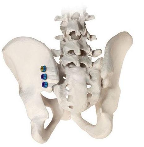
AI in the Operating Room
AI-powered imaging technologies have gradually been introduced to the operating room. Previous studies have harnessed AI technology for augmented reality for intraoperative use.[30] Navigation and robotic technologies in spine surgery are especially interesting avenues of AI disruption, as image processing is a clear strength of AI techniques. Computer-assisted navigation systems use CT imaging with reference clamps to produce 3D renderings of the spine in real-time.[31] AI-powered systems are beginning to be used in operating rooms across the country and have demonstrated better safety profiles, such as increased accuracy in pedicle screw fixation, compared to more traditional technologies.[32] However, these computer-assisted navigation systems and robotic technologies are still relativity new, and the use of AI in improving surgical safety has not been fully explored. Additionally, AI is being used for surgical planning. For example, Lafage et al automated vertebrae selection for ASD surgery. Utilizing preoperative imaging, their model was almost 90% accurate in identifying the upper treated vertebra.[33] Medicrea, a French startup, is also harnessing the power of AI in surgical planning. They developed a modeling tool that similarity utilizes preoperative imaging to create custom 3D-printed rods to eliminate the guesswork of manual manipulation.[30] AI holds great potential in revolutionizing intraoperative imaging, planning, and treatment.
Future Perspectives
AI and its applications within spine imaging, while relatively new, have been well studied. Reports largely demonstrate its efficacy in assisting with radiographic analysis both preoperatively and intraoperatively. However, its actual adoption clinically has been slow. The major obstacle is lack of trust in the technology. More advanced artificial neural networks are “black box” programs, meaning even the developers often do not understand why a specific output was generated from the given inputs. This also introduces ethical and legal concerns with its application clinically. While this is a difficult-to-understand notion, we believe the limitation is not with the technology itself; rather, it is with the data to which it has access to. Therefore, before AI can be trusted in applications that may directly impact patients, large-scale, high-quality data sets should be generated for model training. This next step is close, and as the AI field continues to mature, we will likely see its utility in spine imaging continue to rise.
References
1. Charles YP, Lamas V, Ntilikina Y. Artificial intelligence and treatment algorithms in spine surgery. Orthop Traumatol Surg Res . 2023;109(1S):103456.
2. Hornung AL, Hornung CM, Mallow GM, et al. Artificial intelligence in spine care: current applications and future utility. Eur Spine J. 2022;31(8):2057-2081.
3. Huber FA, Guggenberger R. AI MSK clinical applications: spine imaging. Skeletal Radiol. 2022;51(2):279-291.
4. Gutman MJ, Schroeder GD, Murphy H, Flanders AE, Vaccaro AR. Artificial intelligence in spine care. Clin Spine Surg. 2021;34(4):121-124.
5. Cui Y, Zhu J, Duan Z, Liao Z, Wang S, Liu W. Artificial intelligence in spinal imaging: current status and future directions. Int J Environ Res Public Health. 2022;19(18):11708.
6. Martín-Noguerol T, Oñate Miranda M, Amrhein TJ, et al. The role of artificial intelligence in the assessment of the spine and spinal cord. Eur J Radiol. 2023;161:110726.
7. Jamaludin A, Lootus M, Kadir T, et al. ISSLS prize in bioengineering science 2017: automation of reading of radiological features from magnetic resonance images (MRIs) of the lumbar spine without human intervention is comparable with an expert radiologist. Eur Spine J. 2017;26(5):1374-1383.
8. Langerhuizen DWG, Janssen SJ, Mallee WH, et al. What are the applications and limitations of artificial intelligence for fracture detection and classification in orthopaedic trauma imaging? A systematic review. Clin Orthop Relat Res. 2019;477(11):2482-2491.
9. Jiang D, Dou W, Vosters L, Xu X, Sun Y, Tan T. Denoising of 3D magnetic resonance images with multi-channel residual learning of convolutional neural network. Jpn J Radiol. 2018;36(9):566-574.
10. Yang Q, Yan P, Zhang Y, et al. Low-dose CT image denoising using a generative adversarial network with Wasserstein distance and perceptual loss. IEEE Trans Med Imaging. 2018;37(6):1348-1357.
11. Higaki T, Nakamura Y, Zhou J, et al. Deep learning reconstruction at CT: phantom study of the image characteristics. Acad Radiol. 2020;27(1):82-87.
12. Ouyang J, Chen KT, Gong E, Pauly J, Zaharchuk G. Ultra-low-dose PET reconstruction using generative adversarial network with feature matching and task-specific perceptual loss. Med Phys. 2019;46(8):3555-3564.
13. Plenge E, Poot DHJ, Bernsen M, et al. Super-resolution methods in MRI: can they improve the trade-off between resolution, signal-to-noise ratio, and acquisition time? Magn Reson Med. 2012;68(6):1983-1993.
14. McCollough CH, Leng S. Use of artificial intelligence in computed tomography dose optimisation. Ann ICRP. 2020;49(1_suppl):113-125.
15. Zhou S, Yao H, Ma C, et al. Artificial intelligence x-ray measurement technology of anatomical parameters related to lumbosacral stability. Eur J Radiol. 2022;146:110071.
16. Cho BH, Kaji D, Cheung ZB, et al. Automated measurement of lumbar lordosis on radiographs using machine learning and computer vision. Global Spine J. 2020;10(5):611-618.
17. Watanabe K, Aoki Y, Matsumoto M. An application of artificial intelligence to diagnostic imaging of spine disease: estimating spinal alignment from moiré images. Neurospine . 2019;16(4):697-702.
18. Galbusera F, Niemeyer F, Wilke HJ, et al. Fully automated radiological analysis of spinal disorders and deformities: a deep learning approach. Eur Spine J. 2019;28(5):951-960.
19. Weber KA, Smith AC, Wasielewski M, et al. Deep learning convolutional neural networks for the automatic quantification of muscle fat infiltration following whiplash injury. Sci Rep. 2019;9(1):7973.
20. Sardjono TA, Wilkinson MHF, Veldhuizen AG, van Ooijen PMA, Purnama KE, Verkerke GJ. Automatic Cobb angle determination from radiographic images. Spine (Phila Pa 1976). 2013;38(20):E1256-1262.
21. Kim YJ, Ganbold B, Kim KG. Web-based spine segmentation using deep learning in computed tomography images. Healthc Inform Res. 2020;26(1):61-67.
22. Bae HJ, Hyun H, Byeon Y, et al. Fully automated 3D segmentation and separation of multiple cervical vertebrae in CT images using a 2D convolutional neural network. Comput Methods Programs Biomed. 2020;184:105119.
23. LewandrowskI KU, Muraleedharan N, Eddy SA, et al. Feasibility of deep learning algorithms for reporting in routine spine magnetic resonance imaging. Int J Spine Surg. 2020;14(s3):S86-S97.
24. Wang S, Hu Y, Shen Y, Li H. Classification of diffusion tensor metrics for the diagnosis of a myelopathic cord using machine learning. Int J Neural Syst. 2018;28(2):1750036.
25. Jamaludin A, Fairbank J, Harding I, et al. Identifying scoliosis in population-based cohorts: automation of a validated method based on total body dual energy x-ray absorptiometry scans. Calcif Tissue Int. 2020;106(4):378-385.
26. Vergari C, Skalli W, Gajny L. A convolutional neural network to detect scoliosis treatment in radiographs. Int J Comput Assist Radiol Surg. 2020;15(6):1069-1074.
27. Wang J, Fang Z, Lang N, Yuan H, Su MY, Baldi P. A multi-resolution approach for spinal metastasis detection using deep Siamese neural networks. Comput Biol Med. 2017;84:137-146.
28. Wang SH, Tang C, Sun J, et al. Multiple sclerosis identification by 14-Llyer convolutional neural network with batch normalization, dropout, and stochastic pooling. Front Neurosci. 2018;12:818.
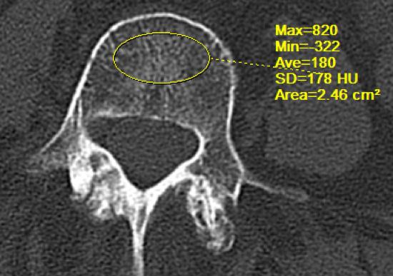
29. Muehlematter UJ, Mannil M, Becker AS, et al. Vertebral body insufficiency fractures: detection of vertebrae at risk on standard CT images using texture analysis and machine learning. Eur Radiol. 2019;29(5):2207-2217.
30. Akosman I, Lovecchio F, Lyons K, et al. The emerging role of artificial intelligence in adult spinal deformity. Semin Spine Surg. 2022;34(4):100986.
31. Rasouli JJ, Shao J, Neifert S, et al. Artificial intelligence and robotics in spine surgery. Global Spine J. 2021;11(4):556-564.
32. Luther N, Iorgulescu JB, Geannette C, et al. Comparison of navigated versus non-navigated pedicle screw placement in 260 patients and 1434 screws: screw accuracy, screw size, and the complexity of surgery. J Spinal Disord Tech. 2015;28(5):E298-303.
33. Lafage R, Ang B, Alshabab BS, et al. Predictive model for selection of upper treated vertebra using a machine learning approach. World Neurosurg. 2021;146:e225-e232.
AUTHORS
Tejas Subramanian, BE
Chad Simon, BS
Marcel Dupont, BA
Joshua Zhang, BS
Tomoyuki Asada, MD
Sravisht Iyer, MD
From the Hospital for Special Surgery in New York, New York.