10 minute read
Advancements in Biomaterials and Implications For Spine Surgery


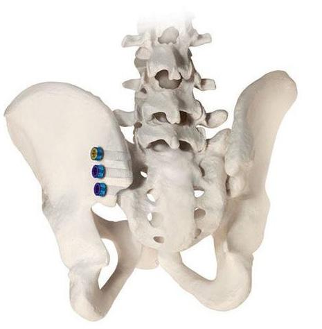
During the 19th century, Dutch surgeon Job Van Merren reported the first autologous graft success with subsequent reports of allogeneic grafts, which are still in use today.[1] In the modern era, more than 2 million bone graft procedures are reported annually worldwide.[2] Autogenic harvesting from the iliac bone remains the gold standard, with allogeneic grafts being accepted as an alternative. Despite its popularity, autologous bone grafts still pose considerable problems, such as a lack of neovascularization and the requirement of multiple surgical sites during harvesting procedures, thus increasing risk of infection.[1] These limitations stem from challenges in creating biocompatible materials that successfully substitute human bone in vivo. There is a current need for osteogenic, osteoconductive, and osteoinductive methods that are equally effective compared to established standards.[2,3]
The progression of biomaterials was marked by changes in use from stainless steel to lighter materials such as titanium.[4] Newer materials have been associated with improved postoperative patient-reported outcome measures (PROMs) such as visual analog scale (VAS) pain scores and Oswestry Disability Index (ODI) scores, among others. Presently, polymers with higher levels of biocompatibility are used because of improved longterm patient outcomes. Looking forward, technologies such as 3-dimensional (3D) printing will enhance the current use of biomaterials by allowing personalized medicine at lower costs. The goal of this article is to review advancements in biomaterials used in spine surgery and discuss their implications.
Stainless Steel/Titanium
Stainless steel is an affordable material with a high mechanical strength that is able to withstand biomechanical forces. Originally one of the first biomaterials established in orthopedic surgery, stainless steel is still utilized in medical device implants today. However, associated nickel and chromium toxicity caused the industry to shift toward other metal compounds and alloys.[5]
The emergence of titanium marked the new age of metals in spine surgery and quickly gained popularity for its low weight, resistance to corrosion, and ability to positively interact with bones. Research has demonstrated that titanium in saline solution was capable of forming a protective coating of TiO2, which further enhances its use. It is now being approached as a material for protective coating.[5] A study conducted by Hasegawa et al compared polyetheretherketone (PEEK) cages with titanium-coated PEEK (TiPEEK) cages in posterior lumbar interbody fusion (PLIF) surgery and found that the TiPEEK cages had significantly better fusion rates at 6 months.[6] This study’s findings suggest that adding titanium can significantly decrease postoperative recovery times and allow faster return to work. While this may be exciting, titanium is limited in use due to its high costs.[5] Future research should explore additional benefits with the use of titanium in addition to decreased toxicity and enhanced fusion rates to provide patients with adequate expectations following spine surgeries.
Calcium Sulfate
Used as early as 1892, calcium sulfate, also known as “gypsum” or “plaster of Paris,” acts as a synthetic bone substitute. It offers many advantages due to its bone-like structure, affordability, and availability in different forms, such as pellets and injectable fluids. It is also non-allergenic and promotes bone capillary infiltration, enhancing osteoconductivity.[1] A study by Hoffman et al found that injectable calcium sulfate cement was noninferior to autologous bone graft when repairing tibial fractures.[7] Furthermore, there were no statistically significant differences seen in scores on the 12-item Short Form physical component summary (PCS) or the VAS between the autologous and calcium sulfate cement groups, and the calcium sulfate cement group showed significantly less blood loss. This study serves as a significant indication of calcium sulfate’s role as a synthetic bone replacement.
Calcium sulfate can also be used in bead forms for the treatment of spondylodiscitis. Due to its flexibility, it can be used to carry antibiotics to help treat osteomyelitis of the spine and reduce infection rates that could result from the associated surgery.[8] Future research should focus on improving the osteoconductivity of the material. Because this material is already cheaper than autologous grafting, improving its osteoconductivity would allow it to become the new gold standard and would increase the accessibility of associated surgeries and lead to further improved postoperative PROMs.
Calcium Phosphate
Because of the morbidity associated with autologous bone grafting, calcium phosphate modalities have remained a popular synthetic alternative to established methods.[9] Hydroxyapatite (HA) stood out due to its osteoconductive properties, as its crystallographic scaffold provided structural support similar to that of bone while being porous enough to facilitate neovascularization.[10,11]
Yoshii et al demonstrated the effectiveness of HA in their study comparing outcomes of patients who underwent anterior cervical corpectomy and fusion with HA treatment versus autologous grafting.[12] The study showed similar effectiveness between groups, with the autologous group showing more blood loss and a higher incidence of donor site pain. Another calcium phosphate alternative is β-tricalcium phosphate (TCP), which has gained a considerable reputation for being both osteoconductive and osteoinductive.[13]
Established literature has highlighted both the benefits and weaknesses of its surgical use. A study by Thaler et al demonstrated favorable clinical outcomes with TCP use in posterior lumbar interbody fusion; however, an increased incidence of pseudoarthrosis was noted.[14] Multiple studies have reported the benefit of using HA and TCP in a composite material termed biphasic calcium phosphate (BCP). Prior research has shown this composite material outperforms both HA and TCP used in isolation; it also promotes TCP degradation, thus mitigating its adverse effects.[15-17]
Future Directions: 3D Printing
The latest progress in the field of spine surgery is 3D printing, which has paved the way for groundbreaking possibilities for biomaterials.[18] In addition to personalized pedicle screw guides, Sheha et al explored the creation of patient-specific 3D-printed vertebrae with the potential to replicate the complex anatomy of the spine.[19] Zhu et al demonstrated that 3D-printed vertebrae were equivalent in strength to natural vertebral discs with acceptable cytocompatibility in rat studies.[20] The discs maintained statistically comparable heights at 2, 3, and 6 months postoperatively with favorable proteoglycan and collagen deposition in the scaffold.
Experimental studies have also highlighted the possibility of 3D-printing technology as a solution to common problems in bonegraft surgery. Plantz et al explored the ability to use a 3D-printed HA-demineralized mone matrix (DBM) to reduce host inflammatory responses. 21 Rats undergoing lumbar fusion in a HA-DBM group demonstrated significantly less edema postoperatively than rats in a bone morphogenic protein (rhBMP-2) control group.[21] Interested in optimizing production, Lai et al demonstrated the benefits of using magnesium in combination with a porous poly lactide-co-glycolide (PLGA) and TCP biodegradable 3D-printed implant in rats, showing favorable osteogenesis and angiogenesis with reduced inflammatory reactions.[22] The involvement of 3D printing is in its nascent stages of transitioning from animal trials to human clinical cases. Lador et al reviewed several case studies in which 3D-printed patient-specific implants were used in complex spine surgeries, all being oncology cases.[23] The patients with custom implants achieved favorable stability, and the study calls for future studies comparing outcomes of 3D-printing implants to other established implants. As this ongoing transition persists, the utilization of 3D printing looks to extend its breadth to various spinal procedures in the realm of clinical practice.
Conclusion
The field of spine surgery has developed significant advancements in the use of biomaterials, specifically in bone grafting. Since the successes of autologous and allogeneic grafts, advancements in biomaterials have improved patient outcomes and reduced associated risks. Limitations in gold standard autologous grafts, such as lack of vascularization and the need for multiple donor sites, have spurred interest in alternative options. The emergence of biomaterials with higher biocompatibility has provided novel avenues for applications in spine surgery that will continue. 3D-printing technology presents an opportunity for personalized implants, better patient outcomes, and fewer complications. With significant advancements in biomaterials thus far and a promising potential for expansion, there is a bright future for the involvement of novel biomaterials in spine surgery.
References
1. Fernandez de Grado G, Keller L, Idoux-Gillet Y, et al. Bone substitutes: a review of their characteristics, clinical use, and perspectives for large bone defects management. J Tissue Eng. 2018;9:2041731418776819.
2. Gillman CE, Jayasuriya AC. FDA-approved bone grafts and bone graft substitute devices in bone regeneration. Mater Sci Eng C Mater Biol Appl. 2021;130:112466.
3. Lementowski PW, Lucas P, Taddonio RF. Acute and chronic complications of intracortical iliac crest bone grafting versus the traditional corticocancellous technique for spinal fusion surgery. Orthopedics . 2010;33(4):240-247.
4. Hofmann A, Gorbulev S, Guehring T, et al; CERTiFy Study Group. Autologous iliac bone graft compared with biphasic hydroxyapatite and calcium sulfate cement for the treatment of bone defects in tibial plateau fractures: a prospective, randomized, open-label, multicenter study. J Bone Joint Surg Am. 2020;102(3):179-193.
5. Choi SR, Kwon JW, Suk KS, et al. The clinical use of osteobiologic and metallic biomaterials in orthopedic surgery: the present and the future. Materials (Basel). 2023;16(10):3633.
6. Hasegawa T, Ushirozako H, Shigeto E, et al. The titanium-coated PEEK cage maintains better bone fusion with the endplate than the PEEK cage 6 months after PLIF surgery: a multicenter, prospective, randomized study. Spine (Phila Pa 1976). 2020;45(15):E892-E902.
7. Hofmann A, Gorbulev S, Guehring T, et al; CERTiFy Study Group. Autologous iliac bone graft compared with biphasic hydroxyapatite and calcium sulfate cement for the treatment of bone defects in tibial plateau fractures: a prospective, randomized, open-label, multicenter study. J Bone Joint Surg Am. 2020;102(3):179-193.
8. Tang X, Li J, Wang C, et al. Antibiotic-loaded calcium sulfate beads in spinal surgery for patients with spondylodiscitis: a clinical retrospective study. BMC Musculoskelet Disord. 2022;23(1):270.
9. Dimitriou R, Mataliotakis GI, Angoules AG, Kanakaris NK, Giannoudis PV. Complications following autologous bone graft harvesting from the iliac crest and using the RIA: a systematic review. Injury. 2011;42 Suppl 2:S3-S15.
10. Litak J, Czyzewski W, Szymoniuk M, et al. Hydroxyapatite use in spine surgery-molecular and clinical aspect. Materials (Basel). 2022;15(8):2906.
11. Spivak JM, Hasharoni A. Use of hydroxyapatite in spine surgery. Eur Spine J. 2001;10(Suppl 2):S197-S204.
12. Yoshii T, Hirai T, Sakai K, et al. Anterior cervical corpectomy and fusion using a synthetic hydroxyapatite graft for ossification of the posterior longitudinal ligament. Orthopedics . 2017;40(2):e334-e339.
13. Bohner M, Santoni BLG, Döbelin N. β-tricalcium phosphate for bone substitution: synthesis and properties. Acta Biomater. 2020;113:23-41.
14. Thaler M, Lechner R, Gstöttner M, Kobel C, Bach C. The use of beta-tricalcium phosphate and bone marrow aspirate as a bone graft substitute in posterior lumbar interbody fusion. Eur Spine J. 2013;22(5):1173-1182.
15. Toth JM, An HS, Lim TH, et al. Evaluation of porous biphasic calcium phosphate ceramics for anterior cervical in - terbody fusion in a caprine model. Spine (Phila Pa 1976). 1995;20(20):2203-2210.
16. Ng AM, Tan KK, Phang MY, et al. Differential osteogenic activity of osteoprogenitor cells on HA and TCP/HA scaffold of tissue engineered bone. J Biomed Mater Res A . 2008;85:301- 312.
17. Garrido CA, Lobo SE, Turíbio FM, Legeros RZ. Biphasic calcium phosphate bioceramics for orthopaedic reconstructions: clinical outcomes. Int J Biomater. 2011;2011:129727.
18. Lo WC, Tsai LW, Yang YS, Chan RWY. Understanding the future prospects of synergizing minimally invasive transforaminal lumbar interbody fusion surgery with ceramics and regenerative cellular therapies. Int J Mol Sci. 2021;22(7):3638.
19. Sheha ED, Gandhi SD, Colman MW. 3D printing in spine surgery. Ann Transl Med. 2019;7(Suppl 5):S164.
20. Zhu M, Tan J, Liu L, et al. Construction of biomimetic artificial intervertebral disc scaffold via 3D printing and electrospinning. Mater Sci Eng C Mater Biol Appl. 2021;128:112310.
21 Plantz M, Lyons J, Yamaguchi JT, et al. Preclinical safety of a 3D-printed hydroxyapatite-demineralized bone matrix scaffold for spinal fusion. Spine (Phila Pa 1976). 2022;47(1):82-89.
22. Lai Y, Li Y, Cao H, et al. Osteogenic magnesium incorporated into PLGA/ TCP porous scaffold by 3D printing for repairing challenging bone defect. Biomaterials . 2019;197:207-219.
23. Lador R, Regev G, Salame K, Khashan M, Lidar Z. Use of 3-dimensional printing technology in complex spine surgeries. World Neurosurg. 2020;133:e327-e341.
AUTHORS
Aayush Kaul, BS
Jacob C. Wolf, BS
Andrea M. Roca, MS
Fatima N. Anwar, BA
Alexandra C. Loya, BS
Srinath S. Medakkar, BS
Vincent P. Federico, MD
Dustin H. Massel, MD
Gregory D. Lopez, MD
Kern Singh, MD
From the Chicago Medical School at Rosalind Franklin University of Medicine and Science in North Chicago, Illinois (Mr Kaul and Mr. Wolf), and Rush University Medical Center in Chicago, Illinois (Mss Roca, Anwar, Loya, Medakkar and Drs Federico, Massel, Sayari, Lopez, and Singh).