13 minute read
The Clinical Significance of Segmental Lumbar Lordosis in Adjacent Segment Disease


The progression of adjacent segment pathology is a leading cause of revision surgery after lumbar spinal procedures, significantly impacting patient outcomes and imposing substantial costs to the healthcare system. The development of adjacent segment disease (ASD) is often attributed to increased mechanical stress at levels adjacent to the fused segments. Adjacent segment degeneration (ASDeg) and adjacent segment disease (ASDi) have been further stratified to help define these pathologies following lumbar spinal fusion. Hilibrand and Robbins referred to ASDeg as radiographic changes seen at levels adjacent to a previous spinal procedure that do not correlate with clinical symptoms, while ASDi referred to the clinical symptoms experienced by patients that coincide with radiographic changes.1 The development of ASDeg and ASDi can severely debilitate patients and cause adverse health outcomes leading to increased revision and reoperations in the future.
Several factors have been investigated to identify the optimal approach for restoration during lumbar fusion surgery to minimize the development of both ASDeg and ASDi. Over the past several years, there is a growing research focus on optimizing segmental lumbar lordosis to mitigate the development of ASDi/ASDeg (ASD). Therefore, the purpose of this article is to explore the recent literature on the impact of segmental lumbar lordosis in lumbar fusion surgeries.
Preoperative Evaluation
Preoperative evaluation of spinopelvic parameters and spinopelvic alignment for patients undergoing lumbar spine fusion surgery is essential to determining adequate restoration. Various studies have been conducted to assess preoperative risk factors for developing ASD, including age, gender, body mass index (BMI), number of fusion segments, and specific lumbar levels.2 Age and BMI are debated risk factors that may not serve as reliable indicators for risk stratification.
Kiss et al saw that the presence of significant degeneration in the adjacent disk level along with the presence of disk bulge/protrusion prior to index surgery significantly increased the risk of developing ASD.3 Similarly, Wang et al confirmed that preoperative Pfirrmann’s classification—a radiological method to assess the severity of intervertebral disc degeneration—greater than 3 was a significant risk factor. This highlights the importance of healthy adjacent disks to prevent adjacent segment disease development and the need for proper operative management if diseased adjacent disks are identified.
The relationship between fusion length and the development of ASD remains a topic of considerable debate. Ghiselli et al reported that patients with a multilevel fusion were significantly less likely to develop symptomatic ASD compared to those with single-level fusions.4 In contrast, Cho et al found no significant difference in ASD development between single- and multilevel fusions, complicating its use as a risk stratification method.5 Meanwhile, Chen et al6 utilized a finite element model to demonstrate that stress on adjacent levels increased with increasing number of fused levels, a finding corroborated by Burch et al,7 who saw higher rates of revision surgeries for ASD with longer fusion constructs. Despite these varying perspectives, there is growing consensus on the importance of limiting surgical fusion to only the necessary levels to minimize the risk of ASD. Despite extensive research aimed at risk stratifying patients and identifying modifiable risk factors for ASD, the data remain controversial, suggesting a multifactorial nature to symptomatic degeneration.
Segmental Lordosis
The lumbar spine plays an important role in maintaining sagittal balance and alignment. The L4-L5-S1 spinal level contributes a large percentage of the total lumbar lordosis, with Bernhardt and Bridwell showing >60% of total lumbar lordosis is attributed to the L4-L5-S1 spinal levels.8 This finding was corroborated by Anwar et al and Pesenti et al, but they also saw that the proximal lumbar segments correlated significantly with pelvic incidence.9,10 This suggests that loss of distal lumbar lordosis, at the L4-S1 levels, is a major contributor to loss of sagittal alignment in the spine.
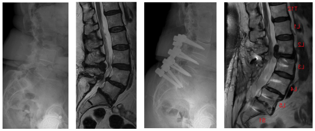
Segmental lumbar lordosis is a spinopelvic parameter that can be adjusted intraoperatively and must be corrected appropriately to match preoperative levels. Kim et al. reported that a low postoperative L4-L5 lordotic angle, specifically less than 20 degrees, was related with the clinical development of ASD.11 Akamaru et al corroborated this finding by showing the effects of hypolordosis in a simulated interbody fusion of L4-L5.12 Their study demonstrated that when L4-L5 was fixed in hypolordosis, the adjacent L3-L4 segment was fixed in flexion, relieving the posterior columns and allowing the facet joints to open contributing to greater degenerative changes. Umehara et al also saw that in 18 patients undergoing L4-L5 fusion, there was a 3-degree decrease in lordosis relative to preoperative level, with an associated 2-degree increase in lordosis at the proximal L2-L3 and L3-L4 segments.13 This increased lordosis at the proximal segments may predispose to sagittal malalignment and eventually could lead to postoperative complications such as lumbar disc herniation and ASD. These findings suggest that a decrease of segmental lordosis postoperatively may induce increased loading at the adjacent segment, which can contribute to lumbar disc herniation, emphasizing the importance of restoring segmental lordosis at the fused segment (Figure 1).14

Degree of Correction
Historically, optimizing the relationship between pelvic incidence and lumbar lordosis was the primary radiographic alignment target. Numerous studies have shown that patients with a pelvic incidence-lumbar lordosis (PI-LL) mismatch greater than 10-11 degrees exhibit higher rates of ASD.15–17 Therefore, achieving an optimal PI-LL mismatch of less than 10-11 degrees was found to be crucial in reducing the risk of adjacent segment disease. This alignment is particularly important for maintaining global sagittal balance in spinal deformity constructs. However, in short-segment lumbar fusions, such as single-level procedures, the PI-LL mismatch may be too broad of a target. In recent years, there has been increasing focus on segmental lumbar lordosis as a more precise parameter for alignment assessment.
In a recent retrospective study, Singh et al examined 168 patients who underwent 1- to 3-level transforaminal lumbar interbody fusions with 2-year follow up. Patients undergoing transforaminal lumbar interbody fusion (TLIF) from the L4-S1 spinal level were assessed for their lordosis correction and risk of developing adjacent segment disease.18 Inadequate correction of L4-S1 lordosis, defined as less than 35 degrees, was compared to adequate restoration (35-45 degrees) and showed significantly higher rates of ASD, instrument-related complications, and revisions in the inadequate correction cohort.18 Despite this relationship between distal lordosis and ASD development, adequate lordosis correction is a patient-specific factor that may require individualized planning.
Proximal segmental lordosis, particularly in the L1-L2-L3 segments, has shown a positive association with pelvic incidence. A retrospective study conducted by Pesenti et al found patients with a larger pelvic incidence also exhibited a greater proximal lumbar lordosis.19 As the pelvic incidence increased, the proximal segments contributed more significantly to the overall lumbar lordosis, emphasizing the relationship between pelvic morphology and global spinal alignment. Similarly, Charles et al conducted a large-scale retrospective radiographic study of 2,599 individuals, demonstrating that global sagittal alignment increased progressively with lumbar lordosis. They also found that pelvic incidence tended to increase with age, underscoring spinal evolvement over time.20 The importance of pelvic incidence in aligning with segmental and global sagittal alignment is reinforced through these studies and in the existing literature.
In another retrospective study, Diebo et al examined 510 patients undergoing adult spinal deformity surgery. Diebo et al observed greater rates of proximal junctional kyphosis and failure when patients were overcorrected at the thoracolumbar level and higher rates of implant failure, particularly rod breakage, when patients were undercorrected.21 Matching normative PI-LL values led to improved surgical outcomes when compared to over- or undercorrection; however, determining the normative values are very patient-specific and must be individualized to the patient. Determining appropriate reconstruction to avoid over- or undercorrection must account for patient-specific alignment targets given that the normal range of lumbar lordosis varies widely, from 18.5 to 72.3 degrees.22
Fusion Construct
There are various fusion constructs that are utilized to help treat lumbar pathologies, including disc degeneration, spondylosis, and spondylolisthesis. Surgical fusion of segments is an effective treatment option to help limit debilitating disease in patients and has become a widely utilized procedure by spinal surgeons within the past few decades. A few of the surgical options include posterior lumbar interbody fusion (PLIF), lateral lumbar interbody fusion (LLIF), transforaminal lumbar interbody fusion (TLIF) and anterior lumbar interbody fusion (ALIF).
The restoration of lordosis is crucial to helping prevent the development of ASD following spinal fusion surgery. The ability to restore lordosis can vary by fusion construct, which may help spine surgeons determine the optimal restoration technique. Watkins et al compared 3 fusion techniques (ALIF, TLIF, and LLIF) in their ability to restore lordosis and increase disk height. The study results found that the ALIF and LLIF procedure significantly improved lordosis from the preoperative state to follow-up when compared to the TLIF procedure.23 Intergroup analysis also demonstrated the greatest amount of restoration with the ALIF technique.23
Given the superiority of ALIF to restore lumbar lordosis, its relationship with ASD development was explored by Lee et al.24 Their study compared the ALIF, LLIF, and PLIF procedure in restoring lordosis and preventing ASD, with results aligning with findings from Watkins et al. Both studies demonstrated the ALIF procedure was associated with greater restoration of lumbar lordosis and a lower incidence of ASD compared to LLIF and PLIF.24 Among fusion techniques, ALIF and LLIF have demonstrated superior lordosis restoration compared to TLIF. However, ALIF and LLIF typically require a dual approach (anterior/lateral and posterior) to achieve sufficient posterior stabilization, increasing surgical complexity and morbidity. In contrast, TLIF is performed entirely from a single posterior approach, but may result in less lordotic correction.
Conclusion
Segmental lumbar lordosis plays a crucial role in the development and management of ASD. With the increasing rates of lumbar spinal fusion surgeries for various spinal pathologies, it is necessary to elucidate proper treatment techniques to help prevent postoperative outcomes, readmissions, and revision surgeries. Proper preoperative evaluation is a multifactorial process that must factor in age, ethnicity, BMI, comorbidities, and preoperative radiographic parameters such as pelvic incidence and lumbar lordosis. Achieving appropriate segmental lumbar lordosis can help mitigate ASD development while avoiding over- or undercorrection. Additionally, determining the type of fusion construct best suited for the individual to help restore sagittal alignment is crucial in preventing future postoperative complications. This study aimed to explore the various surgical techniques and preoperative/intraoperative considerations essential for optimizing patient outcomes with significant spinal pathology. Future research will need large multicentered prospective studies focused on refining patient-specific targets of correction and exploring different fusion techniques to further reduce ASD incidence.
References
1. Hilibrand AS, Robbins M. Adjacent segment degeneration and adjacent segment disease: the consequences of spinal fusion? Spine J. 2004;4(6 Suppl):S190-S194.
2. Liang J, Dong Y, Zhao H. Risk factors for predicting symptomatic adjacent segment degeneration requiring surgery in patients after posterior lumbar fusion. J Orthop Surg. 2014;9:97.
3. Kiss L, Szoverfi Z, Bereczki F, et al. Impact of patient-specific factors and spinopelvic alignment on the development of adjacent segment degeneration after short-segment lumbar fusion. Clin Spine Surg. 2023;36(7):E306-E310.
4. Ghiselli G, Wang JC, Bhatia NN, Hsu WK, Dawson EG. Adjacent segment degeneration in the lumbar spine. JBJS. 2004;86(7):1497.
5. Cho KS, Kang SG, Yoo DS, Huh PW, Kim DS, Lee SB. Risk factors and surgical treatment for symptomatic adjacent segment degeneration after lumbar spine fusion. J Korean Neurosurg Soc. 2009;46(5):425-430.
6. Chen CS, Cheng CK, Liu CL, Lo WH. Stress analysis of the disc adjacent to interbody fusion in lumbar spine. Med Eng Phys. 2001;23(7):485-493.
7. Burch MB, Wiegers NW, Patil S, Nourbakhsh A. Incidence and risk factors of reoperation in patients with adjacent segment disease: a meta-analysis. J Craniovertebr Junction Spine. 2020;11(1):9-16.
8. Bernhardt M, Bridwell KH. Segmental analysis of the sagittal plane alignment of the normal thoracic and lumbar spines and thoracolumbar junction. Spine. 1989;14(7):717-721.
9. Anwar HA, Butler JS, Yarashi T, Rajakulendran K, Molloy S. Segmental pelvic correlation (SPeC): a novel approach to understanding sagittal plane spinal alignment. Spine J. 2015;15(12):2518-2523.
10. Pesenti S, Lafage R, Stein D, et al. The amount of proximal lumbar lordosis is related to pelvic incidence. Clin Orthop. 2018;476(8):1603-1611.
11. Kim KH, Lee SH, Shim CS, et al. Adjacent segment disease after interbody fusion and pedicle screw fixations for isolated L4-L5 spondylolisthesis. Spine. 2010;35(6):625-634.
12. Akamaru T, Kawahara N, Tim Yoon S, et al. Adjacent segment motion after a simulated lumbar fusion in different sagittal alignments: a biomechanical analysis. Spine. 2003;28(14):1560.
13. Umehara S, Zindrick MR, Patwardhan AG, et al. The biomechanical effect of postoperative hypolordosis in instrumented lumbar fusion on instrumented and adjacent spinal segments. Spine. 2000;25(13):1617.
14. Okuda S, Nagamoto Y, Takenaka S, et al. Effect of segmental lordosis on early-onset adjacent-segment disease after posterior lumbar interbody fusion. J Neurosurg Spine. 2021;35(4):454-459.
15. Rothenfluh DA, Mueller DA, Rothenfluh E, Min K. Pelvic incidence-lumbar lordosis mismatch predisposes to adjacent segment disease after lumbar spinal fusion. Eur Spine J. 2015;24(6):1251-1258.
16. Tempel ZJ, Gandhoke GS, Bolinger BD, et al. The influence of pelvic incidence and lumbar lordosis mismatch on development of symptomatic adjacent level disease following single-level transforaminal lumbar interbody fusion. Neurosurgery. 2017;80(6):880.
17. Yoon SG, Lee HC, Lee SM. Pelvic incidence–lumbar lordosis mismatch is predisposed to adjacent segment degeneration after single-level anterior lumbar interbody fusion: a retrospective case-control study. Neurospine. 2023;20(1):301-307.
18. Singh M, Kuharski MJ, Abdel-Megid H, et al. Impact of segmental lordosis restoration during degenerative spinal fusion on two-year adjacent segment disease and revision rates. Spine. Published online September 25, 2024.
19. Pesenti S, Lafage R, Stein D, et al. The amount of proximal lumbar lordosis is related to pelvic incidence. Clin Orthop Relat Res. 2018;476(8):1603.
20. Charles YP, Bauduin E, Pesenti S, et al. Variation of global sagittal alignment parameters according to gender, pelvic incidence, and age. Clin Spine Surg. 2022;35(7):E610.
21. Diebo BG, Balmaceno-Criss M, Lafage R, et al. Lumbar lordosis redistribution and segmental correction in adult spinal deformity: does it matter? Spine. 2024;49(17):1187.
22. Wang SJ, Zhang SB, Yi YY, Xu HW, Wu DS. Estimation of the ideal correction of lumbar lordosis to prevent reoperation for symptomatic adjacent segment disease after lumbar fusion in older people. BMC Musculoskelet Disord. 2020;21:429.
23. Watkins RGI, Hanna R, Chang D, Watkins RGI. Sagittal alignment after lumbar interbody fusion: comparing anterior, lateral, and transforaminal approaches. Clin Spine Surg. 2014;27(5):253.
24. Lee CW, Yoon KJ, Ha SS. Which approach is advantageous to preventing development of adjacent segment disease? Comparative analysis of 3 different lumbar interbody fusion techniques (ALIF, LLIF, and PLIF) in L4-5 spondylolisthesis. World Neurosurg. 2017;105:612-622.
Contributors:
Harmanjeet Singh BA
Daniel Shinn, MD
Nathan J. Lee, MD
From the Department of Orthopedic Surgery at Midwest Orthopedics at Rush in Chicago, Illinois.