Leibniz HealthTech Lecture: Anika Grüneboom Opens the Toolbox of Fluorescence Microscopy Imagine being able to observe the nervous or cardiovascular system from the outside, while it is working. A transparent human being would not only be incredibly exciting, but could certainly also clarify many medical questions about the genesis, early detection or therapy of diseases. After all, biological structures are incredibly complex. They consist of different tissues with various characteristics. Transparency is Prof Dr Anika Grüneboom’s goal. “We need nondestructive and safe methods that allow us to look deep into the
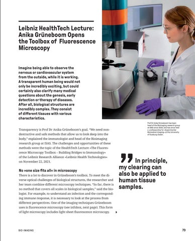
Prof Dr Anika Grüneboom has been head of the Bioimaging research group at ISAS since 2020, and has since held a professorship for »Experimental Biomedical Imaging« at the University of Duisburg-Essen.
body,” explained the immunologist and head of the Bioimaging research group at ISAS. The challenges and opportunities of these methods were the topic of the HealthTech Lecture »The Fluorescence Microscopy Toolbox – Building Bridges to Immunology« of the Leibniz Research Alliance »Leibniz Health Technologies« on November 22, 2021.
No »one size fits all« in microscopy There is a lot to discover in Grüneboom’s toolbox. To meet the diverse optical challenges of biological structures, the researcher and her team combine different microscopy techniques. “So far, there is no method that covers all scales in biological samples,” said the bio-
In principle, my clearing can also be applied to human tissue samples.
logist. For example, to understand an infection and the corresponding immune response, it is necessary to look at the process from different perspectives. One of the imaging techniques Grüneboom uses is fluorescence microscopy (see infobox, next page). This form of light microscopy includes light sheet fluorescence microscopy.
Bio-Imaging
73