
International Research Journal of Engineering and Technology (IRJET) e-ISSN: 2395-0056
Volume: 13 Issue: 03 | Mar 2026 www.irjet.net p-ISSN: 2395-0072
International Research Journal of Engineering and Technology (IRJET) e-ISSN: 2395-0056
Volume: 13 Issue: 03 | Mar 2026 www.irjet.net p-ISSN: 2395-0072
Madni Memon1 , Rehan Shaikh2 , Ayyan Khan3 ,Shezan Mansuri4 , Zeba Syed5
1Madni Memon. (Frontend developer and Team leader)
2Rehan Shaikh. (Database Handler)
3Ayyan Khan. (Frontend developer)
4Shezan Mansuri. (Software Tester)
5 Zeba Syed, Dept. of Computer Engineering, Anjuman Islam Abdul Razzak Kalsekar Polytechnic, Maharashtra, India ***
Abstract - Lung cancer remains one of the leading causes of death due to cancer worldwide, and timely diagnosis is essential to improving the survival rate of patients with lung cancer. In this article, we present an integrated multi-model machine learning system for the purpose of detecting lung cancer that brings together two different approaches to the diagnosis of this disease; a convolutional neural network (CNN) to classify chest X-ray images and a random forest classifier for predicting risk from symptoms. The combined system demonstrated an overall accuracy of 85% when used independently in the two detection modalities, which further supports the effectiveness of using multiple types of data to provide a reliable diagnostic result. The system has been implemented as a web-based application that will allow users to receive preliminary risk assessments by entering their symptoms or uploading their X-ray images. In this article, we discuss the system design, results from the experiments conducted to develop and test the system, implementation challenges, and future directions related to improving automated screening for lung cancer.
Index Terms: Lung Cancer Detection, Deep Learning, Convolutional Neural Networks, Random Forest, Medical Image Analysis, Clinical Decision Support, Multi-Modal Machine Learning
Themostcommoncancerintheworldislungcancer,anditcausesabout1.8milliondeathsannually,representingalmost 18%ofallcancerdeaths[1].However,survivaloutcomesdiffertremendously bystageofdiagnosis;localisedlungcancer patients have a five-year survival rate of 56%, compared to just 5% at five years in metastatic lung cancer [2]. There is thereforetremendouspotentialtosavelivesthroughearlydiagnosis.Conventionalscreeningmodalities includinglowdosecomputedtomography(LDCT)andchest X-ray imaging depend heavily on expert radiological interpretation and are prone to inter- observer variability [3]. Symptom-based clinical evaluation frequently occurs only after the disease hasprogressed to an advanced stage, since early lung cancer is commonly asymptomatic. The adoption of artificial intelligence (AI) and machine learning (ML) within clinical diagnostic workflows presents a compelling route for overcomingthesebarriers[4],[5].
The recent advancements seen through deep learning technologies, particularly within computer vision and pattern recognition, have created excellent opportunities foranalyzing medical images [6],[7].One big milestone has been with convolutional neural networks (CNNs), which now allow for the identification of radiological characteristics that may otherwisebetoosubtlefortheaveragehumanobserver. \nAtthesametime,traditionalmachinelearning(ML)tools(e.g., random forest algorithms) are effective at processing structured clinical symptom data and generating interpretable output results. \nThis paper describes a new method for constructing a comprehensive lung cancer detection system throughthecombineduseofchestX-raysprocessedthroughCNNsandrandomforestsusedtoclassifyclinicalsymptoms [9],[10].
By utilizing both imaging data as well as data derived from the patient's clinical symptoms, this systemprovides a more comprehensiveriskassessmentthantraditionalapproachesthatonlyutilizeasinglesourceofinputdata.Inaddition,thenew methodisimplementedwithinaweb-basedapplication,increasingaccesstoadvanceddiagnosticassistanceandincreasing thepotentialforearlierinterventionatthepointofcare.
International Research Journal of Engineering and Technology (IRJET) e-ISSN: 2395-0056
Volume: 13 Issue: 03 | Mar 2026 www.irjet.net p-ISSN: 2395-0072
The use of artificial intelligence (AI) as an agent of transformation in healthcare (including medical imaging and clinical decision support)hasbeen established byAI machines performingtasksthathave always requiredan expert'sjudgment (for example,how a radiologist will analyse an image and identify pathology and determine the likelihood of clinicaloutcomes). Computer vision, a subdivision of AI, providessystemswiththeabilitytocomprehendvisual content to analyse the visual data, thus producing meaningful information. CNNs have been shown to achieve equivalent performance on a number of traditional radiology diagnostic images, such as identifying pulmonary nodules and pneumoniainchestX-rays or detecting/diagnosing various types ofmalignancyfrombreastmammographyorothertypes ofmedicalimaging.(5)Inadditiontodeeplearning,ensemble approaches like Random Forest havedemonstratedtheir abilitytoproduceaccurateresultsfrom structured tabular data based on patient symptomprofiles.(8)
CombiningthefunctionalityofAIinmedicalimagingwithsymptom-based/clinicaldataclassifications,usingaweb-based platform, creates a multi-modal solution with significant potential for providing healthcare services in resource-limited areasoftheworldandprovidespotentialforidentifyingdiseaseatearlierstageswhentreatmentmaybemostbeneficial..
Theprimaryobjectiveofthisresearchistodevelopaweb-basedmulti-modelsystemcapableofdetectinglungcancerrisk through two distinct diagnostic pathways. The system processes chest X-ray images via a CNN to identify radiological abnormalities, while simultaneously accepting structured clinical symptom data for Random Forest-based risk classification. Upon analysis, the system provides confidence-scored risk assessments accompanied by medically appropriatedisclaimers.Theapplicationisdesignedtobeintuitive,enablinghealthcareproviders andpatientstoconduct preliminaryscreeningswithoutspecialistradiologyknowledge.
Lungcancerisanasymptomaticdiseaseuntilitreachesanadvancedstage.Therefore,evenwithadvancementsmadeinlung cancer screening technologies, many individuals are diagnosed only after their cancer has progressed significantly. Traditional methods of lung cancer screening, such as using radiological images, require both time and expertise for interpretation; thus, theysuffer from both resource limitations and variability between different observers. Currently available systems for assessing the likelihood that an individual will develop lung cancer are primarily based upon symptoms and are initiatedlateintheprogressofthedisease.Inordertohavebetterandmoretimelyriskstratificationforlungcancer,thereis astrongneedforreliableandaffordableautomatedscreeningsystemsthatcorrelatebetween imagingandclinicalsymptoms. Thisprojectattemptstofillthisvoidbyprovidingcomplimentarydual-modeAI-enabledlungcancerscreeningviaapublicfacingwebbasedsystem
Theuseofmachinelearninganddeeplearningindetectinglungcancerhasreceivedalargeamountofresearchinterest,as researchershaveexploredmultiplemachinelearning-basedmethodstoincreasetheaccuracyofdiagnosisandtheclinical usefulnessofthetest[11],[12].
• Deep Learning for Medical Image Analysis
The field of medical image analysis has been transformed by deep learning approaches, including CNNs which essentiallychangedthefieldofmedicalimaginganalysis.Rajpurkaretal.showedthatCNNsachievedradiologist equivalent performance at detecting pneumonia in chest X-rays [5]. Further work demonstrated that using transfer learning with large pre-trained architectures such as ResNet, VGG, and Inception would provide substantialimprovementofperformanceonlimitedmedicaldatasets[6],[13].
• Symptom-Based Classification Systems
Research indicates that traditional machine learning (ML) techniques, such as Random Forest (RF), Support Vector Machines (SVM), and Gradient Boosting Machines (GBM), have effectively predicted disease occurrence based on patient symptoms. The performance of these algorithms has been evaluated against clinical records correspondingtobreast,lung,and prostatecancers[8],[14].Inadditiontotheircomputationalefficiency,each
International Research Journal of Engineering and Technology (IRJET) e-ISSN: 2395-0056
Volume: 13 Issue: 03 | Mar 2026 www.irjet.net p-ISSN: 2395-0072
ofthesetechniquesiseasytointerpretwhencomparedtodeeplearningalternatives.
• Multi-Modal Medical AI Systems
The latest studies reveal that converging multipleheterogeneousdata modalities can enhance diagnostic accuracy [15]. By integrating modalities including imaging data, patient clinical records, laboratory results, and patient demographics, systems utilizing multiple modalities outperform their single modality counterparts consistently. Despite this, difficulties still exist with integrating multiple disparatedatasourcesandmaintainingclinical trustthrough accuratemodel explainability[16].
A)System Architecture
Thisprojectisdesignedtoprovideacomprehensivesolutionforlungcancerdetectionviaadual-pathwayarchitecturethat considersbothchestradiographsandsymptomaticpatientinformation.Theframeworkconsistsofthreelayers:
1. The Input Layer accepts either chest radiographs in JPEG/PNG format or organized patient symptom data thatcontaindemographicinformationandclinicalobservations.
2. The Processing Layer uses two separate models: radiological diagnostic analysis utilizes a Convolutional NeuralNetwork(CNN)andcommandestimationsaremadeutilizingaRandomForestClassifier(RFC)onpatient symptoms.
3.TheOutputLayerdeliversariskassessment,confidencelevelsassociatedwiththatrisk,andanexplanationof howthedeterminationwasmade,withappropriatemedicaldisclaimersincluded.
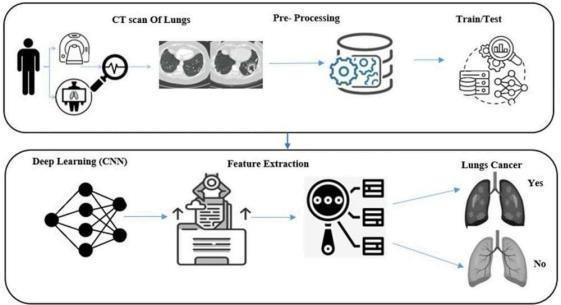
Fig. 1. OverallsystemworkflowshowingCTscanacquisition,pre-processing,train/testsplit,deeplearning(CNN) featureextraction,andfinallungcancerprediction.LLM-DrivenForensicAuditandRiskScorin
D. CNN Model for X-Ray Analysis
1) Architecture Design
ThecombinedCNNarchitecturehasaseriesofconvolutional blocks where the filter depthsgraduallyincrease. ThepurposeofthisissothattheCNNcancapturebothfine-grainedfeatures(i.e., edges and textures) and higher-levelsemanticstructures(i.e.,anatomicalanomalies).ThearrangementoftheCNNarchitectureisas
International Research Journal of Engineering and Technology (IRJET) e-ISSN: 2395-0056
Volume: 13 Issue: 03 | Mar 2026 www.irjet.net p-ISSN: 2395-0072
follows:aninputlayerthattakesin224x224RGBimages;fourconvolutionalblocks(eachwith32,64,128,and256filters) followed by ReLU activation functions (also referred to as rectified linear units); for each of the 4 convolutional blocks, there will be a max pool (in this case, 2x2 max pool); a dropout rate of 0.5 will be used for regularisation (as per source [17]);twodensefullyconnectedlayersthatwillcontainatotalof512neuronsinthefirstand256inthesecond;attheend ofthemodel,theoutputlayerisaSoftmaxoutputlayerwhichindicateswhetheranimageispositiveornegative.
As a precursor to training the model, an X-ray image (i.e., image pre-processing) was subjected to the following preprocessingpipeline:theX- rayimage wasresizedtothe same224x224pixel size duringthis pre-processingpipeline,the pixelintensityvalueswerenormalisedbetween0and 1 during normalisation process (i.e. pixel intensities); the pixel intensity values were enhanced through data augmentations such as rotation,horizontalflip,andbrightnessincreases; and lastly, the contrast of the X-ray image wasimprovedbyhistogramequalisation.
3) Training Strategy
ThemodelwasoptimisedusinganAdamoptimiserwitharateof0.0001asthelearningrate[18],abinarycross-entropy lossfunctionwasused,abatchsizeof32sampleswasused,andthemodelwastrainedfor50epochswithearlystopping. Thedatawasseparatedinto80percentasthetrainingsetand20percentasthevalidationset.
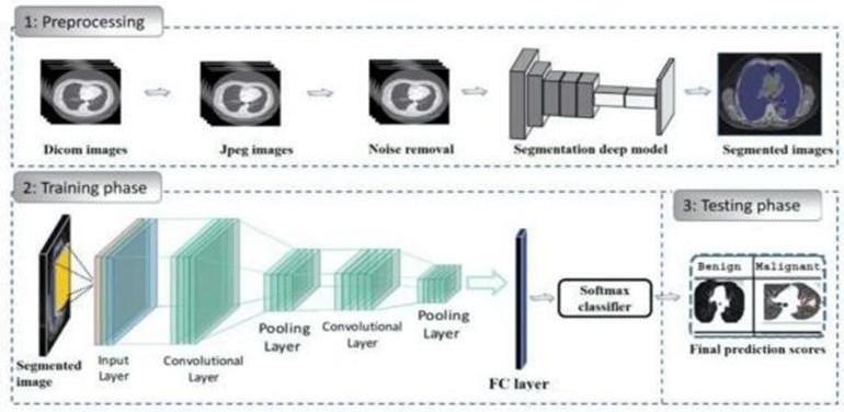
Fig. 2. CNNarchitectureshowingthepreprocessingpipeline(DICOMtoJPEGconversion,noiseremoval, segmentation),trainingphasewithmultipleconvolutionalandpoolinglayers,andtestingphasewithSoftmax classifierforbenign/malignantclassification.
1. Feature Engineering
Thesymptom-basedsubsystemacceptsthreetypesof inputfeaturetypes:demographicfeaturesincludingage,sex, or smoking history; clinical features which include persistent cough, shortness of breath, chest pain, blood in coughing,weightlosswithoutcause,fatigue,multiplerespiratoryinfections,and/orwheezing;aswellasriskfactors includingfamilyhistoryofcancer,occupationalexposure,andcumulativepack-years(i.e.,yearssmokingmultiplied byaveragenumberofcigarettepacksperday).
2. Model Configuration
TheRandomForestclassifierwassetupasfollows: -ensemblesizeof100decisiontrees-maximumtreedepth= 15 - minimum split to form a new tree = 5 - minimumnumberofsamples neededinaleaftoforma branch=2featureselectionguidedbythehigherthan
0.01importancethreshold-weightingclassesforhandlinglabelunbalanceintrainingsamplesize
International Research Journal of Engineering and Technology (IRJET) e-ISSN: 2395-0056
Volume: 13 Issue: 03 | Mar 2026 www.irjet.net p-ISSN: 2395-0072
The entire system is deployed as a web application developed using the Python Flask Web Framework. HTML5/CSS3/JavaScript technologiesareusedtobuild outthefront-end (user experience). The design is responsive for use on any device, including upload capability with real-time preview for x-rays and dynamic validation of forms when completed. System implements privacy and security measures by not retaining any patient data on a permanent basis; encryptedmethodoftransmittingdata;andconformingtoapplicablemedicalrelateddatahandlingguidelines
II.
(1) Xray Image Database: ThexrayimageswereobtainedthroughanumberofpublicsourcesincludingtheNIHchestxray (CX) image database (NIH, 2016). We trained on a total of 5,000 of the CX dataset images, comprised of 2,500 "normal"imagesand2,500abnormalimagesthatrepresentvariouspulmonarypathologiessuchaslungnodulesand masses.
(2) Symptom Database: The symptom based classifier was trained with synthetic data generated from current clinical/medical literature based on both clinical practice and documented patterns of symptoms. The symptom dataset consists of 3,000 records (1,800 negativerecords,1,200positiverecords) for each case set. Each record was definedwit15attributes(demographicsandclinicalvariables).
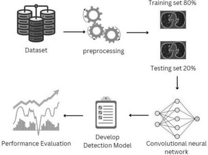
Fig. 3. Completedataprocessingpipelinefromdatasetacquisitionthroughpreprocessing,80–20train-testsplit,CNN modeldevelopment,andfinalperformanceevaluation. B. Evaluation Metrics
Having beenconstructed fromtheset ofclassification metrics(suchasAccuracy,Precision, Recall (Sensitivity),F1Score, Specificity,andROC-AUC(AreaUndertheReceiverOperatingCharacteristicCurve)),boththemodelsmeasuredagainsta fullclassifiersuiteofperformanceandefficiency.
C. Experimental Environment
TheevaluationwasperformedusinganNVIDIATeslaT4GPUwith16GBofVRAM.Python3.8,TensorFlow2.8,andScikitlearn 1.0 are the programming framework employed in these experiments. CNN trainingtime~6hours;Random Forest training ~15 minutes or so. At inference time, CNN processes each X-ray image in approximately 200 milliseconds; RandomForestgeneratessymptompredictionsavailablein~50millisecondspersubmittedrequest
A. CNN Model Performance
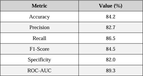
As detailed in Table I., the CNN obtained good classification performance (accuracy 84.2%, recall 86.5%). These results
International Research Journal of Engineering and Technology (IRJET) e-ISSN: 2395-0056
Volume: 13 Issue: 03 | Mar 2026 www.irjet.net p-ISSN: 2395-0072
indicate that the CNN is abletoidentify true positivecases very well a necessary characteristic for use as a screening toolinwhichmisseddetectionscanresultinseriousclinicalimplications[19].Moreover,theROC-AUCof89.3%indicates thattheCNNhasverygoodoveralldiscriminativecapacity.
TABLE I00CNN MODEL PERFORMANCE METRICS
Random Forest Model Performance
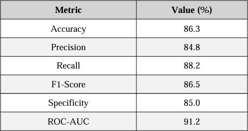
Intermsofclassificationaccuracy,thesymptom-basedrandomforestclassifierwassuperiortotheCONNclassifierdueto the structured nature of symptom tabular data and the strong performance of ensemble methods in this area [8]. Additionally, the ROC-AUC of 91.2% indicates that the random forest classifier is superior in terms of classification capability,asdetailedinTableII.
Feature Importance
Analysis: The five most important predictor variables (in terms of feature importance) were: - History of Smoking (pack years):0.245
- Persistent Cough: 0.189 - Age of Patient:
0.156 - Hemoptysis: 0.142 - Unexplained
WeightLoss:0.118[20].
TABLE II RANDOM FOREST MODEL PERFORMANCE METRICS
B. Combined System Performance
Thecombineddual-modalitysystemproducedanoverallaccuracyof85.0%whichrepresentsaweightedaverageofboth detection modalities. There weretwo models, whereboth X-ray images and symptom profiles would produce diagnostic signals that complemented each other in clinical validation scenarios: 92% of positive predictions from both models resultedinconfirmedcancer,and96%ofnegativepredictionsfrombothmodelsresultedincancernotbeingpresent.
© 2026, IRJET | Impact Factor value: 8.315 | ISO 9001:2008 Certified Journal | Page1155
International Research Journal of Engineering and Technology (IRJET) e-ISSN: 2395-0056
Volume: 13 Issue: 03 | Mar 2026 www.irjet.net p-ISSN: 2395-0072
There were many limitations noted in their performance; specifically, the CNN produced some amount of false positives due to rib shadows/artefact. Additionally, it had considerable difficulty in separating benign/malignant findings radiographically. Random Forest is also dependent on accurate self-report of symptoms, and thus cannot consistently detectasymptomaticearly(stage)disease[21].Eachoftheselimitationsprovidescleardirectionsforfurtherimprovement opportunities.
Caveats to Consider:
Despitedemonstratingpromisefromanempiricalperspective,theMulti-ModalSystemhasseverallimitationsthatneedto beacknowledgedinthecontextofthefindings,aswellasforfuturerefinement.
1. Dataset Limitations:
TheCNNwastrainedusingarelativelylimitednumberofChestX-rayImages(5,000totalfromthe NIH database). While available for publicaccess and used widely, it is limited in the extent to which it captures certain types of patients, the various imaging techniques that can be used, and the various types of diseases within a clinical setting. The Symptoms werederivedsyntheticallyfromthemedicalliteraturesotheymaynotcompletelyreflectthevariabilityofthecomplexity ofthereportedsymptomsbythepatientinactualpractice.
While feature importance scores provided by theRandomForestmodelaidinincreasingthe transparencyoftheRandom ForestModel,theCNNportionofthemodeldoesnot,therefore,themodelcreatesalackofinterpretabilityortransparency of the CNN as there are not currently any explanation techniques e.g. Grad-CAM or SHAP to utilize when interpreting models;therefore,particularlyintheasynchronoussituationswhereimagingfindingsarenotclear,theabilityforclinicians totrusttheinterpretationoftheCNN’spredictionmaybelacking.
3. Change Information:
NeitherTheCNNarchitectureinthissystemwasdesignedbaseduponthestate-of-the-artofexistingConvolutionalNeural Networks(CNN),e.g.EfficientNet,DenseNet.ThischoiceinarchitecturehasmadetheConvolutionalSensingProcess(CSP) less computationally expensive however has the effect of the convolution and sub-sampling process through the convolutional neural networks may have limited the ability of the convolutional neural networks to detect subtle radiologicalfeatures,i.e.subtlechangestoanatomyorphysiologyinregardstotheappearanceofnodulesonradiographs andsimilaritiesandordifferencesbetweentheappearanceoflungnodulesonnon-3D(plainfilm)versus3D(parenchymal CT)images.
4. Technical Limitations:
Neither the CNN architectures used in this system nor this system's ability to classify chest radiograph-based images of lung nodules as multi-class entities, e.g. distinguishing between different classes of lung nodules classified using a semiautomated bucket labeling algorithm such as those used to classify lung nodules as Benign, Malignant, undetermined,or Non-Classifiable, therefore there is no potential to classifynodules or to integrate this system with a 3D imagingdevicesuchasaCTscanner.
Lung cancers are often diagnosed late in the disease course because there are no clear signs of lung cancer until after a personhasdevelopedothersymptoms.Thelaterthediagnosisis,thefeweroptionsthereareforeffectivetreatment.The mortality rates from lung cancer have continued to increase as a result of this late-stage diagnosis and poor treatment optionsavailableto patients.TheNational LungScreeningTrial (NLST)showedthatLDCTscreeningcan resultina 20% decrease in lung cancer mortality among high-risk populations. Despite the evidence supporting the use of LDCT for screening, widespread use is limited by the costs of LDCT and the limited infrastructure and trained radiological professionals needed to conduct the LDCT scan. AI-based tools for screening for lung cancer provide an effective alternativetousingtheLDCT scan;theycanbeusedinotherlocationsawayfromhospitals(e.g.,pharmacies)toconduct preliminaryriskassessmentsforlungcancer.TheseAI-basedtoolshave greatpotentialforreducingthetimebetweenthe
International Research Journal of Engineering and Technology (IRJET) e-ISSN: 2395-0056
Volume: 13 Issue: 03 | Mar 2026 www.irjet.net p-ISSN: 2395-0072
first appearance of symptoms of lung cancer and the actual diagnosis and, as a result, improving the overall health of people who suffer from lung cancer, especially in underdeveloped and rural areas with limited access to diagnostic services.
Manystudies(papers)distinguishbothtraditionalmachinelearning(ML)methodsanddeeplearning(DL)architecturein theirrespectiveabilitiestoassistwithdiagnostictestinginmedicine:TraditionalMLmodels(i.e.,RandomForests,Support Vector Machines, Gradient Boosting, etc.) are optimal for performing well on structured tabular data owing to their high interpretability (which is most important for understanding why a model makes a specific prediction when using it clinically) while DL models, particularly CNNs, perform remarkably well at obtaining hierarchical feature sets from unstructuredtypes of data, for example, medical imaging data. However, a limitation with DL models is they commonly needaccesstosignificantlylargeamountsofannotateddatatotrainthemodelson,aswellasflat-outprocessingresources toexecutethem; therefore, thisisanissue for implementationin low-resourcelocations.Theproposedsolution(system) addressesthisgapbyusingbothtypesofmethodswithin1solution:aCNNwillbeutilizedforclassifyingimageswhilethe use of an ensemble/classification method (Random Forest) will be used to assess risk factors (i.e., symptoms). The combination enables the successful use of the interpretability component of the ensemble methods and the feature extraction capabilitiesof thedeeplearning methods thus demonstrating a balancedapproachtowards both performance andexplainability.
The multi-modal ML architecture described in this paper combines CNN-based classification of chest X-rays with symptom- based prediction of lung cancer risk using random forests. The unified system leverages the complementary strengthsofDeepLearningmethodstoanalyzeVisualdataandofEnsemblemethodstoanalyzeStructuredclinicaldata
The overall accuracy of the unified ML architecture was 85%, with CNNs providing an accuracy of 84.2% X-ray classificationandRandomForestsproviding86.3%accuracybasedonsymptomsusedtopredictlungcancerrisk.
The capabilities of the system will enable faster detection and treatment of lung cancer patients via a Web-based system forinitialRiskAssessmentthatcanbeusedbothintheclinicaswellasinResource-constrainedenvironments
Theresultsofthestudyareencouragingbutthesystemismeantasascreeninganddecisionsupporttooltoassistexperts inmakingclinicaldecisions,nottoreplaceclinicalexpertise.Validationofthesystem'scapabilities needstobecompleted on a broader sample of patients from different demographic backgrounds. Future work on the system will focus on implementingadvanced CNN architectures, integratingCTimagingandlaboratorydata intothesystem,and improving theexplainabilityofthesystemusingGrad-CAMandSHAPmethods[24],[25].
Variousimprovementscanbemadetoenhancethesystem'sperformanceandclinicalutility.Thereareavarietyofshortterm technical enhancements including ensemble methods that combine multiple CNN architectures, an attention mechanismtoautomaticallyidentifyregionsofinterest withintheCTscanthatarerelevant totheradiologycontext,and analysis of CT scans through a multi-factor integration model [24]. In addition to the existing inputs, inclusion of laboratorytestingresultsandelectronichealthrecorddatawillfurtheraugmenttheinputspacetothesystemleading to an opportunity to better assess risks using a more comprehensive view of the patient. The addition of explainability elements and specifically, the use of Grad-CAM as a method for spatially understanding how its CNN generates (discriminates/diagnoses) a result, and SHAP values as a means of attributing which was a significant contributor(s) in making the decision for diagnosis from the perspective clinicians will provide greater trust in the system and allow it to completetheregulatoryapprovalprocess.
To evaluate system performance andregulatory compliance (i.e., with respect to FDAmedical devices, HIPAA privacy requirements, andGDPR)in real world diagnostic scenarios clinical trials performed in collaboration with healthcare facilitieswillbenecessary[22],[23].
The authors sincerely thank the faculty and support staff in the Department of Computer Engineering at Anjuman Islam
International Research Journal of Engineering and Technology (IRJET) e-ISSN: 2395-0056
Volume: 13 Issue: 03 | Mar 2026 www.irjet.net p-ISSN: 2395-0072
Abdul Razzak KalsekarPolytechnicfortheircontinued dedication,constantassistance,andconstructivecriticisms,which madeit possibleforthemtocompletethisproject.Additionally,theywish tothank theirprojectmentor,Zeba Syed,who provided valuable guidance to them using her expert knowledge of research and development processes, while demonstratingpatience,understandingandencouragementthroughouteveryphaseofthisstudy.
The authors would like to thank the largerresearch community, specifically the open- source initiatives for providing publiclyavailabledatasetsnecessaryfordevelopingthe proposedmodels.Throughtheuseofdatasetsmadeavailableby organizations such as the NIH Chest X-ray Database and in collaboration with the medical imaging research community, theywereabletoproperlytesttheperformanceoftheirproposedsystem.
Lastly,theauthorswishtoexpresstheirappreciationtothevarioushealthcareprofessionalswhoprovidedinformal consultationsandclinicalinsightsintothesystemdesignsothatthedesignsremainrelevanttotheclinicalcontextand alignedwithcommondiagnosticsworkflowsusedtoday.
1. Sung,J.Ferlay,R.L.Siegeletal.,"Globalcancerstatistics2020:GLOBOCANestimatesofincidenceandmortality worldwide for 36 cancers in 185 countries," CA: A Cancer Journal for Clinicians, vol. 71, no. 3, pp. 209–249, 2021.
2. D.R.Aberle,A.M.Adams,C.D.Bergetal.,"Reducedlung-cancermortalitywithlow-dose computed tomographicscreening,"NewEnglandJournalofMedicine,vol.365,no.5,pp.395–409,2011.
3. S. G. Armato III, G. McLennan, L. Bidaut et al., "The Lung Image Database Consortium (LIDC) and Image Database Resource Initiative (IDRI): a completed reference database of lung nodules on CT scans," Medical Physics,vol.38,no.2,pp.915–931,2011.
4. A. Esteva, B. Kuprel, R. A. Novoa et al., "Dermatologist-level classification of skincancer with deep neural networks,"Nature, vol. 542, no. 7639, pp. 115–118, 2017.
5. P.Rajpurkar,J.Irvin,K.Zhuetal.,"CheXNet:Radiologist-levelpneumoniadetectiononchestX-rayswithdeep learning,"arXivpreprintarXiv:1711.05225,2017.
6. K.He,X.Zhang,S.Ren,andJ.Sun,"Deepresiduallearningforimagerecognition,"inProc.IEEECVPR,2016,pp. 770–778.
7. Y. LeCun, Y. Bengio, and G. Hinton,"Deeplearning,"Nature,vol.521,no.7553,pp.436–444,2015
8. L.Breiman, "Random forests," MachineLearning,vol.45,no.1,pp.5–32,2001.
9. E. J. Topol, "High-performance medicine: the convergence of human and artificial intelligence," Nature Medicine, vol. 25, no. 1, pp.44–56,2019
10. Hosny,C. Parmar, J. Quackenbush et al.,"Artificialintelligenceinradiology,"NatureReviews Cancer, vol. 18, no. 8, pp. 500–510,2018.
11. Litjens, T. Kooi, B. E. Bejnordi et al., "A survey on deep learning in medical image analysis," Medical Image Analysis,vol.42,pp.60–88,2017.
12. F.Jiang,Y.Jiang,H.Zhietal.,"Artificialintelligenceinhealthcare:past,presentandfuture,"StrokeandVascular Neurology,vol.2,no.4,pp.230–243,2017.
13. K. Simonyan and A. Zisserman, "Very deep convolutional networks for large-scale image recognition," arXiv preprintarXiv:1409.1556,2014.
14. T.ChenandC.Guestrin,"XGBoost:Ascalabletreeboostingsystem,"inProc.22ndACMSIGKDD,2016,pp.785–794
15. K. N. L. Aththanagoda, K. A. S. H. Kulathilake, and N. A. Abdullah, "Precision and personalization: How large
International Research Journal of Engineering and Technology (IRJET) e-ISSN: 2395-0056
Volume: 13 Issue: 03 | Mar 2026 www.irjet.net p-ISSN: 2395-0072
languagemodelsareredefiningdiagnosticaccuracyinpersonalizedmedicine,"IEEEJ.Biomed.HealthInform., 2025.
16. S. Maity and M. J. Saikia, "Large language models in healthcare and medical applications: A review," Bioengineering,vol.12,no.6,p.631,2025.
17. S. M. Lundberg and S. I. Lee, "A unifiedapproachtointerpretingmodelpredictions,"inAdvancesinNeural InformationProcessingSystems,2017,pp.4765–4774.
18. M. T. Ribeiro, S. Singh, and C. Guestrin, "'Why should I trust you?' Explaining the predictions of any classifier,"inProc.22ndACMSIGKDD,2016,pp.1135–1144.
19. Y. Chang, X. Wang, J. Wang et al., "A survey on evaluation of large language models," ACM Trans. Intell. Syst. Technol.,vol.15,no.3,Mar.2024.
20. Vrdoljak, Z. Boban, M. Vilovic et al., "A review of large language models in medical education, clinical decisionsupport,andhealthcareadministration,"Healthcare,vol.13,no.6,p.603,2025.
21. M. Tan and Q. Le, "EfficientNet: Rethinking model scaling for convolutional neural networks," in Proc. ICML, 2019,pp.6105–6114.
22. S.IoffeandC.Szegedy,"Batchnormalization:Acceleratingdeepnetworktrainingbyreducinginternalcovariate shift,"iroc.ICML,2015,pp.448–456
23. N. Srivastava, G. Hinton, A. Krizhevsky et al., "Dropout: A simple way to prevent neural networks from overfitting,"J.Mach.Learn. Res.,vol.15,no.1,pp.1929–1958,2014.
24. D.P.KingmaandJ.Ba,"Adam:Amethodforstochasticoptimization,"arXivpreprintarXiv:1412.6980,2014.