International Research Journal of Engineering and Technology (IRJET)
e-ISSN: 2395-0056
Volume: 08 Issue: 02 | Feb 2021
p-ISSN: 2395-0072
www.irjet.net
Enhanced Optimized MSVM Classification of Lung Cancer in Biomedical CT Image Database Nikita Mahajan1, Sukhdeep Kaur2, Dr. Milanpreet Kaur3, Dapinder Kaur4 1Student,
Department of Computer Science,Chandigarh Group of Colleges, Landran,Punjab,India 2Assistant Professor, Department of Computer Science,Chandigarh Group of Colleges,Landran,Punjab,India 3Assistant Professor, Department of Computer Science,Chandigarh Group of Colleges, LandranPunjab,India 4Assistant Professor, Department of Computer Science,Chandigarh Group of Colleges, LandranPunjab,India --------------------------------------------------------------------------------------------------------------------------------------------------Abstract: Lung cancer is the major tumor, which is
occurred diseases that causes death and it is detected in both the genders above the expected death of about 1.75 million in the year 2018. The unexpected development of the abnormal cells that are not standard in more than one lung is lung cancer. However, the non-standard cells disturb the functioning of the standard lung cell that cause infected tissues in the lungs [1]. Lung cancer is mainly of the two kinds; one is the non-small lung disease (NSLD) [2] and other is the small lung cancer (NLC). Normally, 85% of the lung disease is mainly caused because of the NSLD and 15 % are caused due to NLC [3]. Lung cancer is one of the deadly diseases that caused the countless deaths all over the world. Lung cancer is also called as the lung carcinoma (LC), that is the malignant lung tumor featured by the non-standard growth of the tissues in the lungs. This developed tissue may result in the progression of the metastasis close to tissues and other portions of the body. Mainly the cancers initiate from the lung are called as the primary lung cancers. The major symptoms of the disease are cough with blood, loss of weight, breathlessness, and ache in chest [4].
featured by the regulated cell development of the tissues of the lung. Lung cancer is the main cancer diagnosed globally. Mostly, the large amount of the death takes place due to lung disease as compared to other diseases. However, it is necessary for the early detection and diagnosis of the disease for the maximum survival rate of the cancer patient. The cancer cell identified from the MI (Medical Images), and numerous image processing and computing methods to identify the cancer cells. MRI images have the features of the high resolution, maximum clarity, less noise and distortion value. This existing system presented an effective CNN (EF-CNN) method for the identification of the lung cancer. The previous method contains the convolution layers that include different layers such as; (i) Convolution (ii) Pooling (iii) Maximum pooling (iv) Fully connected, and (v) SoftMax layer. In proposed article used the HoG method to extract the features and Artificial Bee Colony algorithm is a metaheuristic method and used to select the extracted features in the lung images. It finds for high-level artificial feature selection. It finds the best cost parameter vector, which reduces the cost or objective function. MSVM (Multisupport Vector Machine) classification method is used to predict the cancer lung images. The proposed system accurately determines the disease or detection category image. The result shows that the train and test feature is the most important point for classifying the lung images according to the disease. In research model’s performance, accuracy rate value is 97.8 %, recall, value is 98 %, precision rate value is 97%, Processing Time value is 110 milliseconds, and Testing loss value is 0.4150. Key words: Lung Cancer Medical Image, Histogram Orientation Gradient, Artificial Bee Colony algorithm and MSVM Classification Method.
1. INTRODUCTION
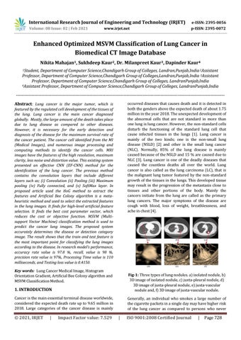
Fig-1: Three types of lung nodules. a) isolated nodule, b) 3D image of isolated nodule, c) juxta-pleural nodule, d) 3D image of juxta-pleural nodule, e) juxta-vascular nodule and, f) 3D image of juxta-vascular nodule.
Cancer is the main essential terminal disease worldwide, considered the expected death rate up to 9.65 million in 2018. Large categories of the cancer disease is mainly
Generally, an individual who smokes a large number of the cigarette packets in a single day may have higher risk of the lung cancer as compared to persons who never
© 2021, IRJET
|
Impact Factor value: 7.529
|
ISO 9001:2008 Certified Journal
|
Page 728