International Research Journal of Engineering and Technology (IRJET)
e-ISSN: 2395-0056
Volume: 07 Issue: 08 | Aug 2020
p-ISSN: 2395-0072
www.irjet.net
Liver Segmentation from CT based on Multi-Scale Candidate Generation and Fractal Residual Network Jasmine J. C Sheeja1 & Dr. B. Sankara Gomathi2 1Assistant
Professor of ECE, ROHINI COLLEGE OF ENGINEERING & TECHNOLOGY 2Professor of EIE, National Engineering College --------------------------------------------------------------------------***---------------------------------------------------------------------------forth. These treatment strategies need the detail data of Abstract — Liver malignant growth is one of the most tumors, for example, the size, shape, and area before treatment so as to build up a _ne treatment program [2].
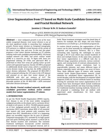
widely recognized diseases. Liver tumor division is one of the most significant strides in rewarding liver malignant growth. Precise tumor division on Computed tomography (CT) pictures is a difficult errand because of the variety of the tumor's shape, size, and area. To this end, this paper proposes a liver tumor division technique on CT volumes utilizing multi-scale candidate generation(MCG), 3D fractal residual network (3D FRN), and active contour model (ACM) in a coarse-to-_ne way. To begin with, livers are fragmented utilizing 3D U-Net and afterward MCG is performed on these liver areas for getting tumor up-andcomers (all superpixel squares). Second, 3D FRN is proposed to additionally decide tumor areas, which is considered as coarse division results. At long last, the ACMis utilized for tumor division refinement. The proposed 3D MCG-FRN C ACM is prepared utilizing the 110 cases in the LiTS dataset and assessed on an open liver tumor dataset of the 3DIRCADb with dice per instance of 0.67. The experimentations and correlations exhibit the presentation favorable position of the 3D MCG-FRN + ACM contrasted with other division techniques.
In routine clinical practices, the segmentation of liver cancer can be done manually by radiologists with good expertise and experience. However, this is a timeconsuming task requiring the radiologist to search through a 3D CT scan which may include hundreds of slices and multiple lesions. At the same time, automatic liver tumor segmentation is a difficult task due to different image acquisition protocols, various contrastagents, and varying levels of contrast enhancements. In addition, dissimilar scanner resolutions lead to unpredictable intensity, and many different types of lesions, especially tumor sub-types, can occur in livers. Thus, these different types of tumors with varying contrast levels (hyper-/hypo-intense tumors) create obstacles for automated tumor segmentation [3]. In recent decades, with the development of computer-aided diagnosis (CAD) [4], several methods based on machine learning for automatic liver tumor segmentation on CT images have been developed which include traditional machine learning methods and deep learning methods. For traditional machine learning methods, Smeets et al. [5] proposed a combining level set method with supervised pixel classification for liver tumor segmentation.
Key Words: Fractal residual network, multi-scale candidate generation method, active contour model, liver tumor segmentation, CT volume. 1. INTRODUCTION As per the World Health Organization (WHO) reports, liver disease is one of the most widely recognized malignant growths on the planet and is a primary driver of death in all tumors. In 2012, 745,000 patients passed on of liver malignant growth overall [1], hepatic cell carcinoma (HCC) represents about 80% of all essential liver tumors and most patients with ceaseless liver ailment have HCC. Identification of HCC at a beginning phase can incredibly improve the fix pace of patients. Computed tomography (CT), included by its high spatial goal and quick checking speed, assumes a critical job in liver disease location and conclusion. The essential treatment techniques incorporate careful resection, interventional treatment, locoregional removal, and so
Š 2020, IRJET
|
Impact Factor value: 7.529
Fig-1 ARCHITECTURE OF PROPOSED METHOD |
ISO 9001:2008 Certified Journal
|
Page 423