International Research Journal of Engineering and Technology (IRJET)
e-ISSN: 2395-0056
Volume: 07 Issue: 04 | Apr 2020
p-ISSN: 2395-0072
www.irjet.net
Identification of Glaucoma Using Convolutional Neural Network Brindha.M1, Harini.D1, Ishwarya.V1
Mr. Carol Praveen.R2
1Department
of ECE, SSM Institute of Engineering and Technology, Dindigul, Tamilnadu-624002
2Assistant
Professor, Department of ECE, SSM Institute of Engineering and Technology, Dindigul, Tamilnadu-624002
---------------------------------------------------------------------***---------------------------------------------------------------------
Abstract -Glaucoma is a chronic eye disease that damages
the eyes optic nerve. The eye produces a fluid called aqueous humor which is secreted by the ciliary body. It is a condition in which the fluid pressure within the eye rises, if left untreated the patient may loss vision or even become blind. Glaucoma cannot be cured but you can keep the condition under control. It is an inherited disorder which affects the people over fourth years of age. In this paper, we have extracted the features from the retinal fundus images using convolutional neural network (CNN). The neuro retinal rim usually follows the normal pattern called ISNT rule where inferior is broader than the superior broader than the nasal broader than the temporal. The alteration of this pattern is a sign of glaucoma. Another non-invasive technique is to calculate the CDR ratio (cup to disc) both horizontally and vertically via optic disc and cup segmentation. This novel technique is implemented on large data set with the accuracy, sensitivity, specificity. Key Words: Glaucoma, Cup to Disc Ratio (CDR), ISNT, Fundus Images.
1.INTRODUCTION Glaucoma is an eye disease of the major nerve of a vision, called the optic nerve and it is often associated with elevated intraocular pressure, in which damage to optic nerve is continuous over a long period of time and leads to loss of vision. Glaucoma is a disease of the eye in which fluid pressure within the eye rises if left unprocessed the patient may lose eyesight, and even grow into blind. The disease generally influences both eyes, although one may have more severe signs and symptoms than the other. Glaucoma cannot be cured, but its development can be slowed down by medicament. Therefore, detecting glaucoma in time is critical to preserve the vision. Since glaucoma continue with few signs or symptoms and the vision loss from glaucoma is irreversible, screening of people at high risk for the disease is vital. There are two types of glaucoma (i)
(ii)
Open-angle glaucoma: The approach to the eye's drainage canals are clear, but a blockage develops within the canal, trapping fluid and causing an increase in pressure in the eye. Eyesight loss is usually slow and gradual Angle-closure glaucoma: The entrance to the canal is either too narrow or is closed completely.
Š 2020, IRJET
|
Impact Factor value: 7.529
|
pressure can rise very quickly. The known tests to detect Glaucoma are Tonometry (inner eye pressure), Ophthalmoscopy (shape and color of the optic nerve) & Perimetry (complete field of eyesight).
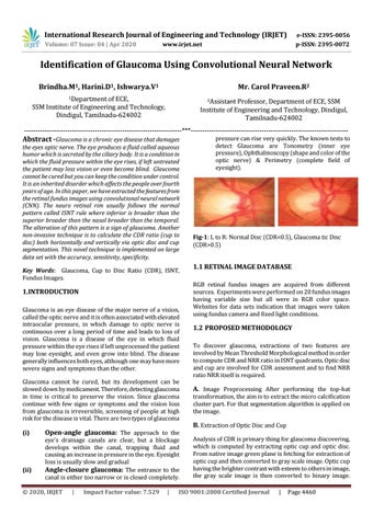
Fig-1: L to R: Normal Disc (CDR<0.5), Glaucoma tic Disc (CDR>0.5)
1.1 RETINAL IMAGE DATABASE RGB retinal fundus images are acquired from different sources. Experiments were performed on 20 fundus images having variable size but all were in RGB color space. Websites for data sets indication that images were taken using fundus camera and fixed light conditions.
1.2 PROPOSED METHODOLOGY To discover glaucoma, extractions of two features are involved by Mean Threshold Morphological method in order to compute CDR and NRR ratio in ISNT quadrants. Optic disc and cup are involved for CDR assessment and to find NRR ratio NRR itself is required.
A. Image Preprocessing After performing the top-hat transformation, the aim is to extract the micro calcification cluster part. For that segmentation algorithm is applied on the image.
B. Extraction of Optic Disc and Cup Analysis of CDR is primary thing for glaucoma discovering, which is computed by extracting optic cup and optic disc. From native image green plane is fetching for extraction of optic cup and then converted to gray scale image. Optic cup having the brighter contrast with esteem to others in image, the gray scale image is then converted to binary image.
ISO 9001:2008 Certified Journal
|
Page 4460