International Research Journal of Engineering and Technology (IRJET)
e-ISSN: 2395-0056
Volume: 07 Issue: 03 | Mar 2020
p-ISSN: 2395-0072
www.irjet.net
HISTOGRAM ANALYSIS FOR MELANOMA DISCRIMINATION IN REAL TIME IMAGE S.Atchaya1, S. Krishna Priya1, M.Preethi1, E. Punitha Valli1, S.Mukesh2 1S.Atchaya,
Agni college of Technology Priya, Agni college of Technology 2Assistant Professor, Department of Biomedical Engineering, Agni College of Technology, Anna University, Chennai, India ----------------------------------------------------------------------***--------------------------------------------------------------------1S.Krishna
Abstract - Melanoma is one of the deadliest forms of cancer, hence, great effort has been put into development of diagnosis methods for this disease. In this paper, we present a novel framework for melanoma image recognition via both K-means clustering, support vector Machine. Automated Melanoma recognition is a very challenging task due to the low contrast of skin lesions, the huge intra class variation of melanomas, the degree of visual similarity between melanoma and non melanoma lesions and the existence of many artifacts in the image. In order to meet these challenges, we propose a method for melanoma recognition by leveraging very support vector machine. Compared with existing methods employing either low level hand-crafts features, our substantially SVM and Kmeans can acquire richer and more discriminative features for more accurate recognition. Key Words: Melanoma, k-means, Support Vector Machine, artifacts in the images, skin lesions. 1. INTRODUCTION Melanoma skin cancer is one of the most rapidly increasing and deadliest cancers in the world, which accounts for 75% of skin cancer deaths[1-3]. Early diagnosis is of great importance for treating this diseases as it can be cured easily at early stage [1-4]. To improve the diagnosis of this disease, dermoscopy has been introduced to assist dermatologists in clinical examination since it is a non-invasive skin imaging technique that provide clinicians.
variation of melanomas in terms of color, texture, shape, size and location in the dermoscopy images as well as the high degree of visual similarity between melanoma and non melanoma Lesions make it difficult to discriminate melanomas from non-melanoma skin lesions. Second, the relatively low contrasts and obscure boundaries between skin lesions (especially at their early stages) and normal skin regions make the automated recognition task even harder. Finally, the presence of artifacts, either natural (hairs, veins) or artificial (air bubbles, ruler marks, color calibration charts, etc.) may blur or occlude the skin lesions and further aggravate the situation. In this paper, we propose a novel method based on K-means and Support vector machine with a set of effective training schemes in order to meet the challenges of automated melanoma recognition. Similar to some previous works, we propose to first segment the skin lesions from dermoscopy images and then classify them into melanoma ones and non-melanoma ones so that the classification stage can extract more specific and representative features within the lesion regions instead of performing it in the whole dermoscopy images. We employ very deep networks (more than 50 layers) for both the segmentation and the classification stages in order to obtain more discriminative features for more accurate recognition.
Fig 2 Less Affected
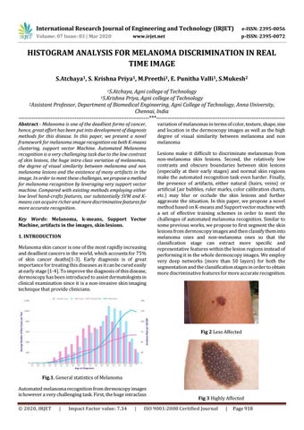
Fig.1. General statistics of Melanoma Automated melanoma recognition from dermoscopy images is however a very challenging task. First, the huge intraclass Š 2020, IRJET
|
Impact Factor value: 7.34
|
Fig 3 Highly Affected
ISO 9001:2008 Certified Journal
|
Page 918