International Research Journal of Engineering and Technology (IRJET)
e-ISSN: 2395-0056
Volume: 07 Issue: 02 | Feb 2020
p-ISSN: 2395-0072
www.irjet.net
Detection and Classification of Breast Cancer from Mammogram Image Vikrant Shitole1, Leena Hulawale2, Nishigandha Bapat3 1,2,3Dept.
of Information Technology, PCET’s NMIET, Maharashtra, India ---------------------------------------------------------------------***----------------------------------------------------------------------
Abstract - Today we know that the deaths of women in the age group 15-54 are increasing due to breast cancer. It is ecognized as the major reason for the deaths of women. Day by day, the number of patients is increasing. As its major factors have not been identified, it is unable to prevent it. So, the possibility of improvement is only an early diagnosis. The purpose of this study is to review the existing approaches of breast cancer detection and pretreatment in mammographic images. The purpose of pretreatment is to improve the image quality and by removing irrelevant noise and unwanted components in the background of the mammogram, there are various methods to pre-process the mammogram image in that process from now on. Discuss their advantages and disadvantages.
This mammogram [3] [4] image examines by radiologists. It detects the abnormalities and to identify whether it is benign or malignant. The radiologist fails many times to differentiate between false positive and false negative. So it’s impossible to detect the correct abnormality by a human radiologist. From digitized mammograms automatically detect the suspicious lesion, for increasing the quality of the image some preprocessing steps have been done. The quality of an image can be increased by removing unwanted regions in the background of the mammogram image. Two very important preprocessing steps are the removal of noise and pectoral muscles. The main goal of removing the noise from the image is to get noise-free data for further preprocessing and detect the breast cancer. The pectoral muscle is another important artifact, because of this disturbs the correct detection of breast cancer. From the mammogram image, hence extracting pectoral plays a very important role in the detection of cancer successfully [5].
Key Words: Breast cancer; Mammogram; Turn count; Classification 1. INTRODUCTION Today, the woman's death has been increased because of breast cancer. The abnormal growth of the cell is the main reason for breast cancer. Generally, cancer is difficult to identify with middle-age women. It cannot prevent completely since its cause remains unknown [1]. To decrease the death rate, the need to detect breast cancer at an early stage and better treatment options available to patients. If breast cancer identified in the earlier stage, the cure rate of breast cancer can be increased. The detection of cancer at an earlier stage can be archived by using mammography. It cannot prevent cancer, but they save the life of a cancer patient. They help to detect intangible tumors and increases the survival rate. In biological and medical field preprocessing is a very important concept.
1.1 Review of Literature This section discusses the literature review in detail about the women's breast cancer prognosis prediction. In biotechnology and medical filed image processing is an important task. Extracting important features of the images they used a texture-based method. Firstly, they apply discrete wavelet transform on the image and the approximation matrix changed into 1-D using zigzag scanning and finally, non-stationary signal features are extracted. These features are more sensitive to light and edge so that they differ from the abnormal and normal individuals [1]. The clustering classification of breast microcalcification into the Malignant and Benign category is a challenging task for the computerized algorithm. In this paper [2] they used Multi-View Classification for the classification of microcalcification and it is implemented using Logistic Regression Classification. This experiment is conducted on the Digital Database for Screening Mammography (DDSM) this dataset includes demographic data of the patients.
It used for diagnosing abnormal cases, image analyzing, and extraction of useful information [2].
Computerized clustered microcalcifications in mammograms, it suffers from the occurrence of falsepositive (FP) results. They investigate the statistical estimation to determine the number of FP that is present in the detected microcalcification. First, they find out a number of true positives by using Poisson-binomial probability distribution of training they used logistic regression models. Three different methods are used for micro calcification (MC) detector namely Context Sensitive Classification Model detector, Support Vector Machine
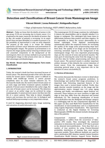
Fig 1: women breast anatomy
© 2020, IRJET
|
Impact Factor value: 7.34
|
ISO 9001:2008 Certified Journal
|
Page 820