International Research Journal of Engineering and Technology (IRJET)
e-ISSN: 2395-0056
Volume: 07 Issue: 12 | Dec 2020
p-ISSN: 2395-0072
www.irjet.net
COVID-19 Detection using Chest X-Ray Images by Artificial Intelligence Amey Sanjay Diolkar1 1B.E
Department of Computer Science and Engineering, G.H.Raisoni College of Engineering-440012, Nagpur, Maharashtra, India ---------------------------------------------------------------------***---------------------------------------------------------------------Abstract – The spreading increase in covid-19 patients is overwhelming healthcare systems all over the world. With limited testing kits, every patient with respiratory illness cannot treat using conventional techniques. Deep Learning has boost multi-fold in recent years, and it has played a significant role in image classification, including medical imaging. Convolutional Neural Networks (CNNs) have performed well in detecting many diseases, including coronary artery disease, malaria, Alzheimer's disease, and different dental diseases. The test also has a long turn-around-time and limited sensitivity. The study reveals that infected patients exhibit distinct radiographic visual characteristics, fever, dry cough, fatigue, dyspnoea. Diagnosing possible covid-19 infections on chest X-ray may help high-risk quarantine patients while test results are waiting. X-ray machines are readily available at all the healthcare centers, with no transportation time involved for samples. This project proposes using a chest x-ray to classify the patient's selection for further testing and treatment. The detection is critical acute respiratory syndrome coronavirus responsible for coronavirus disease 2019 (COVID-19), using chest X-ray images has life-saving importance for both patients and doctors. Also, in countries that cannot purchase laboratory kits for testing, this becomes even more vital. This work shows how a change in convolutional layers and an increase in dataset affect classifying performances. Key Words: COIVD-19, Coronavirus, Pandemic, X-Ray, Neural Network, Convolutional Neural Network, Data Science, Artificial Intelligence 1. INTRODUCTION The ongoing pandemic of Coronavirus or COVID-19 disease 2019-2020 has led to a global health care crisis. The main challenge in this pandemic situation on how to identify COVID-19 patients. Coronavirus or COVID-19 is an infection disease trigged by server acute respiratory syndrome COVID-19 (SARS-COV2). The coronavirus disease was initially identified in December 2019 in Wuhan, China and has spread globally worldwide. The patient with Pneumonia of the mysterious cause was first reported in the WHO country office in china in December 2019. After the month of December 2019, the disease has spread all over the world. The disease spread very fast, and the number of cases increased highly. Then WHO declared it is a Pandemic. As of 27 Nov. 20, there were – 61308116 Coronavirus cases, 1437835 deaths and 42395359 recovered patients. The number is still rising in the world there were.
© 2020, IRJET
|
Impact Factor value: 7.529
|
Although radiological imaging is not recommended for diagnostics as the patient arrives in the clinic. The chest XRay image is useful to observe treatment outcomes and comorbidities in seriously ill patients. The detection of Coronavirus from chest X-Ray and its differentiation from lung disease with indistinguishable opacities is a puzzling function that relies on the accessibility of expert radiologists.
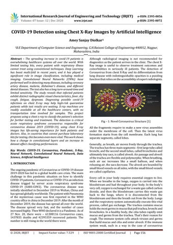
Fig – 1: Novel Coronavirus Structure [1] All the fragments require to make a new virus assembles under the membrane of the cell. Then the latest virus formation starts from the cell membrane. Each lung has separate segments, called lobes. Generally, as breath, air moves freely through the trachea. The trachea has three main segments:- first large tube called bronchi, and the second small tubes, called bronchioles and ultimately tiny sacs, is called alveoli. Air passage and alveoli of the trachea are flexible and polymorphic. When breathing, each air sac increases like a small balloon, and when releasing air, the sacs decrease. The alveoli are bounded by small blood vessels on all sides, with the small blood vessels are called capillaries. Every cell in your body requires essential oxygen to live. When we breathe in the lungs, oxygen is carried into the bloodstream and had throughout your body. In the body's very cell, oxygen is exchanged for a waste gas called carbon dioxide, and then the bloodstream carries this waste gas back to the lungs, where it is disconnected from the bloodstream and then the breath out from the body. Lungs and the respiratory system automatically execute this vital process, called gas exchange. The trachea contains mucus holds the most germs in mucus that pull trachea, bronchi and bronchioles. In a healthy body, the cilia tubes rapidly emit mucus and germs from the trachea. That's their reason for cough. The immune system cells attack viruses and germs that build mucus and cilia and enter alveoli. If the immune system weak, such in a way in the case of coronavirus ISO 9001:2008 Certified Journal
|
Page 156