International Research Journal of Engineering and Technology (IRJET)
e-ISSN: 2395-0056
Volume: 06 Issue: 05 | May 2019
p-ISSN: 2395-0072
www.irjet.net
Automatic detection of Diabetic Retinopathy using R-CNN Athira T R1, Athira Sivadas2, Aleena George3 , Amala Paul4 , Neethu Radha Gopan5 1,2,3,4Student,
Dept. of Electronics and Communication Engineering, Rajagiri School of Engineering and Technology, Cochin, Kerala, India 5Assistant Professor, Dept. of Electronics and Communication Engineering, Rajagiri School of Engineering and Technology, Cochin, Kerala, India ---------------------------------------------------------------------***----------------------------------------------------------------------
Abstract - Diabetic Retinopathy (DR) is an eye
An automatic method for detection of diabetic retinopathy would help people with diabetes to recognize the symptoms at its earlier stage. It can greatly reduce the clinical burden on retina specialists. This also helps to monitor the dynamics of the lesions. Countries with huge population like India, China, Indonesia and Bangladesh contributes to 45% of the global burden in diabetes [2]. Since the counts are expected to move up, an automatic clinical detection would be of much help.
abnormality caused as a result of long term diabetes. As the disease progresses it leads to distortion and blurred vision. The diagnosis of DR using color fundus image requires skilled clinicians to identify the presence of critical features which makes this a difficult and time consuming task. In our paper we propose a R-CNN (Regional Convolutional Neural Network) approach to diagnose DR from digital fundus images. In our research we implemented a new approach were the whole image was segmented and only the regions of interest were taken for further processing. In our method we have used 10 layers for R-CNN, trained it on 130 fundus images and tested on 110 images. All the images were classified into two groups i.e., with DR and without DR. This R-CNN (Regional CNN) approach was found to be efficient in terms of speed and accuracy. An accuracy of approximately 93.8% was obtained from R-CNN.
Key Words: Convolutional neural network, Regional convolutional neural network, Diabetic retinopathy.
1. INTRODUCTION Diabetes has now become a worldwide disease which ultimately leads to complete vision loss. Diabetic Retinopathy is a complication of type 2 diabetes. According to the IAPB (International Agency for the Prevention of Blindness) [1] report published in 2017, there were 422 million people diagnosed with diabetes. 1 in 3 people diagnosed with diabetes will have diabetic retinopathy up to a certain degree and 1 in 10 people will suffer from vision loss. DR results in the damage of blood vessels in the retinal layer of the eye. It forms microaneurysms due to the focal dilation of weakened walls [2]. The capillaries may become leaky forming yellow white flecks which are commonly referred to as exudates.
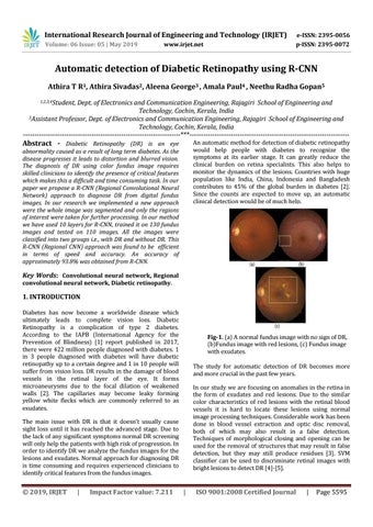
Fig-1. (a) A normal fundus image with no sign of DR, (b)Fundus image with red lesions, (c) Fundus image with exudates. The study for automatic detection of DR becomes more and more crucial in the past few years. In our study we are focusing on anomalies in the retina in the form of exudates and red lesions. Due to the similar color characteristics of red lesions with the retinal blood vessels it is hard to locate these lesions using normal image processing techniques. Considerable work has been done in blood vessel extraction and optic disc removal, both of which may also result in a false detection. Techniques of morphological closing and opening can be used for the removal of structures that may result in false detection, but they may still produce residues [3]. SVM classifier can be used to discriminate retinal images with bright lesions to detect DR [4]-[5].
The main issue with DR is that it doesn’t usually cause sight loss until it has reached the advanced stage. Due to the lack of any significant symptoms normal DR screening will only help the patients with high risk of progression. In order to identify DR we analyze the fundus images for the lesions and exudates. Normal approach for diagnosing DR is time consuming and requires experienced clinicians to identify critical features from the fundus images.
Š 2019, IRJET
|
Impact Factor value: 7.211
|
ISO 9001:2008 Certified Journal
|
Page 5595