International Research Journal of Engineering and Technology (IRJET)
e-ISSN: 2395-0056
Volume: 06 Issue: 04 | Apr 2019
p-ISSN: 2395-0072
www.irjet.net
Lung Cancer Detection Using Grey Level Co-Occurrence Matrix Mahpara Alam1, Bhavika Chaudhari2, Shamal Bangar3, Pratiksha Desale4, Prof. Chaitali Patil5 1,2,3,4,Students,
Department of Computer Engineering, K.K.W.I.E.E.R, Maharashtra, India Department of Computer Engineering, K.K.W.I.E.E.R, Maharashtra, India ---------------------------------------------------------------------***---------------------------------------------------------------------5Professor,
Abstract - Lung cancer is curable disease if detected in its
because of time factor. Image quality and accuracy was the core factor of this research. Image quality assessment as well as improvement depends on the enhancement stage where pre-processing techniques was used.
early stages. In recent years, Image processing techniques are widely used in medical domain for image improvement in earlier detection. For diagnosis of lung cancer, CT image of lung is one of the most common imaging modalities. Manually identifying tumor from hundreds of CT images for any patient may prove to be very tedious and time consuming task. Hence this system aims at detection of cancer through an automated process to minimize human error and making the process more accurate and hassle-free. Lung carcinomas are categorized by the size and appearance of the malignant cells. The growth of epithelial cells on lung parenchyma region shows in CT image which can be used in the system. In this proposed approach image pre-processing techniques are used like noise removal, enhancement and image segmentation then the feature extraction recognize the region of interest of tumor. SVM (Support Vector Machine) algorithm can be used for classification. GLCM (Grey Level co-occurrence Matrix) based textual features detection can be used to search the lung parenchyma region and extract the features.
Ritika Agarwal, Ankit Shankhadhar, Raj Kumar Sagar [2], Many techniques are available to determine the existence of nodule in the lung in its early stage but none of the technique gave the best result. Therefore they implemented the system to give more accuracy and best result they propose a system called as Computer Aided diagnosis System CAD for identifying the presence of nodule in lung. Khinn Mya Mya Tun [3] , The implementation of image preprocessing and image segmentation is carried out to obtain the diagnosis result. Median filter was used for image preprocessing. In feature extraction Gray Level Co-Occurrence matrix (GLCM) was used. In classification, feed-forward neural network was used to classify the lung cancer stages. Mr. Akshay Bhor, Mr. Aditya Likhar, Mr. Azhar Maner, Mr. Aditya Patil, Prof. Dipti Chaudhari [4], The main aim of the system was to automate the classification method for the first detection of carcinoma. It included classification algorithmic program i.e. Neural Network and for improvement GA (Genetic Algorithm) was employed.
Key Words: GLCM, CT images, SVM, Feature extraction, Image processing
1. INTRODUCTION Manually identifying tumours from hundreds of MRI slices for any patient may prove to be very tedious and time consuming task. Cancer is curable disease if it detected in its early stage. To reduce the death rates due to occurrence of lung cancer we need some methodology to detect the cancer in its early stage. Not only the detection of disease in its early stage may reduce the death rate but also proper diet and doctor treatment can be done on time.
Tanushree Sinha Roy, Neeraj Sirohi, Arti Patle[5], focuses their study mainly on the classification of lung images as affected by cancer disease or not. In their system lung CT scan image was used as input image and then some important features was extracted to classify the image. To classify the image Fuzzy Interface System was used. The system used both neural network and fuzzy logic technique.
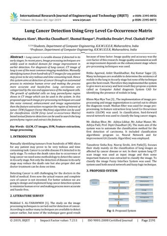
3. PROPOSED SYSTEM
Detecting Cancer is still challenging for the doctors in the field of medical. Even now the actual reason and complete cure of cancer is not invented. For better solution we are going to design and implement lung cancer detection system to minimize human error and making process more accurate and hassle-free.
2. LITERATURE SURVEY Mokhled S. AL-TARAWNEH [1], The study on the image processing techniques is carried out for detection of cancer. According to author many techniques are available to detect cancer earlier, but none of the technique gave good result
Š 2019, IRJET
|
Impact Factor value: 7.211
Fig -1: Proposed System
|
ISO 9001:2008 Certified Journal
|
Page 2191