International Research Journal of Engineering and Technology (IRJET)
e-ISSN: 2395-0056
Volume: 06 Issue: 03 | Mar 2019
p-ISSN: 2395-0072
www.irjet.net
RETINAL FUNDUS IMAGE SEGMENTATION USING WATERSHED ALGORITHM Dr. J. Nithyashri1, S. Divyalakshmi2, N. Mohana Priya3 1Assistant
Professor, Department of Computer Science and Engineering, Jeppiaar SRR Engineering College, Chennai, Tamil Nadu 603103, India. 2B.E., Department of Computer Science and Engineering, Jeppiaar SRR Engineering College, Chennai, Tamil Nadu 603103, India. 3B.E., Department of Computer Science and Engineering, Jeppiaar SRR Engineering College, Chennai, Tamil Nadu603103, India. ----------------------------------------------------------------------***--------------------------------------------------------------------Abstract –Automated optic disk (OD) detection plays a very important role in developing a pc assisted system for eye diseases. The examination of eye disease by ophthalmologists is limiting factor. In this paper, a technique for Optic Disk detection based on structured learning that belongs to a supervised method to avoid creating assumptions. The proposed technique utilizes the edge information of the fundus image to detect the Optic Disk and identifies the presence of cancer. The Sobel edge detector to capture the edge information and segmentation using watershed algorithm. Then circle Hough transform is carried out to approximate the boundary of Optic Disk by a circle. Key Words: Fundus image, Optic Disk, Edge Detection, Watershed Segmentation, Hough Transform. 1. INTRODUCTION Nowadays, some of the most common causes of visual impairment and blindness are diabetic retinopathy, glaucoma, hypertension and macular degeneration. These eye diseases manifest themselves in the retina and all of these diseases can be detected through a direct and regular ophthalmologic examination. Medical image analysis and processing has a great significance in the field of medicine, especially in non-invasive treatment and clinical study. Normally fundus images are manually graded by specially trained clinicians in a time-consuming and resource intensive process. However, several factors, like population growth, aging, are contributing to the risk of the patients with these diseases that makes the quantity of ophthalmologists required for analysis by interrogatory becomes a limiting issue. As a result, a pc assisted diagnosing system which can significantly reduce the burden on the ophthalmologists and may alleviate the inter and intra observer variability is desired. Retinal fundus image segmentation is a fundamental step in retinal image analysis and the follow-up ophthalmic diagnostics. In the process, OD detection plays a very important role, that has attracted intensive attention from clinicians and researchers. OD detection is commonly a key step for the detection of different anatomical structures. As a example, the OD location helps to prevent false positive detection of exudates incurred by diabetic retinopathy, since each OD and exudates are formed by bright regions within the fundus image. To sight these problematic veins that may be done with the segmentation of blood vessels in retinal digital pictures. The retina is a extremely organized structure with the ability to begin the process of visual information before the knowledge is transmitted through the optic nerve to the visual cortex. Layered structure that permits the perform to look restrictions in optical disk function or functional impairment on a layer or cluster of cells. However, the perception of colour, contrast, depth, and form happen within the cortex. OD is one of the most vital from indicator medicine pathology. Image segmentation is a important step in image analysis. Segmentation separates an image into its element components or objects. Here segmentation is critical to separate the optic disc area with blood vessels that exist in the area of the optic disk at the time was not considered in the segmentation of blood vessels.
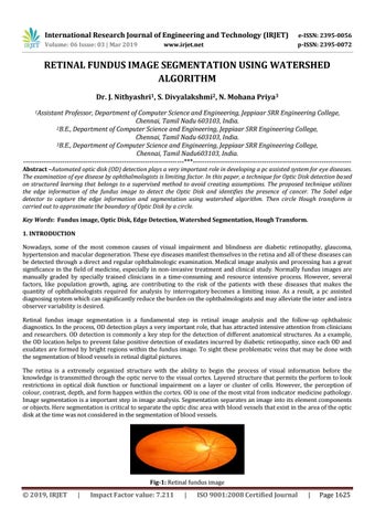
Fig-1: Retinal fundus image
Š 2019, IRJET
|
Impact Factor value: 7.211
|
ISO 9001:2008 Certified Journal
|
Page 1625