International Research Journal of Engineering and Technology (IRJET)
e-ISSN: 2395-0056
Volume: 06 Issue: 02 | Feb 2019
p-ISSN: 2395-0072
www.irjet.net
Diabetic Haemorrhage Detection using DWT and Elliptical LBP Rabul Saikia1, Manish Shankar Kaushik2, Dr. Aditya Bihar Kandali3 1Department
of Electrical Engineering, Jorhat Engineering College, Jorhat, Assam, India of Electrical Engineering, Jorhat Engineering College, Jorhat, Assam, India 3Associate Professor, Dept. of Electrical Engineering, Jorhat Engineering College, Jorhat, Assam, India -----------------------------------------------------------------------***-------------------------------------------------------------------2Department
Abstract - The extraction of exact retinal haemorrhage of
random forest and K-Nearest Neighbor (K-NN) are employed with scale-space features. In Ref. [4], a three- step set up was conferred for revelation of micro-aneurysms by proving filter banks. Firstly whole expedient applicant portion are tabbed for micro-aneurysms. Then, a sort of features is enumerating for each division in the second stage. Finally, a GMM (Gaussian mixture model) and SVM (Support Vector Machine) are combined to perform classification. Moreover in Ref. [5], the authors conferred a methodology for revealing of micro-aneurysms by proving a sparse representation and dictionary learning. In their effort, vicinity with presumed micro-aneurysms is distinguished by proving multi-scale Gaussian correlation filter. At the end most steps Support Vector Machine (SVM) is being employed for training as well as classification.
diabetic patient plays a significant role in medical image processing. This paper presents an accession for detection of both haemorrhage and healthy retina using symmetric Elliptical Local Binary Pattern (ELBP) and Discrete Wavelet Transform (DWT). The features of both haemorrhage and healthy retinal images are extracted using ELBP and further features are reduced by DWT. For classification binary SVM classifier is used here. The approach implemented on HRF database signifies 92.3% rate of accuracy. Key Words: DWT, Elliptical Local Binary Pattern, SVM, Diabetic haemorrhage
1. INTRODUCTION Diabetic retinopathy is one of the paramount topics in the field of medical research. In recent times, automatic tracking down of haemorrhages in diabetic retinal images has become immensely important in the clinical milieu. Indeed, diabetic retinopathy is a disease that deteriorates the retina and can lead to blindness. Haemorrhages in the retina are one of the primeval symptoms of diabetic retinopathy. An early and accurate diagnosis of diabetic retinopathy helps to improve medical treatment and prognosis. To abetment ophthalmologists numerous computer-sustain interpretation setup have been proposed in identifying diabetic retinopathy by detecting exudates, micro-aneurysms, or hemorrhages in the retina.
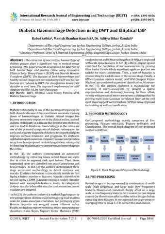
2. PROPOSED METHODOLOGY Our proposed methodology mainly comprises of Preprocessing, Feature extraction, Feature reduction and Classification. The overall block diagram of our proposed method as follows:
In Ref. [1], the authors contemplated an automated methodology by extracting fovea, retinal tissue and optic disc in order to segment dark spot lesions. Then, those segmented spots are classified into micro-aneurysms and hemorrhages. In Ref. [2], the authors contemplated three stage automated methodology to detect exudates and macula. Exudates derivation is conceivably imitate in first lap by a distinct number of features. Macula is identified in second lap by a GMM (Gaussian mixtures model) classifier trained with accomplish features. Lastly, to categorized diabetic macular edema the macular conform and section of exudates are assigned.
Figure 1: Block Diagram of Proposed Methodology
2.1 PRE-PROCESSING Retinal image can be represented by combination of smallscale (high frequency) and large scale (low frequency) features. Illumination variations deeply affect on a large scale i.e. low frequency features. So it is an important step to correct the illumination affects of the retinal images before extracting their features. In our approach we apply mean or averaging filter of mask 5×5 to correct the illumination.
In Ref. [3], the authors conferred a methodology hinge on by extracting regions of interest and normalizing local-maxima scale for micro-aneurysm revelation. For portraying goals Hessian response are assigned across different scales. Finally, to disclose regions with true micro-aneurysm four classifiers: Naïve Bayes, Support Vector Machines (SVM)
© 2019, IRJET
|
Impact Factor value: 7.211
|
ISO 9001:2008 Certified Journal
|
Page 1848