International Research Journal of Engineering and Technology (IRJET)
e-ISSN: 2395 -0056
Volume: 04 Issue: 04 | Apr -2017
p-ISSN: 2395-0072
www.irjet.net
Detection of Macular Edema by Using Various Techniques of Feature Extraction and Classification by SVM Classifier Ajay Advani1, Ashish Thawrani2, Amit Thaware3,Abhinay Gaonkar4,Mrs.Vaishali Kulloli5 1,2,3,4Student,
Information Technology, Pimpri Chinchwad College of Engineering, Maharashtra, India. Information Technology, Pimpri Chinchwad College of Engineering, Maharashtra, India. ---------------------------------------------------------------------***--------------------------------------------------------------------5Assistant Professor,
Abstract - Diabetic retinopathy could be a vision
threatening complication as a results of DM that is that the main cause of impairment and visual defect in diabetic patients. In several cases the patient isn't responsive to the disease till it's too late for effective treatment. The prevalence of retinopathy varies with the age of polygenic disorder and the period of illness. Early diagnosing by regular screening and treatment is useful in preventing visual impairment and visual defect. This paper presents the review of automatic detection of diabetic retinopathy.
1. INTRODUCTION Diabetic Retinopathy could be a complication of polygenic disorder and is an eye illness which may cause loss of sight. It affects almost eightieth of the patients having polygenic disorder for over tenyears. Diabetic Retinopathy is caused by the harm to the blood vessels that cause unseaworthy of blood and different sorts of fluids on the tissue layer. These leakages type patterns like venous loops, laborious exudates, small Aneurysms (MA’s),cotton wool spots, etc. Diabetic macular puffiness (DMA) could be acomplication caused attributable to diabetic retinopathy and is that the true explanation for visual defect and visual loss. Diabetic macularedema can be diagnosed attributable to ECF run from the blood vessels at intervals the macula region. Leakage is caused attributable to the breakdown of epithelium tight junctionspresent within the small aneurysms or retinal vessels. Thus the lipid deposition accumulated within the tissue layer attributable to run is called exudates. Exudates once clinically seen seem as yellow white intra-retinal deposits on digital fundus image. Since the screening of patients affected by diabetes is incredibly slow, thereby abundant effort needs to be placeup for the event of reliable computer aideddiagnosis (CAD) systems strictly acting on colorfundus pictures.
Š 2017, IRJET
|
Impact Factor value: 5.181
|
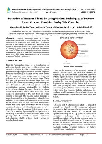
Figure type of diseases [14]
Due to the presence of an outsized variety of patients, the workload of associate specialist is extremely in substantialand automated detection systems square measure a requirement to limit the severity of the illness. There's a requirement to develop associate algorithm to aid ophthalmologists for early diagnosingand remedy of the illness with abundant ease and potency.To build associate economical automatic system, there's a requirement to analyze region, optic disc, common diabetic pathologies like exudates, small aneurysms, hemorrhages, found in large number round the immediate areas round the macula. 2. LITERATURE SURVEY M. Gandhi and R. Dhanasekaran et al. [1] projected method to classify bodily structure pictures victimisation SVM supported exudates and also the difficultness of the lesions. K. SaiDeepak and JayanthiSivaswamy et al. [2] projected newfeature extraction technique to capture the worldcharacteristics of the bodily structure pictures. UmerAftab and M.UsmanAkram et al. [3] gave associate formula for automatic identification of exudates for detection of macular puffiness.Theyused filter bank for candidate exudate detection followed by feature extraction and classification. ISO 9001:2008 Certified Journal
|
Page 1734