International Research Journal of Engineering and Technology (IRJET)
e-ISSN: 2395 -0056
Volume: 04 Issue: 05 | May -2017
p-ISSN: 2395-0072
www.irjet.net
An Automated Approach for the Detection & Analysis of Brain Tumor using Detailed MRI through MATLAB Abhishek Kumar1, Professor Paurush Bhulania2 1Student,
Department of Electronics & Communication, Amity School of Engineering & Technology, Amity University NOIDA, India 2Asst. Professor, Department of Electronics & Communication, Amity School of Engineering & Technology, Amity University NOIDA, India ---------------------------------------------------------------------***---------------------------------------------------------------------
Abstract - It’s very challenging and emerging in modern
opportunity for physicians to support and save lives by identifying the disorder earlier and perform necessary actions. Types of picture processing method are existing to be utilized on some image detection for tumor finding that will detect certain characteristics of the tumors such as the border, shape, calcification and appearance. These characteristics will create the detection procedures extra precise and easier as there are some normal characteristics of each feature for a specific tumor. Magnetic Resonance Imaging (MRI) is an innovative medical imaging method used to produce high-quality images of the organs present in the human body, MRI imaging is frequently used when treating brain tumors. By utilizing these high-resolution MRI images, we can collect in-depth anatomical data to observe human brain's abnormalities.
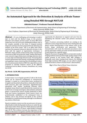
day’s to deal with the Medical image. In this province, detection and analysis of brain tumor [1] from MRI brain image have turn out to be most challenging problems, due to the complex assembly of the brain, A computer-assisted diagnostic technique has been proposed here for detecting and analysis of the brain tumor. This is an effort built which expresses the proposed scheme for detection of tumor in the brain with the support of segmentation techniques in MATLAB; which includes preprocessing phase incorporates conversion of MRI into grayscale image, median filtering for noise removal, image enhancement and edge detection (such as canny edge and Sobel edge detection) [2] . Processing phase involves segmentation like intensity, morphological operation and applying k-means clustering than thresholding to obtain the area of unwanted cells from MRI. Here algorithms are also proposed to calculate Features extract of the different component (such as Mean, Variance, Smoothness, etc.) using the GLCM.
Fig -1: Flow Chart of Brain Tumor detection
Key Words: GLCM, MRI, Segmentation, MATLAB
1. INTRODUCTION A brain tumor is an irregular growth of cells in the brain, which can be cancerous (malignant) or noncancerous (benign). is named as a intracranial tumor produced by irregular and discarded cell partition, classically in the brain itself (neurons, glial cells (astrocytes, oligodendrocytes, ependymal cells, myelin- producing Schwann cells), lymphatic tissue, blood vessels blood) in the cranial nerves, in the brain coverings (meninges), skull, pituitary and pineal gland, or spread from cancers mostly positioned in other body part tumors (metastases). Brain tumors (true) are predominantly established in the subsequent fossa in children and in the forward two-thirds of the cerebral hemispheres in adult human being, although it can influence any motive of the brain. Based on statistics, tumors are the second source of cancerallied deaths in kids (both genders) whose are under the age of 20 and in males whose age 20 to 39 the disarray is also the fifth well-known source of cancer- allied deaths in females ages 20-39. This evidence increase the importance of the studies on the tumor recognition and this will represent the
© 2017, IRJET
|
Impact Factor value: 5.181
|
ISO 9001:2008 Certified Journal
| Page 1806