International Research Journal of Engineering and Technology (IRJET)
e-ISSN: 2395 -0056
Volume: 04 Issue: 05 | May -2017
p-ISSN: 2395-0072
www.irjet.net
Cost Effective Method for Automatic Detection of Diabetic Retinopathy Renu1, Sachin Kumar2, Sumita Mishra3, Pragya4 1Mtech,Dept
of Electronic & Communication Engineering, Amity University Lucknow, Uttar Pradesh, India of Electronic & Communication Engineering, Amity University Lucknow, Uttar Pradesh, India 4Professor,Dept of Science, Mahila Vidyalaya PG College, Aminabad, Lucknow, Uttar Pradesh, India . ---------------------------------------------------------------------***--------------------------------------------------------------------23Professor,Dept
Abstract - Diabetic retinopathy is a condition occurring in
persons with diabetes, which causes progressive damage to the retina, the light sensitive lining at the back of the eye. It is a serious sight threatening complication of diabetes. Over time, diabetes affects the circulatory system of the retina. Diabetic retinopathy is the result of damage to the tiny blood vessels that nourish the retina. These vessels leak blood and other fluids that cause swelling of retinal tissue and clouding of vision. The condition usually affects both eyes. If left untreated, diabetic retinopathy can cause blindness. So the early detection and diagnosis of DR is necessary to preserve the vision. This paper presents a generalized system model for detecting DR, the proposed model consist of camera, Raspberry pi. Further it also summarizes various DR detection techniques that have evolved during last decade.
the gel like fluid that fills the back of the eye. The new blood vessel may leak blood into the vitreous, cloudy vision [2]. Globally, the number of people with DR will grow from 126.6 million in 2010 to 191.0 million by 2030 and we estimate that the number with vision-threatening Diabetic Retinopathy will increase from 37.3 million to 56.3, if prompt action is not taken [3].India is set emerge as diabetic capital of the world. According to WHO, 31.7 million people were affected by diabetes mellitus in India year 2000. This figure is estimated to rise 79.4 million by 2030, the largest number in any nation in the world. Almost two- third of all type2 almost all type1 diabetes are expected to develop diabetic retinopathy [4].
Key Words: optic disc, exudates, fundus images, blood vessel, Retinal images.
1. INTRODUCTION Diabetic retinopathy is a disease that arises in person who has diabetes. It causes gradually damage to the retina, the light sensitive lining at the back of the eye. DR is severe light menacing complication of diabetes. Diabetes is the disease characterized excessive sugar in the body, which can cause damage all over the body, including the eyes. Long time, diabetes damages the blood vessels in the retina. DR occurs when these very small blood vessel leak blood and other fluid [1]. This causes the retinal tissue to swell resulting in blurred or cloudy vision. The disease commonly influences both eyes. The longer person has diabetes, the more likely they will develop diabetic retinopathy. If it leaves untreated, diabetic retinopathy can cause blindness. Diabetic Retinopathy is dividing in two types:Non proliferative diabetic retinopathy (NPDR) is the first stage of the disease in which symptoms will be mild or nonexistent. In NPDR, the blood vessels in the retina are weekend. Small bulges in the blood vessels, known as microaneurysm (MAs), may leak fluid into the retina. The leakage may lead to swelling to the macula. Proliferative Diabetic Retinopathy PDR is the leading condition of the disease. At this stage circulation problems deprive the retina of the oxygen. As a result new weak blood vessel can start to arise in the retina and into the vitreous, Š 2017, IRJET
|
Impact Factor value: 5.181
|
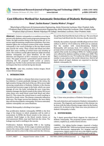
Fig:-1 Zone-wise prevalence of Diabetic Retinopathy The early detection and treatment of diabetic retinopathy is necessary. The diagnosis and treatment of this disease can be done with the help of fundus images taken by the fundus camera [5]. As shown in fig 2 that zone-wise prevalence of diabetic retinopathy in India.
2. SYSTEM MODEL Fig 3 depict generalized block diagram for detection of diabetic retinopathy. The proposed system model consist camera, raspberry pi, ardino board, led etcs. CAMERA - For taking fundus images, required a fundus camera, TOPCON TRC- 50IX tria functional camera using ICG infrared fluoresceine angiography. For imaging with tiff compression, digital camera was set to the highest quality. Images were 1032 X 1320 pixels and 8 bit grey scale [6]. ISO 9001:2008 Certified Journal
|
Page 1172