International Research Journal of Engineering and Technology (IRJET) Volume: 03 Issue: 07 | July-2016
www.irjet.net
segmentation scheme developed in this paper is robust and accurate but can still be improved in several ways, notably by adding rules to handle exceptional situations. Research paper related to finding out abnormalities in chest x- ray using local texture analysis published by B. van Ginneken, S. Katsuragawa, B. ter Haar Romeny, K. Doi, in 2002. In this paper method is presented to find out abnormalities in chest radiographs which are sum into an overall abnormality score. Main aim of method was to find out abnormal texture pattern in chest x-ray. The scheme starts with segmentation of the lung fields, using image appearance models. The segmentation is used to divide the lung fields from background region. [5] S. Candemir, K. Palaniappan, and Y. Akgul (2014) published research paper related to graph cut image segmentation algorithm with the help of energy minimization approximation�. In this paper, author describes a methodology for adaptive parameter learning to improve the segmentation performance using a multi-class classifier approach. Author demonstrated the performance of the system within graph cut segmentation framework. The approaches in literature modulate the regularization parameter using a single feature or a heuristic combination of a few features. Author model the characteristics of the image regions with feature vectors which includes haar feature for edge, local binary patter for texture and hessian for shape information of local regions. Therefore this approach characterizes the image regions better than using single feature. The simple structure of the system allows to incorporate alternative features such as Scale Invariant Feature Transform which is one of the best performing feature descriptors among local descriptors. [2]
3. DATA For this experiment, CXR set of Montgomery County (MC) is used. The MC set, is collected within the tuberculosis control program of the Department of Health and Human Services of Montgomery County (MC), Maryland. The MC set consist of total 138 posteroanterior chest x-rays, out of which 80 Chest x-rays are normal and 58 Chest x-rays are abnormal with TB symptoms. All images of the MC set are in 12-bit grayscale, captured with an X-ray machine. The abnormal Chest x-rays consist of a wide range of TB-related abnormalities, including effusions.
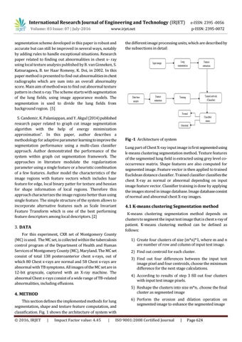
the different image processing units, which are described by the subsections in detail.
Fig -1: Architecture of system Lung part of Chest X-ray input image is first segmented using k-means clustering segmentation method. Texture features of the segmented lung field is extracted using grey level cooccurrence matrix. Shape features are also computed for segmented image. Feature vector is then applied to trained Euclidean distance classifier. Trained classifier classifies the chest X-ray as normal or abnormal depending on input image feature vector. Classifier training is done by applying the images stored in image database. Image database consist of normal and abnormal chest X-ray images.
4.1 K-means clustering Segmentation method K-means clustering segmentation method depends on clusters to segment the input test image that is chest x-ray of patient. K-means clustering method can be defined as follows: 1) Create four clusters of size (m*n)*1, where m and n are number of row and column of input test image. 2) Find out centroid for each cluster. 3) Find out four differences between the input test image pixel and four centroids, choose the minimum difference for the next stage calculations. 4) According to results of step 3 fill out four clusters with input test image pixels. 5) Reshape the clusters into size m*n, .choose the final cluster as segmented image
4. METHOD This section defines the implemented methods for lung segmentation, shape and texture feature computation, and classification. Fig. 1 shows the architecture of system with Š 2016, IRJET
e-ISSN: 2395 -0056 p-ISSN: 2395-0072
|
Impact Factor value: 4.45
|
6) Perform the erosion and dilation operation on segmented image to enhance the segmented image
ISO 9001:2008 Certified Journal
|
Page 624