International Research Journal of Engineering and Technology (IRJET) Volume: 03 Issue: 07 | July-2016
www.irjet.net
e-ISSN: 2395 -0056 p-ISSN: 2395-0072
Medical Imaging: Automatic segmentation of spinal canal Munavar Jasim.K1, Aneesh Kumar B2, 1M.Tech 2Asst.
student,Dept. Of Computer science and Engg., MEA Engg College,Kerala,India Professor,Dept. Computer science and Engg., MEA Engg College,Kerala,India
---------------------------------------------------------------------***---------------------------------------------------------------------
Abstract – Segmentation of spinal canals have a great role in many medical fields. Spinal canal is one of the best way used to diagnosis and analysis. Accurate segmentation of the spinal canals in computed tomography (CT) images is an important task in many related studies. Many research and clinical studies require the automatic segmentation of the spines, which is capable of facilitating disease diagnosis, treatment, and statistical analysis/evaluation. For example, the segmentation of the spine provides spatial reference to locate and identify other neighbouring anatomical structures in abdomens and chests, thus contributing to the understanding of the full-body scan essentially. In terms of image registration, the segmented spines provide important features that are helpful to the correct alignment of corresponding anatomical structures across individual subjects. Furthermore, it becomes easier to conduct disease-oriented analysis given the segmented topologies/shapes of the spines. In the meantime, based on the segmentation of the spinal canal, the entire spinal cord can easily be delineated, making it possible to count radiotherapy dosages which are crucial to the normal functions of the nerve tracts Key Words: Image segmentation, Topology, Random walk, Refinement, Spinal canal, Voxels
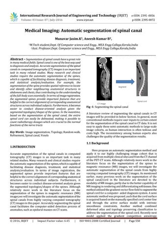
Fig -1: Segmenting the spinal canal A literature review of segmenting the spinal canals in CT images will be provided in below Section. In general, most conventional methods require user inputs to certain extent for the segmentation of the spinal canal in CT data. It is not easy to apply these semi-automatic methods to large-scale image cohorts, as human interaction is often tedious and costs high. The inconsistency among human experts also challenges the quality of the segmentation results.
1.1 Background 1.INTRODUCTION Accurate segmentation of the spinal canals in computed tomography (CT) images is an important task in many related studies. Many research and clinical studies require the automatic segmentation of the spines, which is capable of facilitating disease diagnosis, treatment, and statistical analysis/evaluation. In terms of image registration, the segmented spines provide important features that are helpful to the correct alignment of corresponding anatomical structures across individual subjects. Furthermore, it becomes easier to conduct disease-oriented analysis given the segmented topologies/shapes of the spines. Although relatively more work in the literature focus on the segmentation of the spines in magnetic resonance (MR) images,we will investigate the automatic segmentation of the spinal canals from highly varying computed tomography (CT) images in this paper. Accurately segmenting the spinal canal facilitates the computer-aided detection process of anomalies, such as epidural masses on CT scans.
Š 2016, IRJET
|
Impact Factor value: 4.45
|
Here propose an automatic segmentation method and apply it to our highly challenging image cohort that is acquired from multiple clinical sites and from the CT channel of the PET-CT scans. Although relatively more work in the literature focus on the segmentation of the spines in magnetic resonance (MR) images, we will investigate the automatic segmentation of the spinal canals from highly varying computed tomography (CT) images. As mentioned earlier, many previous work on the segmentation of the spinal canal/cord in the literature are devoted to the modality of MR images, partly due to the better capability of MR imaging in rendering and differentiating soft tissues. One method utilized the gradient vector flow field to segment the spinal cord for a computer- aided diagnosis system. A semi automatic method was proposed ,such that the cord surface is acquired based on the manually specified cord center line and through the active surface model with intrinsic smoothness constraints. Another method combined deformed atlas and topology preserving classification to address the segmentation of the spinal cord. Recently next model applied the gradient competition anisotropy
ISO 9001:2008 Certified Journal
|
Page 550