International Research Journal of Engineering and Technology (IRJET)
e-ISSN: 2395-0056
Volume: 11 Issue: 02 | Feb 2024
p-ISSN: 2395-0072
www.irjet.net
Experimental & ANSYS Stress Analysis of Real Human Femur Bon Pranjal P. Farakte 1, Rohini N. Gaikwad2, Priyanka S. Chavan3 1Assitant Professor, D. Y. Patil college of Engineering and Technology, Kolhapur, Maharashtra, India 2Assitant Professor, D. Y. Patil college of Engineering and Technology, Kolhapur, Maharashtra, India 3Assitant Professor, D. Y. Patil college of Engineering and Technology, Kolhapur, Maharashtra, India
---------------------------------------------------------------------***---------------------------------------------------------------------
Abstract - The human femur is the leg bone that
problems such as: failure of the bone due to stress concentration, fatigue of bone cemented rarely, a failure of the metal fixation device. A common tool in engineering for the prediction of stress, strain and deformations is the finite element method. The purpose of these studies was to predict the mechanical response of the preserved human femur, and validated by experiments on the same.
experiences the most deformation. The femur bone, which is the largest and longest bone in the body, is the most likely to fail, especially in women. In the event of failure, orthopedic implantation is performed. Sometimes implanting a new synthetic femur is necessary following femoral bone loss.Therefore, in order to compare with a synthetic femur, we must see how the real and actual femur behaves. Analysis of the material's properties, size, form, surface treatment, load resistance, and failure probability is required prior to implantation.
2. PROBLEM DEFINITION “Femur is leg bone of the human body undergoing more deformation. Being longest and heaviest in size, failure of femur bone is the most common among bone failures especially in woman. Orthopedic implantation is done in case of failure. After failure of femur bone sometimes it is necessary to implant a new synthetic femur .So that we must check the behaviour of real and actual femur to compare with synthetic femur. Before implantation it is necessary to analyze the perfectness in case of its material property, size and shape, surface treatment, load resistance and chances of failure.’’
Key Words: Femur, bone, resistance, Orthopaedic, skull , Titanium
1. INTRODUCTION The femur, or thigh bone, is the most proximal (closest to the body) bone of the leg invertebrates capable of walking or jumping. The word femur is Latin for thigh. In medical Latin its genitive is always femoris, but in classical Latin its genitive is often feminis, and should not be confused with case forms of femina, which means "woman".
Fig.3.1Real Titanium Femur Bone
3. INTRODUCTION OF FEMUR BONE
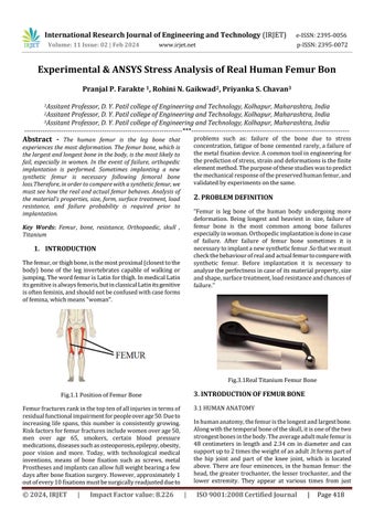
Fig.1.1 Position of Femur Bone
3.1 HUMAN ANATOMY
Femur fractures rank in the top ten of all injuries in terms of residual functional impairment for people over age 50. Due to increasing life spans, this number is consistently growing. Risk factors for femur fractures include women over age 50, men over age 65, smokers, certain blood pressure medications, diseases such as osteoporosis, epilepsy, obesity, poor vision and more. Today, with technological medical inventions, means of bone fixation such as screws, metal Prostheses and implants can allow full weight bearing a few days after bone fixation surgery. However, approximately 1 out of every 10 fixations must be surgically readjusted due to
© 2024, IRJET
|
Impact Factor value: 8.226
In human anatomy, the femur is the longest and largest bone. Along with the temporal bone of the skull, it is one of the two strongest bones in the body. The average adult male femur is 48 centimeters in length and 2.34 cm in diameter and can support up to 2 times the weight of an adult .It forms part of the hip joint and part of the knee joint, which is located above. There are four eminences, in the human femur: the head, the greater trochanter, the lesser trochanter, and the lower extremity. They appear at various times from just
|
ISO 9001:2008 Certified Journal
|
Page 418