
International Research Journal of Engineering and Technology (IRJET) e-ISSN: 2395-0056
Volume: 11 Issue: 02 | Feb 2024 www.irjet.net p-ISSN: 2395-0072
International Research Journal of Engineering and Technology (IRJET) e-ISSN: 2395-0056
Volume: 11 Issue: 02 | Feb 2024 www.irjet.net p-ISSN: 2395-0072
Aryan Galande1 , Suraj Mohite2 , Pavan Thorat3, Prof. Rajani Jadhav4
1Aryan Galande: Student, Dept. of Computer Engineering, PICT Pune.
2Suraj Mohite: Student, Dept. of Computer Engineering, PICT Pune.
3Pavan Thorat: Student, Dept. of Computer Engineering, PICT Pune.
4Prof. Rajani Jadhav: Professor, Dept. of Computer Engineering, PICT Pune.
Abstract - Cancer is one of the largest health problems the world faces today, and early detection is key to better patient outcomes. Traditional tumour detection methods sometimes involve intrusive procedures and have disadvantages. The integration of Artificial Intelligence (AI) and Machine Learning (ML) has led to breakthrough developments in the field of medical diagnostics in recent years, particularly in the identification of malignant tumours. This research provides a thorough analysis of the application of AI and ML techniques in the early diagnosis and detection of cancer. Keywords Cancer Detection, Tumor Detection, Artificial Intelligence, Machine Learning, MedicalImaging,Radiology,Healthcare,EarlyDiagnosis.
Key Words: Cancer Detection, Tumour Detection, Artificial Intelligence, Machine Learning, Medical Imaging , Radiology,Healthcare,EarlyDiagnosis.
A crucial area of medical research and clinical practice is the detection of cancer in the brain. This field focuses on the identification and diagnosis of tumours that originate inside the central nervous system, which includes the brain and spinal cord. Brain tumours, commonly referred toasbraincancer,canbemalignant(cancerous)orbenign (noncancerous). Early brain cancer detection is essential forsuccessful treatmentandbetterpatientoutcomes.The following are some important details about brain cancer detection: Different Brain Tumour Types: Brain tumours are categorized according to their origin, location, and level of malignancy. Whereas secondary brain tumours originate from cancer that has progressed to the brain from other parts of the body, primary brain tumours originate in the brain or spinal cord. Brain cancer symptoms can vary greatly and include weakness, eyesight issues, altered behaviour or cognitive function, chronicheadaches,andseizures.Duetothemildandnonspecific nature of these symptoms, early identification is frequently difficult. Diagnostic Tools: A mix of diagnostic tools and medical imaging techniques, such as magnetic resonance imaging (MRI), computed tomography (CT), and positron emission tomography (PET) scans, are commonly used to detect brain cancer. The presence,
location, and size of brain tumours can be seen with the aid of these imaging investigations. Biopsy: To obtain a conclusive diagnosis, a biopsy often entails taking a tiny sample of the tumour tissue to be examined under a microscope. This aids in identifying the exact type of tumour and whether it is malignant. Blood Tests: New studies are investigating the possibilities of liquid biopsies, which entail testing blood samples for the presence of DNA mutations linked to brain cancer or tumor-specific markers. Developments in Imaging: The capacity to accurately map brain tumours and evaluate their effects on surrounding brain structures has been enhancedbydevelopmentsinmedicalimagingtechnology, including diffusion tensor imaging and functional magnetic resonance imaging. Options for Treatment: Surgery, radiation therapy, chemotherapy, immunotherapy, targeted therapy, or a mix of these may be used to treat brain cancer. Options for less invasive treatmentarefrequentlymadepossiblebyearlydetection. ResearchandInnovation:Developingnoveltreatmentsfor brain tumours and enhancing the precision and noninvasiveness of diagnostic methods are the two main objectivesofcontinuouseffortsinthefieldofbraincancer detection. Early Diagnosis and Prognosis: Early detection of brain cancer is critical to improving patient quality of life and raising the chance of a successful treatment plan. Moreover, accurate prognosis is necessary to tailor treatmentplanstoindividualpatients.
In a 2021 study by Q.D. Buchlak et al., the applications of machine learning in neuroimaging for glioma detection and classification were explored. This research offers valuable insights into the use of AI specifically for glioma diagnosis, a common and aggressive type of brain tumor. Understanding the specific methods and algorithms employed for glioma diagnosis is essential for comprehending the broader landscape of brain tumor detection. M.K. Abd-Ellah et al.'s 2019 review provides a comprehensive overview of brain tumor diagnosis from MRI images, with a focus on the practical implications. This study offers a holistic perspective on the challenges and opportunities associated with using MRI for brain
International Research Journal of Engineering and Technology (IRJET) e-ISSN: 2395-0056
Volume: 11 Issue: 02 | Feb 2024 www.irjet.net p-ISSN: 2395-0072
tumor diagnosis, shedding light on the real-world applications of this technology. The same work by M.K. Abd-Ellah et al. emphasizes lessons learned from the application of MRI in brain tumor diagnosis. The lessons derived from this research are crucial for understanding theevolvinglandscapeofbraintumordiagnosisandserve asvaluableguidanceforfuturestudies.V.P.Groveretal.'s 2015 publication on magnetic resonance imaging principles and techniques is instrumental in providing a fundamentalunderstandingofMRI,whichformsthebasis for brain tumor imaging. Understanding the underlying principlesofMRIisessentialforinterpretingtheresultsof machine learning models applied to MRI data. In a 2000 study by H. Tang et al., MRI brain image segmentation usingmulti-resolutionedgedetectionandregionselection techniques was discussed. This study provides insights into the image processing approaches used in the context of brain tumor detection and their integration with machine learning for more accurate tumor delineation. In a2010studybyK.Somasundarametal.,afullyautomatic brain extraction algorithm for axial T2-weighted MRI images was presented. This addresses a crucial preprocessing step for brain tumor diagnosis. The automatic extraction of the brain region is an essential component of the pipeline, and this study contributes to thetechnicalaspectsoftheprocess.
A. Detecting brain tumors using a Support Vector Machine(SVM)algorithminvolvesseveralsteps,fromdata collection and preprocessing to model training and evaluation. Here's a step-by-step algorithm on how to detectbraintumorusingSVM:
1.Start
2.DataCollection
3.Data Preprocessing:ProcesstheMRIimagestoprepare themfortrainingandtesting.
4. Feature Extraction: Depending on your dataset and the nature of the images, you may want to extract relevant features from the images. Some common feature extraction methods include Histogram of Oriented Gradients(HOG)orLocalBinaryPatterns(LBP).5.Feature Vector Generation: Convert the processed or extracted featuresintofeaturevectorsthatcanbefedintotheSVM. Each image should be represented as a set of numerical features.
6.Labeling:Assignlabelstoyourfeaturevectors,suchas0 for non-tumor and 1 for tumor. Ensure that your labels matchtheimagesinthetrainingset.
7. SVM Model Selection: Choose the appropriate type of SVM (linear, polynomial, radial basis function, etc.) based on your dataset and problem. You can experiment with
differentkernelfunctionstoseewhichoneworksbestfor yourproblem.
8. Model Training: Train the SVM model on the labeled training data. The SVM algorithm will learn the decision boundary that best separates the tumor and non-tumor classes.Youcanuselibrarieslikescikit-learninPythonfor SVMimplementation.
9.Model Evaluation:Assess the performanceofyour SVM model using appropriate evaluation metrics. Common evaluation metrics for binary classification tasks include accuracy, precision, recall, F1-score, and ROC curve analysis.
10. Hyperparameter Tuning: Fine-tune the hyperparameters of the SVM model, such as the regularization parameter (C) or the kernel parameters, to optimizethemodel'sperformance.Youcanusetechniques likecross-validationforthis.
11. Testing: Use the trained SVM model to predict tumor vs.non-tumorlabelsonyourtestdataset.
12. Post-processing: You can apply post-processing techniques to improve the model's output, such as thresholdingtoreducefalsepositivesorfalsenegatives.
13. Visualization: Visualize the SVM's decision boundary or important features, which can help understand the model'sdecision-makingprocess.
14.Deployment
15. If the model performs well, we can deploy it for real worldbraintumordetection,possiblyinamedicalsetting. 16.End
Using the above algorithm, we can detect samples containingbraintumour.
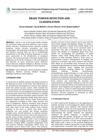
For this experiment, we utilized a diverse dataset comprising MRI images of the brain, including both cancerous and non-cancerous cases. The dataset was carefully curated to encompass a wide range of tumor types,sizes,andlocations,ensuringthediversitynecessary fortrainingandevaluatingtheSVMmodeleffectively. Fig.2.Samplestestedfornotumor.
International Research Journal of Engineering and Technology (IRJET) e-ISSN: 2395-0056
Volume: 11 Issue: 02 | Feb 2024 www.irjet.net p-ISSN: 2395-0072
Thechosendatasetincludestwotypesofdata:-
1)TrainingData
2)TestingData
Eachofthesedivisions of datasets includes MRI Scansof Human Brain having no tumor and MRI Scans of Human BrainhavingPituitaryTumor
The SVM Model is trained on the training data of chosen dataset,Fig2showsresultsoftestingofSVMmodelonthe testingdataofchosendatasetfornotumor.
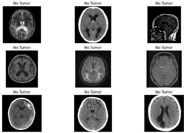
Fig.3.Samplestestedforpituitarytumor.
Fig 3 shows results of testing of SVM model on the testingdataofchosendatasetforpituitary tumor.

Fig.4.TestingResults.
After testing the model, we got the training score of 0.9887andtestingscoreof0.9592.
3. CONCLUSIONS
Inconclusion,apromisingdirectioninthefieldofmedical diagnostics is the application of Support Vector Machines (SVM) for brain tumour detection. SVMs are a useful tool foraccurateclassification,butthereareissuesthatneedto be resolved, including issues with generalization, data limitations,andethics.
Investigating deep learning methods, multimodal data fusion, and real-time detection systems, among other possibilities, can significantly improve the efficiency and
accuracy of tumour detection. Moreover, collaboration between AI and medical experts is necessary for the successful integration of SVM-based tumour detection in clinical settings. These developments herald a bright future for improving brain tumour identification, which willeventuallyleadtobetterpatientoutcomesandcare.
[1] Q.D. Buchlak et al. Machine learning applications to neuroimaging for glioma detection and classification: an artificial intelligence augmented systematic review J. Clin. Neurosci.(2021)
[2]M.K.Abd-Ellahetal.Areviewonbraintumordiagnosis from MRI images: practical implications, key achievements, and lessons learned Magn. Reson. Imag. (2019)
[3]M.K.Abd-Ellahetal.Areviewonbraintumordiagnosis from MRI images: practical implications, key achievements, and lessons learned Magn. Reson. Imag. (2019)
[4] V.P. Grover et al. Magnetic resonance imaging: principlesandtechniques:lessonsforcliniciansJournal of clinicalandexperimentalhepatology(2015)
[5]. H. Tang et al. MRI brain image segmentation by multiresolution edge detection and region selection Comput.Med.Imag.Graph.(2000)
[6] K. Somasundaram et al. Fully automatic brain extraction algorithm for axial T2-weighted magnetic resonanceimagesComput.Biol.Med.(2010)