Sol-gel auto-combustion produced gamma irradiated Ni1-x
xFe2O4 nanoparticles:TGA, UV-Vis spectra, and Raman spectroscopy
aDepartment of Physics, Deogiri College, Aurangabad 431001 INDIA
Cd
bDepartment of Physics, Dr. Babasaheb Ambdekar Marathwada University Aurangabad431001 INDIA
Abstract
This work discusses the thermo gravimetric analysis (TGA), UV-Vis spectroscopic absorption, and Raman active modes of gamma-irradiatedNi1-xCdxFe2O4NPswithatotaldoseof50Mrad.Ni1-xCdxFe2O4NPsweremadeusingthewet-chemicalsol-gel assistedauto-combustionmethod(x=0.0and0.1).Ni1-xCdxFe2O4NPs'thermalstudyandphysicalcharacteristicsrevealedthat theywereexothermicbetween150and600 °Candendothermicatthattemperature.Tosupportthephaseformationstudy, thespecificactivemodesT2g(1),T2g(2),T2g(3),andA1gintheRamanspectrawereexamined.Ni1-xCdxFe2O4NPs(x=0.0and 0.1) were reported to have UV-Vis spectral absorption values of at λ = ~292.4 Å; ~340.2 Å; and 340.2 for NiFe2O4 NPs and ~340.77 Å; ~563.28 Å for Ni1-xCdxFe2O4NPs exposed to gamma radiation. Using a Tauc's plot and the "Kubelka-Munk function,"whichwasclaimedtobebetween1.41and1.5eV,theopticalbandgapwascalculated.
Keywords: TGA; Ferrite; NPs; Raman spectra; UV-Vis spectra, gamma irradiation

1. Introduction
Ever since humans first began to evolve, "iron" and its oxides have been associated with human culture[1, 2]. Since antiquity,ferriteshave been recognizedasthestonesthatmayattractandmagnetizewhen exposedtoanexternal magnetic field[3] The researchers have been examining the cubic spinel ferrite's structural, morphological, electrical, and magnetic propertiesaswellasallofitspotentialuses.Thecrucialsubgroupofmagneticmaterialsismadeupofmixedtransitionmetal oxides,whicharefrequentlyusedforcatalyticactivitiessuchasselectiveoxidation,selectivereduction,dehydrogenation,semi conductive characteristics etc.[4].Studies has focused on nano crystalline materials because they offer a fresh and superior physicalandchemicalprospectivethatisnotyetexpressedwiththebulk.Thesematerialsrangeinsizefrom1nmto100nm. Spinel,whichrepresentferrites'significanceinallpotentialsectors,havebeengroupedintothreemajorclassesbasedontheir crystal structure, like ZnFe2O4, NiFe
O
, Zn/NiFe
, CoFe
MnFe
, and MgFe
O4, etc.[5]The general specification of the ferrites'structuralarchitectureisasfollows[6,7];
It's interesting to note that ferrite still has the best magnetic and electrical characteristics, which makes them adaptable materialswithhighresistivity,minimaldielectriclosses,mechanicalhardness,andhighCurrietemperature. Theseferritesare suited for high-frequency applications because to their chemical stability. The spinels also possess stunning physical and chemical characteristics, including better permeability, fewer eddy current losses, and higher saturation magnetization[8, 9]etc Owingto their prospectiveuses in storage devicesand high-frequencydevice applications, interest in nano crystalline ferrite has continued to grow[10, 11] multiple imaging methods, pharmaceutical, the spatial and temporal resolution of diagnostic techniques [12], antibacterial, surgical implants, genetic engineering [13], medical appliances, particularly in the sterilization process [14], thus they have a significant potential for applications in the field of biomedicine [15, 16]The surface's magneto crystalline anisotropy, spins' canting, a super paramagnetic motion, and shape recovery[17]Nickel ferrite aresuitableforcatalyticactivities,transformercore[18,19],etc.inductors,electricmotors,andmanymore.Amongtherange ofspinel-typeferrites,nickelferrite [ ] hasbeenfocusedonduetovariousapplications.NiFe2O4 belongs to an inverse type spinel structure with on [B]-site; and distributed equally at [B]-site and [A]-site. It was aimed to expand the unit cell of NiFe2O4; or increasing lattice constantby doping of the divalent cadmium (Cd2+) into the intersticesbyformingNi1-xCdxFe2O4(x=0.0and0.1)asthe(Cd2+) haslargerionicradius0.97ÅascomparedNi2+ 0.72Å[20] TheincrementallatticeparametercanbestudiedaccordingtotheVegard’slaw[21]
2. Preparation of Ni1-xCdxFe2O4NPs (x = 0.0 and 0.1)
Thewet-chemicalapproachandthesolgelroutewereusedtocreatetheNi1-xCdxFe2O4 NPs(x=0.0and0.1).Toproducea homogenoussolpreparation,theAR-gradenickelnitrate,cadmiumnitrate,andferricnitratewerethoroughlycombinedinDI water. Citric acid was consumed as a complexant in a 1:3 metal nitrate solution. The blended "sol" was held on a magnetic hotplateandswirledatamoderatespeedwhileat80°C.TokeepthepHofthesolutionat7,ammoniasolution(NH4OH)was gently introduced into the mixture. Eventually, a "sol" transformed into a "gel" and in the beaker, a sticky brown gel was created.Thesolutionwasraisedtothenextintermediatestageandtheself-ignitiontookplaceasaresultofthetemperature beingslightlyraisedto120°C.Theself-ignitedconstituentsarepropelledtoburnoutautomaticallyaccordingtothepropellant chemistry,andeventuallyitgivesafinepowderofNi1-xCdxFe2O4 NPs(x=0.0and0.1)thatwascrushed,sintered, andusedfor furthercharacterization.Theacquiredsamplesreceivedatotaldoseof50Mradofgammaradiationinthegammachamber..
3. Characterization techniques
ThethermalanalysisfortheobtainedNi1-xCdxFe2O4 (x=0.0and0.1)NPswascarriedoutbyTGA-DTAcurve;atatemperature regionof0 -1000°C(withaheatflowrate5°C⁄min)byThermal (TGA)Analyzer (TG-DTA-DSC)TAInc;SDT- 2790.TheUVVis spectra of γ-irradiated Ni1-xCdxFe2O4 (x= 0.0 and 0.1) NPs were carried out by using a UV-Vis spectrophotometer: Make: JASCO make V-750; Serial No. D084261799 from 1000 Å to 200 Å In the nanometer scale; photometric mode: absorbance unit; with a measuring range 900 - 190 along X-axis; the first point obtained at 0.01473 max. at 0.20956 and min at0.34010;Data interval= 0.2 nm;Bandwidth=2.0 nm;Response=0.96sec;Scan mode: Continuous;Scanspeed: 400nanometer perminute.RamanspectroscopywascarriedoutbyHORIBALabRAMSoleilTM
4. Results and characterization output
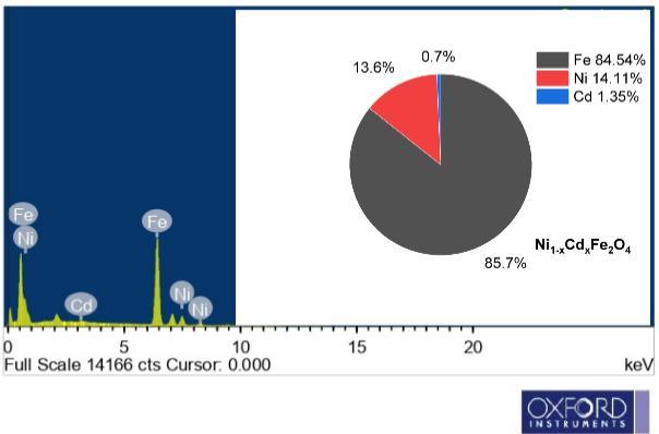
4.1. Thermal analysis(TGA)
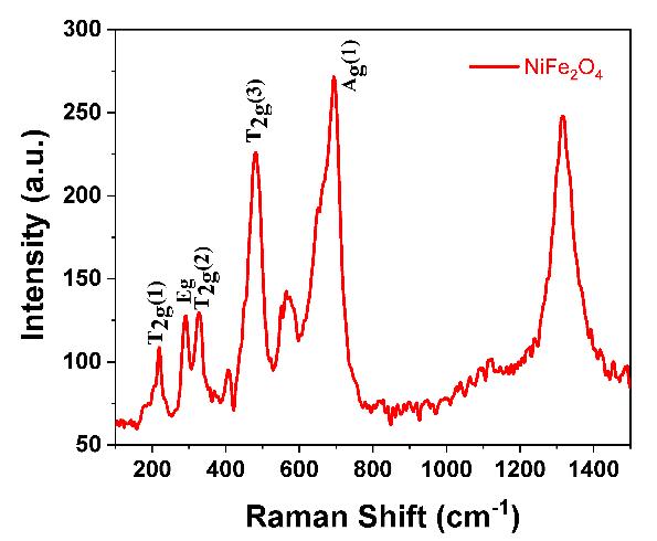
To determine the processing temperature for the compositional stoichiometry created for the experiment, the TGA of γirradiatedNi1-xCdxFe2O4(x=0.0and0.1)wasperformed.Thermo gravimetricisemployedtospecifytemperaturedependence, weightloss,andheatflowforawiderangeofmaterials,aswellastoprovidesupplementaldataforthemostpopularthermal technique,DSC.Inthismethod,thesampleistypicallyheatedsteadily undersyntheticairornitrogen(N2),andthedifference in sample mass is assessed. A drop in sample mass suggests that the material under investigation has deteriorated. The material'sreactionwiththeoxygeninsyntheticair,however,mightleadtoamassgain.Thistechniqueiscapableofanalyzing samples that exhibit mass gain or loss as a result of breakdown, oxidation, or volatile loss. The thermal analysis of the prepared Ni1-xCdxFe2O4 (x= 0.0 and 0.1) is depicted in Fig. 1(a). SDT- 2790 in atmosphere at a temperature region of ~25~1000C(5°C⁄min)aimedtoestimatethethermalprocessingtemperaturesfortheproductionof Ni1-xCdxFe2O4 (x=0.0 and 0.1). The decomposition of the precursors takes place up to 800 °C. The Ni1-xCdxFe2O4 (x=0.0 and 0.1) specimen has releasedH2Omolecules,asseenbytheendothermicpeakat275°C.Themultipleprocesses,includingoxidationandtheentire productionofthefinalproduct,wereresponsiblefortheexothermicpeakthatwasfoundbetween150 °Cand600°C.Several different types of literature claim that the precursors underwent thermal breakdown at temperatures around 600 °C. However,itwasanticipatedthatproductionimpuritiescouldoccurwithinthisrange. Fig. 1(b) depictstheEnergydispersive X-rayAnalysisofγ-Ni1-xCdxFe2O4 (x=0.1),andtheelementalconstituencyhasbeenidentified.
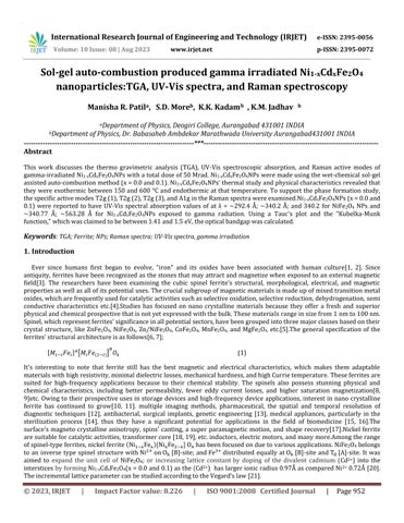
The Raman spectroscopic analysis of -irradiated Ni1-xCdxFe2O4 (x=0.0 and 0.1) was carried out for a number of intense vibrationally active Raman modes, including 3T2g1, T2g2 , T2g2,Eg, and A1g modes; of the molecular structure that could be computedtobefortheformationof(Fd3m-1)cubicspinelstructure[22].In Fig. 2 (a) and (b),depictstheactiveRamanmodes forthe(A)-siteassociatedwiththewavenumber˃600cm-1;thatbelongstoFd. The[B]-siteisassociatedtothewavenumber thatarebelongstotheD3d pointgroup[23].ItcouldbeseenthatA1g reflectionliesat660±10cm 1 indicatingthestretchingof Fe3+trivalentionandO2-divalentionsinthe(A)-sites[24].Theotherreflectionconsumeslowerenergy;thatraisedduetothe veering of O2- ions in comparison to that of the Fe ion at (A)-sites; and . the stretching complexants of the Fe-O and Ni-O in NiFe2O4 NPsarenotprominentinthisspectra[25].

International Research Journal of Engineering and Technology (IRJET) e-ISSN: 2395-0056
Volume: 10 Issue: 08 | Aug 2023 www.irjet.net p-ISSN: 2395-0072
TheUltra Violet range-Vis spectra of γ-irradiated Ni1-xCdxFe2O4 (x= 0.0 and 0.1) was taken using a UV-Vis spectrophotometer:JASCOfrom900Å-190Å. Fig. 3 (a) and (b) depictstheUVabsorbancespectra;highlightingamaximum absorbancelayinUVbandofelectromagneticspectrumoflight.TheUVabsorbance ofNiFe2O4 rangesatλ=~292.4Å;~340.2 Åhighpeakedat~340.2Å;peakheightat0.06882hasbeendepictedin Table 1.Similarly,forNi
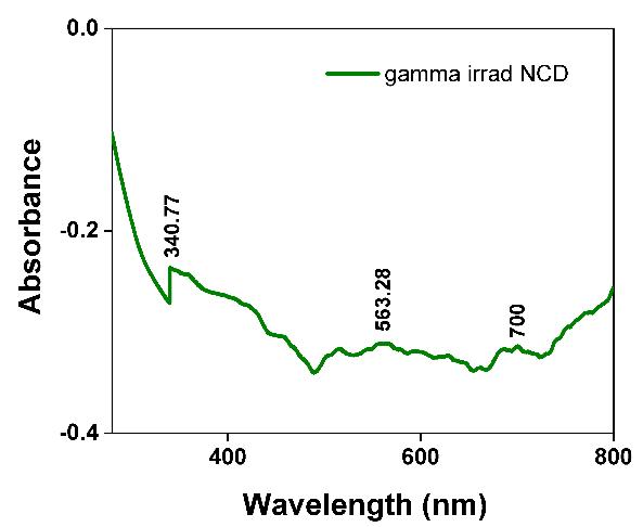

Å;~563.28Å;~700Åhighpeakedat~340,77Å;peakheight~0.087.
Table 1. PeakdetailsoftheofUV-Visabsorbancespectraγ-irradiatedNi1-xCdxFe
0.1)
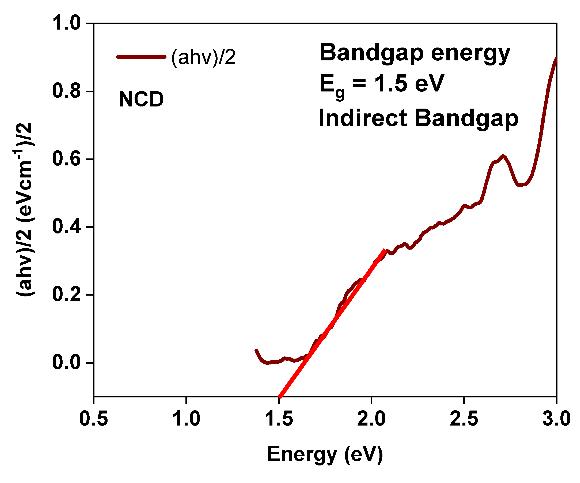
TheUtilizingtheKubelka-MunkfunctionF(R),whichisdenotedbytheformula,thedirectbandgapenergy(Eg)of -irradiated Ni1-xCdxFe2O4 (x=0.0and 0.1)wascomputed: ;[26, 27];.where,α =absorptionconstant equivalent totheTauc’sequation;R=defusedreflectance;The extrapolatedplotof[αhν]2 isshownin fig. 4(a) and (b);thatrepresents theopticalbandgap(Eg)parameterswerefoundtobe1.41eVforγ-irradiatedNiFe2O4and1.5eVfor-irradiatedNi1-xCd
2O4; tostudythequantumconfinementeffectwithintheγ-irradiatedNi1-xCdxFe2O4 (x=0.0and0.1)
International Research Journal of Engineering and Technology (IRJET) e-ISSN: 2395-0056
Volume: 10 Issue: 08 | Aug 2023 www.irjet.net p-ISSN: 2395-0072
5. Conclusions
ThisstudyusedwetchemicalsynthesistocreateNi1-xCdxFe2O4 NPsforx=0.0and1.0,utilisingthesol-geltechniqueandusing asafuelwhilemaintainingpH7.Ni1-xCdxFe2O4 hasanexothermicpeakintheTGAthatrangesfrom275 °Cto600 °C,andan endothermicpeak at150 °C. The existence ofthe active Raman modes 3T2g1,T2g2,T2g2,Eg;andA1g reflection inthe Ramanspectraoftheγ-irradiatedNi1-xCdxFe2O4 NPshassupportedtheferritephase'screation.The(A)-site'sA1g activeRaman mode is located at a distance of 660 ±10 cm 1. Peaks in the UV-Vis spectroscopic absorption of Ni1-xCdxFe2O4NPs exposed to gamma radiation were observed at ~340.77 Å; ~563.28 Å, and the bandgap determined by Touc's plot using the KubelkaMunkfunctionF(R)wasreportedinthespecificrangebetween~1.41eVand~1.5eV

Acknowledgments
Dr.BabasahebAmbedkarMarathwadaUniversity,Aurangabad,hasprovidedfinancingfortheproject,andtheresearcherDr. Manisha Patil is grateful. Likewise, I would like to thank Prof. K.M. Jadhav, who served as the former head of the physics departmentattheDr.BabasahebAmbedkarMarathwadaUniversityinAurangabad,forhisinvaluableadvice
Reference
[1]D. Salazar,D. Jackson, J.L.Guendon, H. Salinas,D. Morata,V. Figueroa,G. Manríquez, V. Castro, Early evidence (ca.12,000 BP)forironoxideminingonthePacificcoastofSouthAmerica,CurrentAnthropology,52(2011)463-475.
[2] R. Balasubramaniam, A.R. Kumar, P. Dillmann, Characterization of rust on ancient Indian iron, Current Science, (2003) 1546-1555.
[3]P.Thakur,A.Thakur,Nanomaterials,theirTypesandProperties,in: SynthesisandApplicationsofNanoparticles,Springer, 2022,pp.19-44.
[4] V. Kirankumar, S. Sumathi, A review on photodegradation of organic pollutants using spinel oxide, Materials Today Chemistry,18(2020)100355.
[5]K.D.Martinson,I.B.Panteleev,A.P.Shevchik,V.I.Popkov,EffectoftheRed/Oxratioonthestructureandmagneticbehavior ofLi0.5Fe2.5O4nanocrystalssynthesizedbysolutioncombustionapproach,LettersonMaterials,9(2019)475-479.
International Research Journal of Engineering and Technology (IRJET) e-ISSN: 2395-0056
Volume: 10 Issue: 08 | Aug 2023 www.irjet.net p-ISSN: 2395-0072
[6]N.Rezlescu,E.Rezlescu,P.Popa,E.Popovici,C.Doroftei,M.Ignat,Preparationandcharacterizationofspinel-typeMeFe2O4 (Me=Cu,Cd,NiandZn)forcatalystapplications,MaterialsChemistryandPhysics,137(2013)922-927.
[7] R. Anu, K. Vinod, Study of magnetic and optical transitions in MFe2O4 (M= Co, Zn, Fe, Mn) with spinel structure, Наносистемы:физика,химия,математика,12(2021)481-491.
[8] N. Kumari, S. Kour, G. Singh, R.K. Sharma, A brief review on synthesis, properties and applications of ferrites, in: AIP ConferenceProceedings,AIPPublishingLLC,2020,pp.020164.
[9] R. Jabbar, S.H. Sabeeh, A.M. Hameed, Structural, dielectric and magnetic properties of Mn+ 2 doped cobalt ferrite nanoparticles,JournalofMagnetismandMagneticMaterials,494(2020)165726.
[10] T. Dippong, E.A. Levei, O. Cadar, Recent Advances in Synthesis and Applications of MFe2O4 (M= Co, Cu, Mn, Ni, Zn) Nanoparticles,Nanomaterials,11(2021)1560.
[11]N.Grimes,Thespinels:versatilematerials,PhysicsinTechnology,6(1975)22.
[12] H.A. Adeola, S. Sabiu, T.A. Adekiya, R.T. Aruleba, C.E. Aruwa, B.E. Oyinloye, Prospects of nanodentistry for the diagnosis andtreatmentofmaxillofacialpathologiesandcancers,Heliyon,6(2020)e04890.
[13] X. Zhao, Z. Meng, Y. Wang, W. Chen, C. Sun, B. Cui, J. Cui, M. Yu, Z. Zeng, S. Guo, Pollen magnetofection for genetic modificationwithmagneticnanoparticlesasgenecarriers,Natureplants,3(2017)956-964.
[14] S. Dutz, S. Wojahn, C. Gräfe, A. Weidner, J.H. Clement, Influence of sterilization and preservation procedures on the integrityofserumprotein-coatedmagneticnanoparticles,Nanomaterials,7(2017)453.
[15] K.R. Reddy, P.A. Reddy, C.V. Reddy, N.P. Shetti, B. Babu, K. Ravindranadh, M.V. Shankar, M.C. Reddy, S. Soni, S. Naveen, Functionalized magnetic nanoparticles/biopolymer hybrids: synthesis methods, properties and biomedical applications, Methodsinmicrobiology,46(2019)227-254.
[16] Y. Tao, H.F. Chan, B. Shi, M. Li, K.W. Leong, Light: a magical tool for controlled drug delivery, Advanced Functional Materials,30(2020)2005029.
[17]M.Bengisu,M.Ferrara,Materialsthatmove:smartmaterials,intelligentdesign,Springer,2018.
[18]C.Murugesan, K.Ugendar,L. Okrasa,J.Shen,G.Chandrasekaran,Zinc substitutioneffectonthestructural,spectroscopic andelectricalpropertiesofnanocrystallineMnFe2O4spinelferrite,CeramicsInternational,47(2021)1672-1685.
[19] S. Shaat, H. Dawoud, Influence of variation of structural parameters on magnetic properties of Al-substituted Ni spinel ferrite,JournalofMaterialsScience:MaterialsinElectronics,32(2021)11536-11546.
[20] P.A. Rao, V. Raghavendra, B. Suryanarayana, T. Paulos, N. Murali, P.P. Varma, R.G. Prasad, Y. Ramakrishna, K. Chandramouli,Cadmiumsubstitutioneffectonstructural,electricalandmagneticpropertiesofNi-Znnanoferrites,Resultsin Physics,19(2020)103487.
[21]M.Rahimi,M.Eshraghi,P.Kameli,StructuralandmagneticcharacterizationsofCdsubstitutednickelferritenanoparticles, Ceramicsinternational,40(2014)15569-15575.
[22]H.Liu,Z. Yu,B.Fu,M. Ran,C.Wu, X.Jiang,R.Guo, Z.Lan,K.Sun,Anisotropicgrowthandmagneticpropertiesof nickel–zincferritethinfilmbyspinspraydeposition,CeramicsInternational,47(2021)1318-1324.
[23] M.P. Ghosh, S. Sharma, H.K. Satyapal, K. Tanbir, R.K. Singh, S. Mukherjee, Tuning the microstructural, optical and superexchange interactions with rare earth Eu doping in nickel ferrite nanoparticles, Materials Chemistry and Physics, 241 (2020)122383.
International Research Journal of Engineering and Technology (IRJET) e-ISSN: 2395-0056
Volume: 10 Issue: 08 | Aug 2023 www.irjet.net p-ISSN: 2395-0072
[24] M. Anupama, N. Srinatha, S. Matteppanavar, B. Angadi, B. Sahoo, B. Rudraswamy, Effect of Zn substitution on the structuralandmagneticpropertiesofnanocrystallineNiFe2O4ferrites,CeramicsInternational,44(2018)4946-4954.
[25]H. Kardile, S.B. Somvanshi,A.R.Chavan,A. Pandit, K. Jadhav,Effect ofCd2+ doping onstructural,morphological,optical, magneticandwettabilitypropertiesofnickelferritethinfilms,Optik,207(2020)164462.
[26]S.A.Jadhav,S.B.Somvanshi,M.V.Khedkar,S.R.Patade,K.Jadhav,Magneto-structuralandphotocatalyticbehaviorofmixed Ni–Zn nano-spinel ferrites: visible light-enabled active photodegradation of rhodamine B, Journal of Materials Science: MaterialsinElectronics,31(2020)11352-11365.
[27] T. Vidya Sagar, T. Subba Rao, N. Raghuram, Temperature dependent structural, morphological, FTIR, optical, magnetic propertiesofNiMgZnferrites,Наносистемы:физика,химия,математика,11(2020)434-443.