PHONOCARDIOGRAM HEART SOUND SIGNAL CLASSIFICATION USING DEEP LEARNING TECHNIQUE
Nishant Sanjay Indalkar 1 , Shreyas Shrikant Harnale2 , Prof. Namrata3
1Computer Engineering, Pimpri Chinchwad College of Engineering Pune, India
2 Computer Engineering, Pimpri Chinchwad College of Engineering Pune, India
3Computer Engineering, Pimpri Chinchwad College of Engineering Pune, India
Abstract Most common reason for human mortality in today’s world that causes almost one-third of deaths is especially due to heart disease. It has become the most common disease where in every 5 people 4 of them are dealing with this disease. The common symptoms of heart diseases are breath shortness, loss of appetite, irregular heartbeat, chest pain. Identifying the disease at early stage increases the chances of survival of the patient and there are numerous ways of detecting heart disease at an early stage. For the sake of helping medical practitioners, a range of machine learning& deep learning techniques were proposed to automatically examine phonocardiogram signals to aid in the preliminary detection of several kinds of heart diseases. The purpose of this paper is to provide an accurate cardiovascular prediction model based on supervised machine learning technique relayed on recurrent neural network (RNN) and convolutional neural network (CNN). The model is evaluated on heart sound signal dataset, which has been gathered from two sources:
1. From general public via I Stethoscope pro iPhone app.
2. From clinical trials in the hospitals. Experimental results have shown that number of epochs and batch size of the training data for validation metrices have direct impact on the training and validation accuracies. With the proposed model we have achieved 91% accuracy.
Keywords CNN, RNN, Epochs, Deep Learning
I. INTRODUCTION
Heart Disease is an illness that causes complications in human being such as heart failure, liver failure, stroke. Heart disease is mainly caused due to consumption of alcohol, depression, diabetes, hypertension [2]. Physical inactivity increase of cholesterol in body often causes heart to get weaken. There are several types of heart diseases such as Arrhythmia, congestive heart failure, stroke, coronary artery disease and many more. Identification of cardiovascular disease can be done by usingthewidelyknownauscultationtechniquesbasedon echocardiogram, phonocardiogram, or stethoscope. Machine learning and deep learning is a widely used method for processing huge data in the healthcare domain. Researchers apply several different deep learning and machine learning techniques to analyze huge complex medicaldata,topredicttheabnormalityin
heart disease. This research paper proposes heart signal analysis technique based on TFD (Time Frequency Distribution) analysis and MFCC (Mel Frequency Cestrum Coefficient). Time Frequency Distribution represents the heart sound signals in form of time vs frequency simultaneouslyand the MFCCdeterminesa soundsignal in the form of frequency coefficient corresponding to the Mel filterscale[3].AquiteHelpfulmethodwasusedtoimprove theaccuracyofheartdiseasemodelwhichisabletopredict the chances of heart attack in any individual. Here, we present a Deep Learning technique based on Convolutional Auto-Encoder (CAE), to compress and reconstruct the vital signs in general and phonocardiogram (PCG) signals specifically with minimum distortion [4]. The results portray that the highest accuracy was achieved with convolution neural network with accuracy 90.60% with minimum loss and accuracy achieved through recurrent networkwasabout67%withminimumlosspercentage.
II. LITERATURE REVIEW

Ryu et al. [5] Studied about cardia diagnostic model using CNN. Phonocardiograms(PCG) were used in this model. It can predict whether a heart sound recording is normal or not. First CNN is trained to extract features and build a classification function. The CNN is trained by an algorithm called back propagation algorithm. The model then concludesbetweennormalandabnormallabels.
Tangetal.[6]Combinedtwomethodsi.e.deeplearningand feature engineering algorithms for classification of heart sound into normal and abnormal. Then features were extracted form 8 domains. Then, these features were fed intoconvolutionneuralnetwork(CNN)insuchawaythatthe fullyconnectedlayersofneuralnetworkreplacestheglobal average pooling layer to avoid over fitting and to obtain global information. Theaccuracy,sensitivity and specificity observed on the PhysioNet data set were 86.8%, 87%, 86.6%and72.1%respectively.
JiaXinetal.[7]Proposedasysteminwhichheartsoundsare segmented and converted using two classification method: simplesoftmaxregressionnetwork(SMR)andCNN.Features were determined automatically through training of the neuralnetworkmodelinsteadofusingsupervisedmachine learningfeatures.AfterworkingonbothSoftmaxregression and Convolutional neural network(CNN) they found out CNN gave the highest accuracy. The accuracy achieved
throughCNNmodelis93%.
Volume: 10 Issue: 08 | Aug 2023
Mehrez Boulares et al. [8] developed a model based for cardiovascular disease(CVD) recognition based on unsupervised ML and supervised ML methods based on CNN.ThedatasetsaretakenfromPASCALandPhysioNet. They have worked on PCG signals mainly and enhanced their focus on denoising and preprocessing. For feature selection they have used MFCC as it is the best out there now-a-days and similarly for classification they’ve gone with CNN. Classification results of defined models had overallaccuracyof0.87,andoverallprecisionof0.81,and overallsensitivity0.83.
SuyiLietal.[9]proposedapapertoprovideanoverview ofcomputeraidedsounddetectiontechniques.Theyhave worked on PCG signals and characteristics of heart sounds introduced first. They did a thorough review on preprocessing and analyzing techniques that have developedover the lastfiveyears.They’vefurtherdonea deep research on denoising, feature extraction, segmentation, classification and most importantly computeraidedheartdetectiontechniques.
Raza et al. [10] Proposed a framework for denoising the heart sound by applying band filter, them the size of samplerateofeachsoundisfixed.Thenthefeatureswere extracted using sampling techniques and reduce the dimension of frame rate. RNN method is used for classification. RNN using Long Short-Term Memory (LSTM), Dropout, Softmax and Dense layers used.Hence, themethodismoreaccuratecomparedtoothermethods.
Perera et.al [11] developed a software tool to predict heart abnormalities which can be recognized using heart sounds.
The audio inputs are taken through e- stethoscope and then entered into a database with symptoms of each patient. Feature extraction is done using MATLAB “MIR toolbox” and prominent features and statistical parametersare extracted.
Segmentationisdonebytallpeakandshortpeakprocess followed by classification of S1 and S2 systole and diastole square of wavelet fourth detail coefficient methodwasusedforfurtherclassificationprocess.
Yadav et al. [12] They proposed a model to extract discriminatory features for machine learning which involves strategicframingandprocessingofheartsound. They trained a supervised classification model based on most prominent features for identification of cardiac diseases. The proposed method achieved the accuracy 97.78% with error rate of 2.22% for normal and abnormalheartdiseaseclassification.
Khan et al. [13] Proposed a model based on different classifiers such as (KNN, Bagged Tree, Subspace,

subspaceDiscriminant,LDA, FineTreeandQuadraticSVM) toobtainandaccuracyandresults.Kaggledatasetwasused to extract features from the sets of different domains i.e. frequency domain, Time domain and statistical domain to classifytheheartsoundsintwodifferentclassesi.e.normal and abnormal. Out of 6 classifiers the highest accuracy of 80.5%wasobtainedusingBaggedtree.
III. PROPOSED MODEL
The study aims in classifying the heart sounds of heart disease patients into normal and abnormal heart sound basedonPhonocardiogramsignals.
The dataset was obtained from Physionet website which contains physionet challenge heart dataset which was providd publicly. There were two challenges related with this competition. Dataset contains heart sounds of 3 to 30 sec in length [4]. The proposed model described ahead is divided into three main parts. These three parts are preprocessing,train-testandclassification.
The classification was performed using the most robust techniques which gave the highest accuracy among CNN and RNN.Themain motiveofthispaperistopredictwhetherthe heartsoundisnormal orabnormal withitsconfidence value. The first step is preprocessing which includes data compression, feature extraction where denoising was performed in order to remove the unwanted noise and were enhanced to remove the unwanted frequencies. The dataset used for training and testing in this model consist of phonocardiogram sound signal files which contains normal and abnormal heart sounds. These files are audio recordings ofheartsoundsatvariousdifferentstagesofheartbeat.
Volume: 10 Issue: 08 | Aug 2023
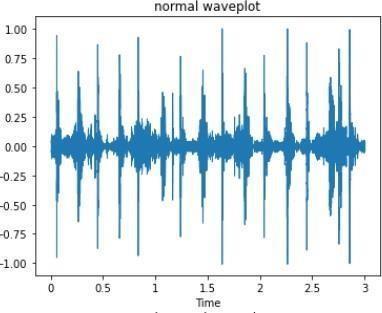
The denoising and compression of heart sound in this modeltakesplacewhilebuildingthemodelbyusingconvo 2D autoencoder[18]. Using the functional API convolution autoencoder was build and once the model was build autoencoderwastrainedusingtrain_dataasbothourinput dataandtargetdata[14].Theconvolutionencoderconsists ofMaxPooling2Dlayersand2Dconvostackformaxdown sampling.Belowshownarethegraphsoflossandaccuracy achivedafterusingconvo2Dautoencoderwith100epochs.
c. ApplyingDFT:DFTisappliedonwindowedframe toconvertitintomagnitudespectrum.
d. Mel-spectrum: Fourier transformed is applied to Melspectrumsignalthroughasetoffiltersknown as Mel-filter bank and by applying inverse DCT frequenciesarewrappedonamel-scale.
e. DCT: As sound signals are smoothened; the energy levels are correlated. so a set of cepstral coefficients are produced by Mel frequency coefficients.
f. Dynamicfeatures:Ascepstral coefficientscontain information from a given frame they are referred asstaticfeatures.Theextrainformationaboutthe temporal dynamics of the signal is obtained by computingfirstandsecondderivativesofcepstral coefficients.
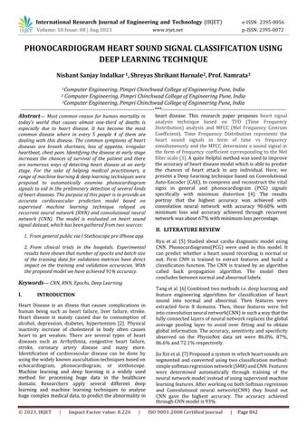
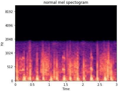
Librosa.display method was used to display the audio signalsindifferentformatssuchaswaveplot,spectrogram. Thewaveplotrepresentsthegraphofheartbeatsignalsas shownbelow:
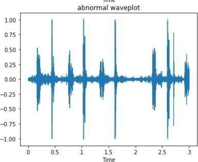
X- axis:Timein(Sec)
Y-axis:Frequencyin(Hz)
Fig3:Accuracycurvewith100epochs

In feature extraction the informative and relevant features were extracted from PCG signals using Mel-scaled power spectrogram and Mel-frequency cepstral coefficients (MFCC) which are then fed into a classification model to classify each PCG signal into an abnormal or normal heart sound [15]. The MFCC which was introduced by Mermelstein in the 1980s is widely used in automatic speechrecognition.Themel-frequencyanalysisisbasedon human acoustic perception and experimental results have shownthathumanbeingsearactsasafilterthatfocuseson certain level of frequency components. It transmits audio signal of certain frequency level and directly ignores the unwanted and undesired signals. In mfcc it converts the audio signal from analog to digital format with sampling frequency.Itbasicallyincludes:
b. Windowing the signal: The sound signals of time varying signal. For sound, signal needs to be examined over a short period of time. Therefore, speech analysis is to be carried out on short segments across which the speech signal is assumed to be stationary. Short-term spectral measurements are typically carried out over 20 mswindows,andadvancedevery10ms.
a) Mel-Scaled Power Spectrogram


Time period vs. Frequency representation of a sound signal is said to be spectrogram of signal. It graphically represents the change in frequency of a sound signal w.r.ttime,whichhelpsthebuildingmodeltounderstand the sound accurately. The Mel-scaled filters present in Mel-scales are placed non- uniformly to mimic human earpropertiesinfrequencyaxis[15].
b) Mel-Frequency Cepstral Coefficients
Mel-frequency cepstrum is found by taking Discrete CosineTransformofalogpowerspectrumonanonlinear Mel-scaleoffrequency.ItistherepresentationoftheMelscaledpowerspectrogram[15][16].Mostoftheextracted features for PCG heart signal are computed mainly using time,frequency.
Inclassificationstagethepreprocesseddataisfedto CNN [19] [20] [25] [26] and RNN [21] [29] [30], forTraining and testing. The model was trained using CNN and RNN and was build based on accuracy comparison of both the techniques. By comparing the accuracy and losspercentageofRNNandCNNasCNNhas greater accuracy and less loss percentage than RNN thus usingCNNfortheprediction model was preferred. The model has anaccuracy of 90.6% and test loss of 0.29 with 350 epochsand 128 batch size. The less test loss indicates that the model performs better after each iteration. The final prediction of model is categorized into normal and abnormal heart sound. Previous referred models fromdifferent researchers have had the same output whichpredictesifheartsoundisnormalor abnormal [20], what makes this model different from other researchers is theconfidence value of normal or abnormal heart sound shown on the UI of classification model. Refer belowfigure
TheconfidencevalueofheartsoundPCGsignalwillhelpthe medical practitioners to identify the patients withgreater risk of heart disease. The actual confidence value of heart sound makes a huge difference on the resultsbecausethe higher or lower percent could help the physician to makebetterdecisions.


IV. RESULT
Performance of the proposed Heart Sound Classification Techniques is shown in table 1. We applied Convolution neural network (CNN) and Recurrent neural network(RNN) in order to classify heart sound dataset described in above section. The dataset used for this project consists of Phonocardiogram signals of heart sounds containing heart sounds from 3 to 30 seconds in length. Preprocessing was performed in order to filter out the noisy data using auto-encoder and relevant features were extracted using mfcc. In order to test and train the proposed model the dataset was split into training and testing in the proportion of 80%- 20%. The primary objective of this paper is to examine the effects of the hidden layers of a CNN and RNN to check the overall performance of the neural network. To demonstrate this, we have applied CNN and RNN with different number of epochs on the given dataset and also to observed the variations in accuracy of both the techniques based on different number of epochs and batchsize.
Table1:AccuracyandlosspercentageConvolutionneural networkoutperformedrecurrent
neural network having same number of epochs and batch size with accuracy 90.82% and 90.60% with 300 and 350 epochs. The precision, recall and F1 score was calculated for normal and abnormal heart sounds using CNN. Precision,recall,F1ofnormalheartsoundswere0.83,0.96, 0.89 and those for abnormal heart sounds were precision 0.97,recall0.89andf10.93
V. CONCLUSION
In this paper, we have compared the accuracies of two different neural networks i.e. Convolution Neural Network and Recurrent Neural Network based on the implemented model and proposed a model based on the technique which has performed well i.e. CNN with 90.60%.Themodelwastrainedusingdifferentnumberof epochs to ensure the model was not overfitted or underfittedandaconstantnumberofepochswerechosen to train both the algorithms where they have performedwell with having highest accuracy and less loss percentagewithhighestprecisionvalue.
Fig10:Proposedmodel(GUI) Fig11:Normalheartsoundwith99.97%confidencevalue Fig12:Abnormalheartsoundwith99.99%confidence valueV. REFERENCES
[1] M. Tschannen, T. Kramer, G. Marti, M. Heinzmann and T. Wiatowski, "Heart sound classificationusing deep structured features," 2016 Computing in CardiologyConference(CinC),2016,pp.565-568.
[2] Rohit Bharti, Aditya Khamparia, MohammadShabaz, Gaurav Dhiman, Sagar Pande and Parneet Singh, “Prediction ofHeart Disease Usinga Combination of MachineLearning and Deep Learning”,Volume 2021 ArticleID8387680.
[3] I. Kamarulafizam, Shussain Salleh, Mohd Najeb Jamaludin, " Heart Sound Analysis Using MFCC and Time Frequency Distribution" doi: 10.1007/978-3540-68017-8_102.
[4] W. Chen, Q. Sun, X. Chen, G. Xie, H. Wu, C. Xu, “Deep LearningMethods for Heart SoundsClassification:A SystematicReview”Entropy(Basel).2021;23(6):667 Published2021May26doi:10.3390/e23060667.
[5] Heechang Ryu, Jinkyoo Park, and H. Shin, "Classification of heart sound recordings using convolution neural network," 2016 Computing in CardiologyConference(CinC), 2016,pp.1153-1156.
[6] Hong Tang, Ziyin Dai, Yuanlin Jiang, Ting Li and Chengyu liu, “PCG Classification Using Multidomain Features and SVM Classifier” Volume 2018 |Article 4205027| doi: 10.1155/2018/4205027.
[7] Jia Xin L and Keng Waah Choo, “Classification of Heart Sounds Using Softmax Regression and Convolutional Neural Network” ICCET '18: Proceedingsofthe2018InternationalConferenceon Communication Engineering and Technology February2018Pages18–21.
[8] Mehrez Boulares, Reem Al-Otaibi, Amal Almansour and Ahmed Barnavi, “Cardiovascular Disease Recognition Based on Heartbeat Segmentation and Selection Process” October 2021 International Journal of Environmental Research and Public Health 18(20):10952. Suyi Li, Feng Li , Shijie Tang and Wenji xiong, “A Review of Computer-Aided Heart Sound Detection Techniques” Volume 2020 ArticleID5846191.
[9] Suyi Li, Feng Li, Shijie Tang and Wenji Xiong, “A Review of Computer-Aided Heart Sound Detection Techniques”JF-BioMedResearchInternationalPB–Hindawi.
[10] A. Raza, A. Mehmood, S. Ullah, M. Ahmad, G. S. Choi, andB.-W.On,“Heartbeat SoundSignal Classification
Using Deep Learning,” Sensors, vol. 19, no. 21, p. 4819,Nov.2019,doi:10.3390/s19214819.
[11] I. S. Perera, F. A. Muthalif, M. Selvarathnam,M.R. Liyanaarachchi and N. D. Nanayakkara, "Automated diagnosis of cardiac abnormusing heartsounds,"2013IEEEPoint-of-CareHealthcare Technologies (PHT), 2013, pp. 252-255, doi: 10.1109/PHT.2013.6461332.
[12] A. Yadav, A. Singh and M. K. Dutta,“Machine learning-based classification of cardiac diseases from PCG recorded heart sounds” Neural Comput & Applic 32, 17843–17856 (2020) doi: 10.1007/s00521-019-04547-5.
[13] Younas Khan, Usman Qamar, Nazish Yousaf,Aimal Khan, “Machine Learning Techniques for Heart Disease Datasets: A Survey”, ICMLC '19: Proceedings of the 2019 11th International Conference on Machine Learning and Computing February2019Pages27–35.
[14] Ying-Ren Chien, Kai-Chieh Hsu and Hen Wai Tsao, “Phonocardiography Signals Compression with Deep Convolutional Autoencoder for Telecare Applications” Appl. Sci. 2020, 10, 5842; doi:10.3390/app10175842.
[15] T. H. Chowdhury, K. N. Poudel and Y. Hu, "TimeFrequency Analysis, Denoising, Compression, Segmentation,andClassificationofPCGSignals,"in IEEEAccess,vol.8,pp.160882-160890,2020,doi: 10.1109/ACCESS.2020.3020806.
[16] M.Rahmandani,H.A.NugrohoandN.A.Setiawan, "CardiacSoundClassificationUsingMel-Frequency Cepstral Coefficients (MFCC) and Artificial Neural Network (ANN)," 2018 3rd International Conference on Information Technology, Information System and Electrical Engineering (ICITISEE), 2018, pp. 22-26, doi: 10.1109/ICITISEE.2018.8721007.
[17] Pronab Ghosh, Sami Azam, Asif Karim, Mirjam Jonkman, MD. Zahid Hasan, “Use of Efficient MachineLearningTechniquesintheIdentification of Patients with Heart Diseases“, ICISDM 2021: 2021 the 5th International Conference on Information System and Data MiningMay2021 Pages14–20.
[18] Abeer Z. Al-Marridi, Amr Mohamed and Aiman Erbad, "Convolutional Autoencoder Approach for EEGCompression andReconstructioninm-Health Systems," 2018 14th International Wireless Communications & Mobile Computing Conference (IWCMC), 2018, pp. 370-375, doi: 10.1109/IWCMC.2018.8450511.
[19] Ximing Huai, Siriaraya Panote, Dongeun Choi,and Noriaki Kuwahara,“Heart Sound Recognition TechnologyBasedonDeep Learning”Digital Human Modeling and Applications in Health, Safety, Ergonomics and Risk Management.Posture, Motion and Health: 11th International Conference, DHM 2020, Held as Part of the 22nd HCI International Conference, HCII 2020, Copenhagen, Denmark, July 19–24,2020.
[20] Fan Li, Hong Tang, Shang and Klaus Mathaik,“ Classification of Heart Sounds Using Convolution Neural Neural Network” June 2020 Applied Sciences10(11)3956.
[21] M.F.Khan,M.AtteeqandA. N.Quereshi, “Computer Aided Detection of Normal and Abnormal Heart Sound using PCG” ICBBT'19: Proceedings of the 2019 11th International Conference on Bioinformatics and Biomedical Technology May 2019Pages94–99doi:10.1145/3340074.3340086.
[22] Suyi Li, Feng Li, Shijie Tang and Wenji Xiong, “A Review of Computer-Aided Heart Sound Detection Techniques”JF-BioMedResearchInternationalPB–Hindawi.
[23] I. Grzegorczyk, M. Solinski, M. Lepek, A. Perka, J. Rosinski, J. Rymko, K. Stepein and J. Gieraltowski "PCG classification using a neural network approach," 2016 Computing in Cardiology Conference(CinC),2016,pp.1129-1132.
[24] K.K.Tseng,C.Wang,Y.F.Huang,G.R.Chen,K.L.Yung and W. H. Ip, “Cross-Domain Transfer Learning for PCG Diagnosis Algorithm”Biosensors2021, 11,127. doi.org/10.3390/bios11040127.
[25] Noman Fuad, Ting Chee-Ming, S. Salleh and H. Ombao, "Short-segment Heart Sound Classification Using an Ensemble of Deep Convolutional Neural Networks," ICASSP 2019 - 2019 IEEE International Conference on Acoustics, Speech and Signal Processing (ICASSP), 2019, pp. 1318-1322, doi: 10.1109/ICASSP.2019.8682668.
[26] Sunjing,L.Kang,W.WangandSongshaoshaui,“Heart SoundSignalsBasedonCNNClassificationResearch” ICBBS '17 Proceedings of the 6th International Conference on Bioinformatics and Biomedical Science June 2017 Pages 44–48 doi: 10.1145/3121138.3121173.
[27] Low, Jia Xin and Keng Wah Choo. “Automatic Classification of Periodic Heart Sounds Using Convolutional Neural Network.” World Academyof Science, Engineering and Technology, International JournalofElectrical,Computer, Energetic,Electronic andCommunicationEngineering12(2018):96-101.
[28] D. R. Megalmani, S. B. G, A. Rao M V, S. S. JeevannavarandP.K.Ghosh,"UnsegmentedHeart Sound Classification Using Hybrid CNN-LSTM NeuralNetworks,"202143rdAnnualInternational Conference of the IEEE Engineering in Medicine & Biology Society (EMBC), 2021, pp. 713-717, doi: 10.1109/EMBC46164.2021.9629596.
[29] Y.Chen,Y.Sun,and J.Lv,“End-to-endheartsound segmentation using deep convolutional recurrent network” Complex Intell. Syst. 7, 2103–2117 (2021).doi:10.1007/s40747-021-00325-w.
[30] C.ThomaeandA.Dominik,"UsingdeepgatedRNN with a convolutional front end for end-to-end classification of heart sound," 2016 Computing in CardiologyConference(CinC),2016,pp.625-62