Diabetic Retinopathy detection using Machine learning
Ratan Tirkey Cazal1Student,JAIN(Demmed-To-Be University), Bengaluru, Karnataka, India
2Student, CHRIST(Deemed-To-Be University), Bengaluru, Karnataka, India
3Researcher, Bengaluru, Karnataka, India
4Researcher, Bengaluru, Karnataka, India ***
Abstract - According to the International Diabetes Federation, there are currently over 470 million individuals diagnosed with diabetes globally, and by 2050, that number might rise to 700 million. There are two types of it: Type 1 and Type 2. Type 1 diabetes is chronic and incurable, however Type 2 diabetes can be cured if caught early. Identification of anomalies in retinal pictures is tough and complex in the medical sector since these signs of diabetes that affect the eyes appear to be very modest. Therefore, a non-invasive technique to uncover these abnormalities was required.
After reviewing a number of research projects and developments, we attempt to highlight and explain the various methodologies used, their benefits and limitations, the general goal of the results, and the significance of a DR detection system. The survey also highlights the significance of early detection and the need to eliminate the factors that obstruct timely discovery.
Key Words: Diabetic Retinopathy (DR), Non-Invasive, DigitalImageProcessing


1.INTRODUCTION
About 470 million people worldwide are diagnosed with diabetes. During the period spanning 2000-2016, a 5% increase in deaths related to diabetes was observed, with 1.6milliondeathseachyear.Diabetescanbeconsidereda major public health problem in India. Its symptoms are indistinct,onecangrowthirstierorbecomehungrierthan usual, or we might not figure out why we’re more tired than usual. The meteoric rise in urbanization and industrialization,along with ourlifestylechoicesprovides an ideal scenario for the prevalence of diabetes and its associatedcomplications.
It has been noted that there are variances in the ratio of ophthalmology-trained medical professionals to patients, which makes it difficult to diagnose and treat diabetic retinopathy. This is owing to the steady and gradual increase of diabetes around the world. As a result, problems with screening time, greater prices, decreased device sensitivity and specificity, etc., needed to be addressed. As a result, the introduction of automated detection systems based on standalone algorithms was suggested. The screening and diagnosis of DR are importantstudyareas,andmanyresearchersareworking to advance these fields. Methodologies for image processing have made it simple and advantageous to examine acquired images, including their traits, behaviours,andprocessing.
Additionally, the development of new image processing techniques,classificationalgorithms,andneuralnetworks has helped computer-aided detection systems perform quickly and has been recognised as a reliable option for applying analysis on retinal pictures. The aforementioned system models would seek to determine the necessity of referralforadditionaltreatmentandongoingeyecare.We won't be too far off from being able to apply these techniques on our cell phones given the speed at which technology is developing. To identify DR, extensive study has been done and numerous methods/processes have been developed. The implementation of automated screening systems and the outcomes obtained are characteristics of success and a potential failure. The effectivenessandcomplexityoftheexistingalgorithmsfor the identification of DR employing the various image processing processes and their associated methodologies can yet be improved. Here, we want to provide you a general overview of how to create an application that can identifyanomaliescausedbyDRintheretinaoftheeye.
2. EXISTING SYSTEM
The author of the research paper [1] employs feature extractiontolocateandextractthedetailsthatarespecific totheimage.Speeded-UpRobustFeatures(SURF)detects blobfeatures.Itisakindofdetectoralgorithmthatdraws attentiontoparticularlocationsonimagesthathavebeen transformed into a coordinate system. The MSER (maximally stable extremal region) is used for matching. Extremal pixels are those that are either more or less
intense than the pixels on the MSER's outer perimeter. Comparingtheextractedimageandthecategorizedimage to their counterparts in the input image, observations are made.
Theprocedureforremovingbloodvesselsandopticaldisc detection are both thoroughly detailed in this research paper [2]. They act as a starting point for identifying further traits. Candidate area selection, Sobel edge detection, and Estimation are the three steps in the detection process here. The Kirsch method is used to detectthebloodvesselsbyedgedetection.Thistechnique convolutions the image with eight template impulse response arrays to calculate the gradient. After enhancement, a reference point or threshold is set up to determineifapixelisanedgepixelornot.
For the purpose of detecting diabetic retinopathy, the authors of the research [3] employ morphological operations. To reduce false positive exudates, the blood vessels and optical disc are first retrieved and removed. Thisisessentialfortheremovaloflesions.Here,theuseof procedures like erosion and dilation for the removal and detection of blood vessels is also noted. The structuring elementcausesapropercirculardilationoftheopticdisc's fragmentedparts.Inordertolocatethecircleintheimage that corresponds to the optical disc, the circular Hough transformisused.
Inthepaper[5],featureextractionisdoneinthreephases. Initially, feature extraction is done because it's crucial to distinguish between different stages of diabetic retinopathy, and for this to happen, the binary image's totalsumofwhitepixelsmustbedefinedas1.Theconcept of mean is then introduced to equalize both photos when they are compared to one another and lessen the likelihood that the images will be of different resolutions or sizes. The classification of disease, which draws a conclusion because an exudate is noticeable in NPDR, considerstheentireareaofexudatesseenintheimage.
The Gabor filter is used by the author of this research work [7] to carry out automatic detection and classification. This will be utilized for texture detection andhasthepotentialtobeusedforimageretrieval.When used, this procedure operates in the Fourier domain. Multiple tests revealed that large blood arteries with anomalies only occurred and were visible at high frequency or smaller scales in the finer scale output generated from the filters. They thus hardly ever showed upintheoutputoflowpassfilters.
The authors of the research paper [8] suggest a novel method to enhance the prognosis. The severity and treatment strategy for treating DR depend on the detection of Retinal vessels and Microaneurysms. As the sensitivity is easily optimized using evolutionary algorithms, they discover that the matched filter (MF)

technique is more effective than the Sobel operator or Morphologicaloperatorinthiscase.Atop-hatalterationof the image is taken into consideration for the microaneurysm detection method. Real-time detection and ground truth retinal images provided by clinical specialists are used to train the algorithm to distinguish betweentheoriginalandalteredimages.
Theauthorsoftheresearcharticle[9]provideaneffective method for localizing characteristics and lesions. Geometric properties and correlations are employed to separatetheintensitiesofoverlappingfeatures.Here,it is noticedthatanoriginalrestrictionforlocatingoculardiscs (OD) is based on the intersection of retinal blood vessels to approximation its position, and further localized utilizing colour features. The Hough Transform is used to triangulatethedataandcreateanintersectionmap.
3. PROBLEM STATEMENT
Earlydiagnosisandtreatmentofdiabeticretinopathy(DR) can be aided by the identification of the condition's first signs. The current methods of DR detection are generally manual, expensive, and possibly time-consuming, necessitating the assistance of professional employees. Although some experimental research have made advances, they have not yet been implemented on a wide basis.
Here, we put the same little model into practise to find certainabnormalitiesintheimpactedretinalpictures.
4. METHODOLOGY
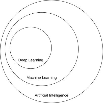
Machine learning, which is a subfield of artificial intelligence(AI),includesdeeplearning.Figure2provides agraphicalrepresentationofthisrelationship.
The main objective of AI is to develop a set of algorithms and methods that can be used to issues that people intuitively and almost automatically solve but that are otherwise exceedingly difficult for computers to handle. Interpretingandcomprehendingthecontentsofanimage is a fantastic example of this category of AI challenges; whereas a human can perform this task with little to no effort,robotshavefoundittobeincrediblychallenging.
ImageAcquisition:
The obtained image is in digital form hence requires scalingfollowedbycolorconversioni.e.,eitherRGBto greyorviceversa.Theseareobtainedfromthepublic open-source or privately owned data repositories. SomeofthemhavebeenmentionedinTable1.
ImageProcessing:

This comprises extracting or enhancing the specific features of the image based on the requirement, reducing the effects that degrade the image, keeping theresolutiontoanacceptablelevel,etc.
• BackgroundSubtraction:
Here, we detect blood vessels and optical disks to eliminate them. It is the crucial step to simplify the process of identification of the exudates and microaneurysms.
• LesionDetection:
Microaneurysms and Hard exudates are symptoms usually observed in the mild and moderate nonproliferativestagesof retinopathy.Thehardexudates are the yellow flecks of lipid residues which are clear lesions. Microaneurysms appear as tiny red dots. The top-hatmorphologicalprocedureisutilizedhere.
• Classification:
Assigning a label to the output image, indicating whether the image is healthy or not, based on descriptors.
5. METHODOLOGY
One of the major reasons of road accidents in real world has solution now. The system is one step towards safeguarding precious lives by avoiding accidents in real world. Proposed system is based on DLIB & SOLVE PNP Models.
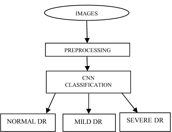
1) Data collection: Compile a sizable dataset of retinal pictures, including both images with and without diabetic retinopathy. The existence and degree of retinopathy should be noted on these images.
2) Datapreprocessing:Enhancethepropertiesofthe photos and eliminate any noise or artefacts that can obstruct the detection procedure. Resizing, normalization, and contrast correction are frequentpreprocessingtechniques.

3) Data augmentation: Enhance the dataset by subjecting the photos to various transformations, including rotation, scaling, flipping, and cropping. This increases the diversity of the data and strengthens the generalization abilities of the model.
4) Model selection: Select a deep learning architecture like convolutional neural networks (CNNs) that is suited for the task. CNNs are frequently employed for image-related applications because of their efficiency in capturingspatialdata.
5) Modeltraining:Createtrainingandvalidationsets from the dataset. Utilize the training set to train the deep learning model while adjusting its settings to reduce the difference in expected and

actual labels. Toavoidoverfitting, keepaneye on themodel'sperformanceonthevalidationset.
6) Model evaluation: Use a different test set that wasn'tutilizedfortraining orvalidationtoassess the trained model. To evaluate the model's effectiveness in diagnosing diabetic retinopathy, compute pertinent assessment metrics including accuracy,precision,recall,andF1score.
7) Optimisation and fine-tuning: If the initial model performance is unsatisfactory, think about optimizing the model by modifying the architecture, including transfer learning from trained models, or adjusting the model's hyperparameters. Repeat this procedure as necessarytoattainthedesiredresults.
8) Deployment:Themodelcanbeusedtocategorise retinal images for the identification of diabetic retinopathy once it satisfies the appropriate performance criteria. For simple access and usability, make sure the model is integrated with theappropriateuserinterfaceorapplication.

Thequalityanddiversityofthedataset,aswellastheskill in choosing and optimizing the deep learning model, are critical components in making the process successful. Working with medical data also necessitates getting ethicalpermissionandfollowingdataprivacylaws.
6. IMPLEMENTATION
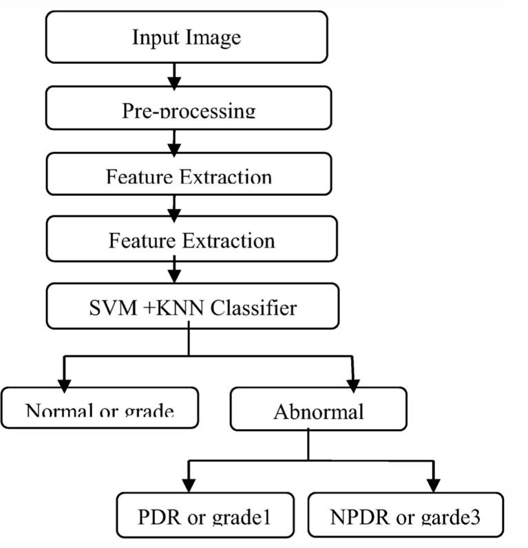
Pre-processingtechniquesareusedinthegivenproposed methodology, as shown in Fig. 2, to obtain input images from the given data set. After that, morphological operationsarecarriedouttoidentifyexudatesandmicroaneurysms. Finally, a multiclass SVM and KNN classifier are applied to determine the degree of irregularity. The input photos for the approach are collected from MESSIDORandDiabeticretDB1.

Fig -3 Implementationofproposedsystem
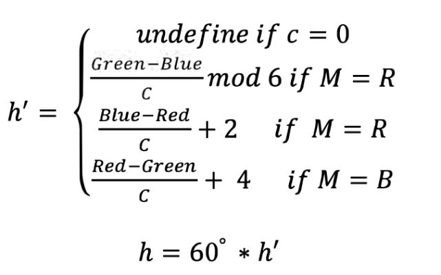
The pre-processing stage is used to prepare the input image, which is 2240 x 1488 pixels in size. The segmentation method is then applied. During the preprocessing stage, issues with blurring, clarity, and size are fixed in the image. This stage involves image resizing, followed by colour space conversion issues, image restoration, and image enhancement. The input colour fundus image is turned into a hsi model during colour spaceconversion. HueSaturationandIntensity,orHSI. It isthebesttoolforimageprocessingbecauseinthiscolour model space, the intensity component is divorced from colour,whichcontainsinformation(hueandsaturation)in colour images. All futures are taken from fundus photos that are grey in colour for processing purposes. The intensity (adjustment problem) of a grey image is more suited than that of a colour image. It's trans and the saturationcomponentisgivenby,

Finallyintensitycomponentisgivenby: I=II[3*(Red+Green+Blue)]
Using a hybrid median filter, the transformed images are subsequentlyfilteredtoremovenoiselikepepperandsalt that appeared during image acquisition. The hybrid median filter improves edge comer prevention, reduces noisecausedbythickandnarrowfeatureboundaries,and smooths the image quality. The CLACHE acronym stands for contrast limited adaptive histogram equalisation, which is carried out after contrast enhancement filtering to boost image quality. The image and histogram equalisation after preprocessing are shown in the figure below.
7. CONCLUSION
Both exudates and micro-aneurysms are found using the suggested approach. To prevent ophthalmologists from treatingspuriousproblems,opticaldiscsandbloodvessels are removed for exudate detection. Morphological operations like closure are carried out for the purpose of detectingexudates.Operatorsfor erosionanddilationare used. Count the number of micro-aneurysms that appeared in the image during micro-aneurysm detection so that we can determine the system's grade. Once featureshavebeendetermined,theyaresentintotheSVM and KNN classifiers. SVM classifiers are superior to KNN classifiers. Inferring the disease grade as normal, moderate,andseverestraightfromtheretrievedfeature.
REFERENCES
[1] R.S. Mangrulkar, “Retinal Image Classification Technique for Diabetes Identification”, InternationalConferenceonIntelligentComputing and Control (I2C2) 23-24 June 2017, DOI: 10.1109/I2C2.2017.8321873.
right
DeepLearning
Our classifier uses a training set to "learn" the characteristics of each category by generating predictions on the input data and then correcting itself when predictions are incorrect. We can assess the classifier's performanceonatestingsetafterithasbeentrained.
[2] Surbhi Sangwan1, Vishal Sharma2, Misha Kakkar3, “Identification of Different Stages of Diabetic Retinopathy”, 2015 International Conference on Computer and Computational Sciences (ICCCS) 27-29 Jan. 2015, doi:10.1109/ICCACS.2015.7361356.

[3] Chethan. N and Nisha. K.C, “Identification and Classification of Retinal Lesions for Early Detection of Diabetic Retinopathy using Fundal Image”, 2018 3rd IEEE International Conference on Recent Trends in Electronics, Information & CommunicationTechnology (RTEICT)18- 19May 2018,DOI:10.1109/RTEICT42901.2018.9012591.

splits
It is crucial that the training set and the testing set are distinct from one another and do not overlap! Your classifier will have an unfair advantage if your testing set isincludedinyourtrainingdatabecauseithaspreviously seen the testing instances and "learned" from them. Instead, you must completely maintain this testing set apart from your training procedure and just utilise it to assessyournetwork.
For training and testing sets, typical split sizes are 66:6%33:3%,75%=25%,and90%=10%,respectively.

[4] Kranthi Kumar, Palavalasa and Bhavani Sambaturu, “Automatic Diabetic Retinopathy Detection Using Digital Image Processing”, 2018 International Conference on Communication and Signal Processing (ICCSP) 3-5 April 2018, DOI: 10.1109/ICCSP.2018.8524234.
[5] H. Narasimha-Iyer, B. Roysam, V. Stewart, H.L. Tanenbaum, A. Majerovics, H. Singh, “Robust Detection and Classification of Longitudinal Changes in Color Retinal Fundus Images for Monitoring Diabetic Retinopathy”, IEEE Transactions on Biomedical Engineering (Volume:53,Issue: 6, June 2006), DOI:10.1109/TBME.2005.863971.
[6] Huiqi Li and Opas Chutatape, “Fundus Image FeatureExtraction”,2020 IEEE 3rd International Conference on Automation, Electronics and
ElectricalEngineering(AUTEEE)20-22 Nov. 2020, DOI: 10.1109/AUTEEE50969.2020.9315604.
[7] Deepika Vallabh Ramprasath, Dorairaj Kamesh Namuduri, and Hilary Thompson,” Automated Detection and Classification of Vascular Abnormalities in Diabetic Retinopathy”, Conference Record of the Thirty-Eighth Asilomar Conference on Signals, Systems and Computers, 2004,DOI:10.1109/ACSSC.2004.13994
[8]
DipikaGadriyeandGopichandKhandale,”Neural network- based method for diagnosis of diabetic retinopathy “, 2014 International Conference on Computational Intelligence and Communication Networks 14-16 Nov 2014, DOI: 10.1109/CICN.2014.225.
[9] Sai prasad Ravishankar, Arpit Jain, Anurag Mittal, “Automated Feature Extraction for Early Detection of Diabetic Retinopathy in Fundus Images”,IEEEConferenceonComputerVisionand Pattern Recognition 20-25 June 2009, DOI: 10.1109/CVPR.2009.5206763.
[10] KarkhanisApurvaAnant,TusharGhorpade,Vimla Jethani, “Diabetic Retinopathy Detection through Image Mining for Type 2 Diabetes”, 2017 International Conference on Computer Communication and Informatics (ICCCI) 5-7 Jan. 2017,DOI:10.1109/ICCCI.2017.8117738
[11] C.I. Sanchez, R. Hornero, M.I. Lopez, J. Poza, “Retinal Image Analysis to Detect and Quantify Lesions Associated with Diabetic Retinopathy”. The 26th Annual Internationa l Conference of the IEEE Engineering in Medicine and Biology Society,1-5 Sept. 2004, DOI: 10.1109/IEMBS.2004.1403492.
[12] Saumitra Kumar Kuri, Automatic, “Diabetic Retinopathy Detection Using Gabor Filter with Local Entropy Thresholding”, 2015 IEEE 2nd International Conference on Recent Trends in Information Systems (ReTIS), 978-1- 4799-83490/15/$31.00

