International Research Journal of Engineering and Technology (IRJET)
e-ISSN: 2395-0056
Volume: 10 Issue: 11| Nov 2023
p-ISSN: 2395-0072
www.irjet.net
Deep Learning for Pneumonia Diagnosis: A Comprehensive Analysis of CNN and Transfer Learning on Chest X-rays Rohith Singhu1 1 Student, Department of Computer Science and Engineering (SCOPE),
VIT-AP University, Amaravathi, Andhra Pradesh, India. ---------------------------------------------------------------------***---------------------------------------------------------------------
Abstract - Pneumonia is a condition characterised by
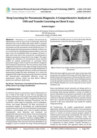
conditions of a healthy person as well as the lungs affected by pneumonia is shown in the following figure
inflammation in the air sacs of one or both lungs, where the affected areas may be filled with either fluid or purulent material, such as pus. Particularly in infants, young children, and people over 60, pneumonia can be fatal.Every year, 2.5 million people die from pneumonia worldwide. A chest x-ray is a crucial test for identifying pneumonia.Chest x-rays can show areas of consolidation that are opaque. However, it takes more time and is less accurate for a professional to diagnose pneumonia using chest X-ray images. The paper suggests the utilisation of convolutional neural network models by medical professionals to accurately detect pneumonic lungs in chest X-rays for the effective diagnosis and treatment of pneumonia. The two primary steps of the suggested framework are the stage of image preprocessing and the stage of feature extraction and image classification. A model that can distinguish between normal and abnormal (pneumonia) images has been developed. As a result, the suggested CNN model-based pneumonia detection method has demonstrated considerable efficiency across all performance metrics, which can help deliver effective patient care and lower death rates.
Fig -1: Chest X-ray images without pneumonia and with pneumonia White dots that might be seen in the chest cavity show that pneumonia has impacted the lungs. [2] Early detection of a deadly disease like pneumonia is always preferable, and if a bacterial infection is likely to be the cause of mild pneumonia, it may be typically treated at home with rest, antibiotics, and lots of fluids. Hospital care can be required in more severe situations. [3]
Key Words: Pneumonia, Convolutional Neural Network (CNN), Chest X-ray, Image classification
Deep convolutional neural networks are being used in a variety of current research projects related to medical picture processing. Deep learning employs a variety of models to extract information from pictures presented to the model. Deep learning is currently frequently used in the field of medicine to identify and diagnose diseases and then classify them into a specific disease group. A convolutional neural network is the most extensively used model for medical image processing.
1.INTRODUCTION An infection known as pneumonia causes inflammation in one or both of the lungs' air sacs. The air passages may enlarge with liquid or mucus (purulent material), which can cause a high temperature, chills, a cough that produces pus or phlegm, and difficulty breathing. Pneumonia may be caused by a wide range of organisms, such as pathogens, viruses, and fungal infections. [1] Most occurrences of pneumonia happen in developing and underdeveloped countries because of adverse environmental circumstances, pollution, overcrowding, and a lack of medical facilities. Consequently, early detection and treatment can significantly help prevent the illness from becoming fatal. Radiological inspection of the lungs using computerised tomography (CT), magnetic resonance imaging (MRI), or radiography (X-rays) is commonly used for the identification of lung problems. Non-intrusive and fairly affordable X-ray imaging is used to evaluate the lungs. Demonstrating the
© 2023, IRJET
|
Impact Factor value: 8.226
[4] Furthermore, DL models outperformed standard approaches utilising chest X-ray pictures from pneumonia patients. [5]. Deep convolutional neural networks are being used in a variety of current research projects related to medical picture processing. Consequently, the need for an automated pneumonia detection technique arises. The quantity of our data, which allows us to test and evaluate our models in various situations, is a crucial strength of our work in comparison to earlier studies. Transfer learning is a remedy for this hurdle. This approach makes use of the
|
ISO 9001:2008 Certified Journal
|
Page 704