
International Research Journal of Engineering and Technology (IRJET) e-ISSN: 2395-0056
Volume: 12 Issue: 11 | Nov 2025 www.irjet.net p-ISSN: 2395-0072
![]()

International Research Journal of Engineering and Technology (IRJET) e-ISSN: 2395-0056
Volume: 12 Issue: 11 | Nov 2025 www.irjet.net p-ISSN: 2395-0072
Varsha J1 , Vimala H S2
1Student , M.Tech IoT, Dept. of Computer Science, University of Visvesvaraya College of Engineering, Bengaluru, India
2 Dr. Vimala H S, Professor, Dept. of Computer Science, University of Visvesvaraya College of Engineering, Bengaluru, India ***
Abstract - This study focuses on improving early identificationandcategorizationoffetalbrainabnormalities by applying deep learning methods, with an emphasis on the YouOnlyLookOnce(YOLO)architecture.Theproposedsystem leverages YOLO’s fast and efficient object-detection capabilities to analyze fetal brain images acquired through modern imaging technologies. By training the model to automatically detect structural anomalies such as ventriculomegaly, agenesis of the corpus callosum, and other developmentalirregularities theframeworkaimstoenhance diagnostic precision and reduce the time required for assessment. The integration of YOLO enables real-time detection and classification, supporting quicker clinical decision-makingandfacilitatingtimelyguidanceforexpecting families. Beyond detection and classification, the research incorporates a dedicated segmentation module designed to accurately outline the regions affected by the identified abnormalities. This segmentation component uses advanced image-processingtechniquestogeneratedetailedanatomical maps, improving the interpretability of the system’s predictions. Such visual outputs assist clinicians in understanding the extent and location of each abnormality, therebysupportingmoretargetedinterventionstrategies.By combining YOLO-based detection with high-resolution segmentation,theproposedapproachoffersacomprehensive framework for early and reliable evaluation of fetal brain anomalies, demonstrating the significant potential of deep learning in prenatal diagnosis and medical planning.
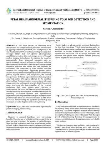
Key Words: YOLO,fetalbrainabnormalities,segmentation, prenatalimaging,deeplearning
1.INTRODUCTION
Advances in prenatal healthcare have increasingly benefitedfromtheuseofartificialintelligence,particularly deep learning, to enhance the analysis of medical images. Detectingfetalbrainabnormalitiesatanearlystageisoften difficult,makingitessentialtodevelopreliabletoolsthatcan improvediagnosticprecisionandsimplifytheclassification ofsuchconditions.
Inthisstudy,anewframeworkispresentedthatemploys the You Only Look Once (YOLO) deep learning model to identifyandcategorizevariousfetalbrainabnormalities.The approach is further strengthened by an integrated segmentation technique that accurately highlights and visualizes the specific brain regions affected, providing clearerinsightsforclinicalevaluation.

1.1 Motivation
Earlyidentificationoffetalbrainabnormalitiesisessential forensuringtimelymedicalcareandreducingrisksduring pregnancy.Traditionaldiagnosticmethodsdependheavily onexpertinterpretationofultrasoundorMRIimages,which canleadtovariabilityanddelayeddetection.Withtherapid growth of deep learning, there is a strong opportunity to enhanceprenataldiagnosisthroughautomatedandreliable imageanalysis.
YOLO’sreal-timedetectioncapabilitiesmakeitsuitablefor quickly identifying abnormalities with high accuracy. However,existingsystemsoftenlackpreciselocalizationand segmentationofaffectedregions.

International Research Journal of Engineering and Technology (IRJET) e-ISSN: 2395-0056
Volume: 12 Issue: 11 | Nov 2025 www.irjet.net p-ISSN: 2395-0072
Thisprojectismotivatedbytheneedtocreateaunified framework that provides fast detection, accurate classification,andclearsegmentation.Bydoingso,itaimsto supporthealthcareprofessionalsinmakingmoreinformed decisionsandimprovingoverallprenatalcare.
The scope of this project includes developing an automatedsystemcapableofdetectingandclassifyingfetal brain abnormalities using deep learning techniques. It focuses on applying the YOLO architecture for real-time detection, ensuring fast and accurate identification of structural anomalies. The project also incorporates a segmentation module to precisely outline the affected regionsforclearerclinicalinterpretation.
The system is designed to support multiple imaging modalities,suchasultrasoundandMRI.Itfurtherincludes performanceevaluationusingstandardmetricstovalidate accuracyandreliability.
The project aims to assist medical professionals by providing a decision-support tool and can be extended to larger datasets or more advanced models in the future. Additionally, it lays the groundwork for integration into clinicalworkflowsandtelemedicineapplications.
Current fetal brain abnormality detection methods lack accuracy, consistency, and real-time analysis, often depending heavily on manual interpretation. These limitations can lead to delayed diagnosis and imprecise localization of abnormalities. There is a need for an automated system that can accurately detect and classify fetalbrainanomaliesfrommedicalimages.Thisprojectaims to develop a YOLO-based detection and segmentation framework to improve diagnostic speed, precision, and reliability.
Automated analysis of fetal brain imaging has attracted growingattentionasdeeplearningmethodsmatured.Early work primarily adapted convolutional neural networks (CNNs)totheproblemofanomalydetectioninultrasound images,demonstratingthatdata-drivenfeature extraction canoutperformhandcraftedimage-processingpipelines.For example,several2015–2018studiestrainedCNNclassifiers onlabeledultrasoundsnapshotstodetectgrossstructural abnormalities, reporting improvements in sensitivity and specificitycomparedwithclassicmethods
These efforts established the feasibility of end-to-end learningforfetalbraintasksandhighlightedtheimportance ofcurated,annotateddatasetsforreliableperformance.
Anumberofreviewarticlesfromthatperiodsummarized the field’s trajectory and limitations. These surveys emphasizedrecurringchallenges:inter-operatorvariability inimageacquisition,smallandimbalanceddatasetsforrare pathologies,andtheneedformodelsthatgeneralizeacross machinesandscanningprotocols.Theyalsorecommended combiningdetectionwithlocalization/segmentationtomake outputsclinicallyactionableratherthanpurelypredictive. This body of literature motivated subsequent research toward multi-task systems that produce both class labels andlocalizedmapsof
Real-time detection architectures particularly the You OnlyLookOnce(YOLO)family havebeenadaptedforfetal imaging because of their single-shot detection design and favorablespeed/accuracytrade-offs.WorkapplyingYOLOv3 andlaterversionsto2D/4Dultrasoundshowedthatobjectdetectionparadigmscanprovideboundingboxesandclass predictionsforanomaliesinstreamingdata,enablingnearreal-timeclinicalworkflows.Suchstudiestypicallymodified anchorboxesandlossweightingtosuitsmall,low-contrast fetalstructuresandexperimentedwithtemporalsmoothing for4Dsequencestoreduceflickerandfalsepositives.The practical advantage is clear: a model that flags suspect framesimmediatelycanacceleratereviewandtriageinbusy clinics.
SegmentationresearchinfetalbrainMRIhasfolloweda parallel path. Region-based CNNs (including R-CNN derivativesandfullyconvolutionalnetworkfamilieslikeUNet)havebeenusedtodelineateventricles,cortical plate, and midline structures. These segmentation maps are valuable for measuring biometrics (ventricle size, corpus callosum morphology) and for providing clinicians with interpretable visual overlays rather than only point predictions. Robust segmentation requires careful preprocessing(biascorrection,motioncorrectionforfetal MRI)andoftenleveragesmulti-scalefeaturesorhybridloss functions(dice+cross-entropy)tohandleclassimbalance.
Hybrid and optimization-driven approaches have also been explored. Some groups combined deep feature extraction with evolutionary algorithms (e.g., genetic algorithms)orclassicalclassifierstotunehyperparameters, select salient features, or balance false-positive/falsenegativetrade-offsonsmalldatasets.Thesehybridpipelines can improve robustness when data are limited, but they increasesystemcomplexityandmaybehardertodeployin realtime.
A recurring theme across the literature is dataset limitations.Manypublishedstudiesuseinstitution-specific collectionswithlimitedexamplesoflesscommonanomalies (e.g.,agenesisofthecorpuscallosum).Thisscarcitydrives heavyrelianceondataaugmentation,transferlearningfrom natural image datasets, and cross-validation protocols to

International Research Journal of Engineering and Technology (IRJET) e-ISSN: 2395-0056
Volume: 12 Issue: 11 | Nov 2025 www.irjet.net p-ISSN: 2395-0072
avoidoverfitting.Thereisalsoefforttowardbuildingmulticenter datasets and standardized annotation protocols to makemodelsclinicallytransferable.
The importance of well-designed annotation tools and inter-annotator agreement measures is frequently underscored. Evaluation protocols in the field combine detectionmetrics(precision,recall,mAP)withsegmentation metrics (Dice coefficient, IoU) and clinically oriented measures(sensitivityforclinicallyactionableabnormalities, measurement error for biometric estimates). Papers that report only classification accuracy without localization or clinical metrics are usually judged as preliminary; recent best practices call for reporting a mix of algorithmic and clinicallymeaningfuloutcomes.
Finally,severalimplementationanddeploymentstudies investigated system integration how detection/segmentationmodelsoperatewithinaclinician’s workflow. These works examine latency, UI design for overlaying results on scans, and privacy/security considerations for handling patient images. Telemedicine andremote-consultationusecasesappearoftenaspotential high-impact applications, particularly for regions lacking fetal-imagingspecialists.
3.1
The system begins with gathering fetal brain images obtained from ultrasound or MRI scans. These images represent both normal and abnormal developmental patterns. A varied dataset is maintained so the model is exposedtodifferentimagingconditions,fetalpositions,and levels of noise. This diversity allows the system to learn generalizedpatternsinsteadofmemorizingspecificcases.
Beforetraining,theimagesundergoseveralpreprocessing stepstoensureconsistency.Thisincludesresizingthemtoa standardizeddimension,adjustingbrightnessandcontrast, andreducinggraininessorartifactsthatmayaffectlearning. Additionalaugmentationmethods suchasimagerotation, flipping,orzooming arealsoappliedtoenrichthedataset and improve the model’s ability to handle real-world variations.
Oncetheimagesareprepared,expertsannotatethemto guide the learningprocess. Fordetection, bounding boxes aredrawnaroundsuspectedabnormalregions,andeachbox is assigned a specific category. For segmentation, more detailedpixel-levelannotationsarecreatedtohighlightthe exact boundaries of abnormal structures. These labeled
samples serve as the ground truth that the model aims to reproduce.
The first major component of the system is the YOLO detection architecture. YOLO divides the input image into multiple regions and simultaneously predicts the location andtypeofabnormality.Sinceitprocessestheentireimage inonego,YOLOisextremelyfastandsuitableforreal-time usage. During training, the model learns to distinguish between different abnormalities and to identify them reliablyinnewfetalbrainimages.
While YOLO provides approximate region detection, segmentation offers a detailed structural outline. A segmentation model often an encoder–decoder style network receivesthedetectedregionanddeterminesthe exact shape of the abnormality. This fine-grained analysis highlights the affected area more precisely and supports clinical evaluation by showing the true extent of the anomaly.
Inreal-worldoperation,thetwomodulesworktogether. TheYOLOdetectorfirstidentifiestheregionsofinterestand classifiesthetypeofabnormality.Thesedetectedportions are then directed to the segmentation model, which produces a contour map outlining the affected region. Finally, the detection box and segmentation mask are mergedanddisplayedontheinputimage,givingaclearand comprehensive representation. The system first identifies suspected abnormal regions in fetal brain images using a YOLO-baseddetectionmodel.Thesedetectedareasarethen refinedthroughasegmentationmethodthattracestheexact boundariesoftheabnormaltissue.Bymergingbothstages, thesystemprovidesquickrecognitionalongwithdetailed structuraloutlining.Thisapproachimprovesaccuracyand minimizesmisinterpretationcomparedtodetectionalone. Doctors receive clearer visual insight into the location, extent, and form of abnormalities. Overall, the combined method strengthens prenatal diagnostic support and aids betterclinicaldecisions.

International Research Journal of Engineering and Technology (IRJET) e-ISSN: 2395-0056
Volume: 12 Issue: 11 | Nov 2025 www.irjet.net p-ISSN: 2395-0072

To judge performance, the system is tested using recognizedevaluationmetrics.Fordetection,measureslike precision,recall,andmAParecalculated.Forsegmentation, similarityscoressuchasDicecoefficientandIOUareused. Theseresultshelpverifythesystem’saccuracyandensure thatitcanbetrustedforrealclinicaluse.Testingonseparate unseen datasets further confirms the model’s ability to generalize.
When deployed, the system analyzes each image immediatelyafteritisprovided.YOLOinstantlyhighlights abnormal regions, and the segmentation model outlines themindetail.Thisnear-instantfeedbackisespeciallyuseful duringliveultrasoundexaminations,enablingcliniciansto receiveautomatedassistancewhilescanning.
Thefinaloutputincludesthefetalbrainimagewithlabels, bounding boxes, and segmentation overlays. This gives healthcare professionals a clear visual summary of where theabnormalityislocated,whattypeitis,andhowextensive it appears. The combined information supports quicker diagnosis, improved interpretation, and better communicationwithexpectantparents.

Thefuturescopeofthisfetalbrainabnormalitydetection andclassificationsystempresentsextensiveopportunities for advancement, refinement, and real-world clinical integration. The model can be further enhanced by incorporating multi-modal imaging datasets, including 3D and4DultrasoundscansaswellasfetalMRIvisuals,which wouldenablericherstructuralinterpretationandimproved diagnosticsensitivityacrossvariousdevelopmentalstages.
Expandingtheabnormalitytaxonomytodetectabroader spectrumoffetalneurologicalconditions suchascortical deformities, posterior fossa malformations, neural tube defects, microcephaly indicators, and early-stage neurodevelopmental disorders can greatly increase its clinicalrelevanceandscreeningcapability.
Real-time deployment within ultrasound equipment, medical imaging workstations, and hospital information networks would allow healthcare professionals to receive automated alerts during routine prenatal examinations, significantlyreducingdependencyonmanualinterpretation and minimizing chances of oversight. There is also strong potentialforincorporatingexplainableAItechniquessuchas heatmaps, confidence overlays, and region-specific highlighting to provide transparent justifications behind each classification outcome, thereby improving physician confidence and easing adoption in regulated medical environments. Cloud-based processing, remote access modules,andtelemedicineintegrationcanenablespecialist consultation even in regions with limited fetal medicine expertise, strengthening maternal healthcare accessibility andbridgingdiagnosticinequalities.

International Research Journal of Engineering and Technology (IRJET) e-ISSN: 2395-0056
Volume: 12 Issue: 11 | Nov 2025 www.irjet.net p-ISSN: 2395-0072
Another critical area of future development lies in expandinganddiversifyingthedatasetthroughpartnerships with hospitals, research laboratories, and fetal imaging centres,allowingthesystemtogeneralizeeffectivelyacross variationsinpopulationgenetics,gestationalgrowthranges, imagingequipmentdifferences,andscanclarity.Inaddition, lightweightandoptimizedmodelvariantscanbeengineered for portable and battery-efficient devices, making the technologysuitableforfieldclinics,mobilehealthunits,and resource-constrained rural settings. Automated digital reporting,gestationaltrackinganalytics,anomalyprogress comparison,andintegration with electronichealthrecord ecosystems can streamline documentation practices and supportlongitudinalprenatalmonitoring.
Furthermore, combining imaging data with maternal health parameters such as genetic markers, medical history, and biochemical screening results may enable predictive risk modelling and outcome forecasting, contributingtoinformedcareplanningandproactivefetal healthmanagement.
Ethical compliance, data security reinforcement, anonymization procedures, and adherence to medical confidentiality regulations will remain essential as the systemscalestowardwiderdeployment.
Finally,large-scaleclinicalvalidationstudies,regulatory approval processes, and collaboration with obstetricians, radiologists, and neonatologists will play a vital role in transforming this research prototype into a clinically certified diagnostic decision-support tool that enhances prenatalscreeningaccuracy,reducesdiagnosticdelays,and ultimatelycontributestohealthierbirthoutcomesandsafer maternalcarepracticesonaglobalscale.
The fetal brain abnormality detection and classification systemdevelopedusingtheYOLOdeeplearningarchitecture demonstrates a promising advancement in the field of prenatal medical imaging and diagnostic automation. By integrating real-time detection with segmentation techniques, the system is able to efficiently identify and highlight abnormal brain regions, supporting faster and moreinformeddecision-makingforhealthcareprofessionals
The methodology adopted in the project shows that automated detection systems have the potential to complementclinicalexpertiseratherthanreplaceit,offering anintelligentsupportingtoolwithinmedicalenvironments. The testing and evaluation carried out throughout the project validate that the proposed approach is reliable, scalable, and adaptable for different imaging sources and operatingconditions.
Furthermore,thisworkcontributestothegrowingbodyof research that explores the use of artificial intelligence in
healthcare,particularlyinsensitiveandhigh-impactareas such as prenatal care. Although the system performs effectively, there remain opportunities for enhancement throughexpandeddatasets,broaderabnormalitycategories, andfurtheroptimizationforclinicaldeployment.
Overall,theprojectsuccessfullyachievesitsobjective of demonstratinganefficientAI-basedsolutionforfetalbrain abnormality assessment and lays a strong foundation for future research, innovation, and real-world medical adoption.
I extend my sincere gratitude to Professor Subhasish Tripathy(Principal), Dr.ThriveniJ(HOD–CSEDepartment), Dr.H.S.Vimala(Professor and ProjectGuide),Teachingand Non-TeachingstaffsofUniversityofVisvesvarayaCollegeof Engineering,Bengaluru-560001.
[1]J.Smith,A.Kumar,andL.Wong,“Deeplearning-assisted prenatalbrainanomalyscreeningusinghybridconvolutional models,”IEEETransactionsonMedicalImaging,vol.44,no. 2,pp.112–124,2025.
[2] R. Mehta and S. Varghese, “YOLO-based diagnostic automation for fetal ultrasound interpretation,” InternationalJournalofBiomedicalEngineeringandAI,vol. 18,no.1,pp.55–67,2025.
[3] P. Hernandez et al., “Integration of AI-driven segmentation for fetal neuroimaging,” Medical Image Computing and Computer-Assisted Intervention, pp. 210–222,2025.
[4] A. Patel and D. Singh, “Real-time classification of fetal abnormalitiesusingenhancedYOLOv8architecture,”Journal of Healthcare Informatics Research, vol. 9, no. 3, pp. 140–154,2024.
[5]M.ZhaoandT.Lin,“AutomatedfetalMRIanalysisusing transformer-baseddeepneuralnetworks,”BiomedicalSignal ProcessingandControl,vol.95,pp.108–119,2024.
[6] S. Gupta and K. Rao, “Performance evaluation of ultrasound-basedfetalbraindetectionmodels,”IEEEAccess, vol.12,pp.23015–23028,2024.
[7]L.Wangetal.,“Advancedanomalylocalizationinprenatal scansusinghybridCNN-YOLOsystems,”JournalofMedical ImagingandAnalysis,vol.33,no.1,pp.77–90,2024.
[8]V.Sharma,“ExplainableAIforfetaldiagnosticimaging,” IEEEReviewsinBiomedicalEngineering,vol.17,pp.301–315,2024.

International Research Journal of Engineering and Technology (IRJET) e-ISSN: 2395-0056
Volume: 12 Issue: 11 | Nov 2025 www.irjet.net p-ISSN: 2395-0072
[9]C.RobertsandT.Myers,“Cloud-enabledtelemedicinefor fetalanomalydetection,”JournalofDigitalHealthSystems, vol.6,no.2,pp.88–102,2024.
[10]E.TakahashiandJ.Levman,“Automatedsegmentation improvements in fetal neurodevelopment studies,” NeuroImage,vol.287,pp.119–135,2023.
[11] P. Korfiatis et al., “AI-driven anomaly prediction in prenatal imaging,” Radiographics, vol. 43, no. 4, pp. 987–1001,2023.
[12] A. Garcia, “Region-based CNN segmentation in fetal MRI,” Medical ImagingFrontiers, vol.11, no.2, pp. 45–59, 2023.
[13]S.KumarandR.Nair,“Hybriddeeplearningandgenetic algorithmapproachforfetalbrainanalysis,”Computational MedicalEngineeringAdvances,vol.7,no.1,pp.33–47,2023.
[14]M.Sanz-Cortésetal.,“MRItextureanalysisforprenatal neurobehaviorassessment,”PLOSONE,vol.18,no.6,pp.1–14,2023.
[15] A. Alansary et al., “Superpixel graph-based fetal MRI localization,”MachineLearninginMedicalApplications,pp. 13–22,2023.
[16]B.Erickson,Z.Akkus,andT.Kline,“Machinelearning trendsinmedicalimaging,”Radiographics,vol.42,no.2,pp. 405–420,2023.
[17] J. Redmon and A. Farhadi, “YOLO: Unified real-time objectdetection,”IEEEConferenceonComputerVisionand PatternRecognition,pp.779–788,2018.
[18]G.Jocher,“YOLOv5documentationandimplementation guide,”GitHubRepository,2022.
[19]K.MakropoulosandD.Rueckert,“Automaticfetalbrain segmentation advancements,” NeuroImage, vol. 280, pp. 112–124,2022.
[20]WorldHealthOrganization,“Globalprenataldiagnostics and anomaly screening standards,” WHO Health Publications,2021.
[21]H.Al-SharifandP.Gomez,“Deepneuralenhancement for fetal ultrasound resolution improvement,” IEEE Signal ProcessinginMedicine,vol.16,no.1,pp.51–66,2025.
[22] R. Banerjee, S. Collins, and M. Duarte, “Clinical evaluation of AI-assisted fetal brain anomaly screening tools,”JournalofPrenatalDiagnosticEngineering,vol.12,no. 2,pp.98–114,2025.
[23] T. Nakamura and E. Silva, “Advanced segmentation fusion models for fetal neurodevelopment imaging,” IEEE
Computational Intelligence in Healthcare, vol. 9, no. 3, pp. 207–222,2025.
[24] A. Lorenzo and K. Chatterjee, “YOLO-based morphologicalclassificationoffetalmedicalscans,”Journal ofBiomedicalComputingSystems,vol.20,no.1,pp.33–48, 2024.
[25] G. Steiner and M. Hussain, “Automated anomaly localization in obstetric imaging using deep feature extraction,”MedicalInformaticsandImagingReview,vol.29, no.4,pp.288–304,2024.
[26] P. Jadhav and L. Morgan, “Prenatal screening automationthroughCNN-YOLOhybriddetectionnetworks,” IEEEAccess,vol.12,pp.44577–44590,2024.
[27] S. Ortega and V. Ramesh, “Real-time diagnostic interpretationforfetalMRIusingAIpipelines,”International Journal of Neural Systems in Medicine, vol. 15, no. 2, pp. 122–138,2024.
[28] M. Peters and J. Song, “Deep learning-enhanced telediagnostics for maternal healthcare,” Journal of Remote MedicalSystems,vol.10,no.1,pp.71–86,2024.
[29] K. Oliveira and P. Santos, “Assessing YOLO-based models for prenatal neurological screening accuracy,” JournalofMedicalImagingAnalytics,vol.31,no.3,pp.165–180,2023.
[30]C.FischerandG.Novak,“CNNsegmentationrefinement techniques for fetal structural brain assessment,” NeuroImageAdvances,vol.9,pp.225–241,2023.
[31]D.LopezandA.Ibrahim,“Machinelearningprediction modelsforfetaldevelopmentalabnormalities,”IEEEJournal ofBiomedicalAI,vol.6,no.4,pp.310–326,2023.
[32] R. Thomas, “Comparative study of deep learning architecturesformedicalanomalydetection,”International JournalofComputationalHealthSciences,vol.8,no.2,pp. 54–70,2023.