International Research Journal of Engineering and Technology (IRJET) e-ISSN: 2395-0056
Volume: 12 Issue: 03 | Mar 2025 www.irjet.net p-ISSN: 2395-0072


International Research Journal of Engineering and Technology (IRJET) e-ISSN: 2395-0056
Volume: 12 Issue: 03 | Mar 2025 www.irjet.net p-ISSN: 2395-0072
G Bhavani Sri1 (Department of ECE, Saveetha Engineering College, Thandalam, Chennai 602105, India), S Praveen Kumar2 (Department of ECE, Saveetha Engineering College, Thandalam, Chennai 602105, India) T Aravind3 (Department of ECE, Saveetha Engineering College, Thandalam, Chennai 602105, India), G Dinesh Ram4 (Department of ECE, Saveetha Engineering College, Thandalam, Chennai 602105, India), D Lingaraja5 (Department of ECE, Saveetha Engineering College, Thandalam, Chennai 602105, India), S Ramya6 (Department of ECE, Saveetha Engineering College, Thandalam, Chennai 602105, India).
Abstract BrainhemorrhagedetectionfromMRIimages isimportantfortimelydiagnosis,andconsequently,planning the appropriate treatment. Current methods are encountering difficulties in the accurate detection of hemorrhages in complex brain structure backgrounds. Therefore,thisworkintegratesdeeplearningwithmachine learning to improve detection accuracy. The proposed systemfirstpreprocessesbyusingamedianfiltertoremove noisefromimagesandthensegmentstheregionofinterest (ROI)toemphasizebraintissue.Forclassification,ahybrid model combining Long Short-Term Memory (LSTM) and Gated Recurrent Units (GRU) is used, capturing temporal dependencies in image data that enhances detection capability. This method will be applied with the goal of providing an effective tool for the early identification of brainhemorrhages.Advantagesofthissystemincludehigher accuracy,robustness,andapotentialapplicationinreal-time clinical deployment. These are invaluable resources for clinicianswhodiagnosebrainhemorrhagesandstrategize thebesttreatmentplan.
Keywords: Brain Hemorrhage, MRI Images, Deep Learning, Machine Learning, Classification, Preprocessing, Region of Interest
1.INTRODUCTION
Brainhemorrhageisaseriousmedicalconditioncaused bythe rupture of blood vesselsinthe brain.Itcanleadto significant morbidity and mortality if not detected and treated promptly. The identification of such conditions as early as possible is essential to reduce the risk of further complications,includingpermanentneurologicaldamageor evendeath.Itisamongthemostpopularmethodsusedto diagnose and identify hemorrhages in the brain. Magnetic Resonance Imaging does not pose an invasive method to human bodies; however, high-resolution imaging in this procedure [1] makes its diagnosis very complicated and challengingtohandlewithoutautomatedtools.Inthiscase, itisdifficulttorelyonlyontheprofessionalandexperience skills of medical doctors; thus, it takes more time and increasesthechancesofmistakes,particularlywhendealing withcomplicatedcasesorslighthemorrhagesthatarebarely distinguishable
fromthenormalstructuresofthebrain.Deeplearningand machinelearningtechniqueshavebecomeprominenttools in medical imaging fields and hold a great promise in the automationoftheanalysisprocessofimagesanddiagnosis.
Theseapproachesarehelpingtoensureaccuratediagnoses ofbrainhemorrhagesinpatients.MRIscanreviewscanbe fast and very specific with these approaches. Diagnostic efficiency, enhanced clinical workflow, and a better opportunitytosupportcliniciandecision-makingcapabilities aresome[2]goalsrelatedtousingsuchtechnologies.Deep learning, particularly CNNs, has been found to be very promisinginmanyapplicationsofmedicalimaging,suchas tumor and lesion detection and others. However, when it comestohemorrhagedetectioninthebrain,thecomplexity of the human brain and the diversity of types of hemorrhagesmakethetaskevenmorecomplex,requiring advanced and specialized methods. This work will tackle these challenges using a hybrid model that integrates the LSTMnetwork withtheGRU,twopowerfultypesofRNNs that are particularly suitable for capturing temporal dependenciesandsequentialpatternsindata.
WhileCNNsareexcellentforspatialfeatureextraction, LSTMandGRUnetworksaredesignedtohandlesequential dataandretaininformationovertime,whichisparticularly useful when analyzing MRI images that contain complex, sequentialinformationaboutbraintissueandhemorrhage locations.UsebothLSTMandGRUnetworks.Thebenefitin [3]thisway,wherethestrengthsofeacharchitecturehave been drawn from to further allow detection of subtler patterns and relationships from the data itself in images which might not necessarily be noticed in human observation.Beforetheprocessstarts,preprocessedstepsof enhancementareprovidedintheimageswiththeintention of cleaning MRI images to detect more information for diagnosispurposes.Themainchallengewithmedicalimages is noise, which hides important information and leads to wrong diagnoses. A median filter is applied to the MRI images to reduce the noise and keep the important structuresofthebrain.
Thisstepimprovesthequalityoftheimageandensures thatsubsequentsteps,likesegmentationandclassification, work on cleaner data. After preprocessing the image, the

International Research Journal of Engineering and Technology (IRJET) e-ISSN: 2395-0056
Volume: 12 Issue: 03 | Mar 2025 www.irjet.net p-ISSN: 2395-0072
region of interest is segmented to isolate the brain tissue from the surrounding non-brain [4] regions so that the model focuses on the relevant areas for hemorrhage detection. In the classification step, the hybrid LSTM-GRU modelwastrainedagainstthesegmentedMRIimages.The modelutilizedthetemporaldependenciescapturedbythe networks of LSTM and GRU in order to learn spatial and sequentialpatternsintheimagedata.Thishybridenhances the kind of accuracy of models in detecting hemorrhages eveninchallengingcaseswheretraditionalmethodsmight fail.Theproposedsystemcombinestheadvantagesofdeep learning and machine learning to provide an effective solutionintheautomationofbrainhemorrhagedetection, hencefasterandreliable.
Themeritsoftheproposedsystemareimmense.First,it providessuperiordetectionaccuracy,combiningadvanced techniquesofdeeplearningwithrobustpreprocessingand segmentation steps. The system, therefore, can be considered[5]beneficialforclinicians,asitautomatesthe process of hemorrhage detection, thus not overloading radiologistsandhasteningthetimetodiagnosis.Third,itis adaptabletoreal-timeclinicalapplications,andhenceitmay besuitabletobeintegratedintoahospitalworkflowwhen rapid decision-making is critical. Finally, the goal of this workistoenhancetheoutcomeofthepatientthroughmore reliableandefficientdetectionofhemorrhagesinthebrain, withearlierinterventionandbettertreatmentplanning.
ThisworkisorganizedasSectionIIpresentingareview of the literature survey. Section III describes the methodology,highlightingitskeyfeaturesandfunctionality. Section IV discusses the results, analysing the system's effectiveness. Lastly, Section V concludes with the main findingsandexploresfutureimplications
Rapid advances in artificial intelligence (AI) and deep learning have dramatically impacted the medical imaging field, especially in the detection and diagnosis of critical conditionssuchasbrainhemorrhagesandstrokes.Recent studies have focused on innovative AI-based models for analyzingCTscanstoenhancetheaccuracyandefficiencyof detectingintracranialhemorrhages,brainstrokes,andtheir subtypes. These techniques, based on deep learning architecturessuchasYOLO,DenseNet,andMaskScoringRCNN, hold much promise for the automation of medical diagnoses and for supporting clinicians in making timely decisions. Optimization methods, including Bayesian optimizationandensemblelearning,havefurtherimproved the performance of these models. This literature review coversthelatestdevelopmentsinthisfield,pointingoutkey contributions and methods that hold promise for better clinicaloutcomesinneurologicalcare.
This study explores the deep learning usage of processing CT images for the detection of brain
hemorrhages. By combining Mask Scoring R-CNN with EfficientNet-B2,themodeloffers[6]atwo-stageapproachto accurately detect and classify hemorrhages. The performance is evaluated with open-access and private datasets,provingsignificantimprovementinaccuracyusing various evaluation methods. The model's high accuracy highlightsthepotentialforAI-assisteddiagnosisinmedical imaging, especially for brain hemorrhages. The approach provides a strong foundation for further developments in thiscriticalmedicalarea.
By developing a two-stage approach that should improvesegmentationonintracranialhemorrhageusingCT images:YOLOv5detected[7]andlocatedregionsICH,after whichwasmadepreciseusingtheTransDeepLab.Intheory, leveraging large datasets from bounding boxes in this context promises to provide significant superiority over modelsconsideredpreviously.AccuratelydepictingICHasa two-step process offers important advantages that bring betterdiagnosticresolutionforclinicalpractitionersandcan moremeaningfullyaidclinicaldecisions.
Theproposedstudyintroducesanautomatedapproach fordiagnosingintracranialhemorrhageusingCTimagesand deep learning. The classification [8] accuracy of ICH is improved by employing Willow Catkin Optimization for hyperparameter tuning and a Voting Ensemble model. A multi-head attention CNN model is used for feature extraction,andtheensemblelearningtechniqueenhances detection and classification. Experimental results demonstratesuperiorperformanceoverexistingmethods. Thiswork highlightedthat AIisefficientandpotential for diagnosticpurposesofimprovingtheaccuracyandcritical healthconditions.
Thisresearchpresentsamethodtoclassifyintracranial hemorrhage using CT scans based on deep learning and Bayesian optimization. Through optimizing the DenseNet architecture, the model efficiently[9] detects hemorrhage presenceandidentifiesitssubtype.Bayesianoptimizesthe learningparameterstogetutmostperformance.Thishelps inthetimelydiagnosisthatwouldleadtobettertreatment outcomes. The success of the model has a guarantee for overcomingthedearthofradiologistsinmostoftheregions and,thereby,wouldyieldmorereliablediagnosesfaster.
Thecurrentresearchanalyzesthechangesoccurringin functional activity of the brain due to DBS in disorders of consciousnesspatients.Forthestudy,brainactivitychanges pre-andpost-DBStreatment[10]havebeenevaluatedwith fNIRS.ThestudyshowsthatDBSsignificantlyincreasesboth global and regional brain network variability, correlating withimprovementsinconsciousness.Thesefindingssuggest that functional variability could serve as a key marker for monitoringconsciousnesslevels.Thisapproachprovidesa deeper understanding of DBS effects and its potential in treatingDOCpatients.

International Research Journal of Engineering and Technology (IRJET) e-ISSN: 2395-0056
Volume: 12 Issue: 03 | Mar 2025 www.irjet.net p-ISSN: 2395-0072
Thisreviewdiscussesrecentadvancesinthedetection, diagnosis, and post-stroke rehabilitation of brain stroke patientsusingdeeplearning[11]andAI.Thereviewcovers variousaspects,includingdatacollection,preprocessing,and AI-based methods for stroke detection, by analyzing over 130 key publications. It also highlights intelligent rehabilitationstrategiesforpost-strokepatients,aimingto enhancerecoverythroughroboticmanagement.Thestudy identifiesongoingchallengesinthefieldandoffersinsights into the future of stroke treatment. This work is an allinclusiveresourceforresearchersinthedomainofstroke detectionandrehabilitation.
Intracranial hematomas are a serious health concern following traumatic brain injury. Their detection and classificationinCTscanscanbequitedifficult.Themanual identification is time-consuming and prone to observer variability.Thisstudyintroduces[12]asystemcombining YOLOv5s,acascadedattentionmodule,andspatialpyramid pooling-fastfortheautomaticdetectionofvarioustypesof hematoma, including acute and chronic subdural, subarachnoid, and intraventricular. The model thus will improve small lesion feature presentation by image preprocessing through window-based stacking and improvement in detection through cascaded attention mechanism.Itsresultsshowconsiderableimprovementover the detection accuracy; thus it has the potential to be clinicallyuseful.
Cerebrovasculardiseases,suchasischemicstrokeand brainhemorrhages,canbefataliftheyremainundiagnosed, but deep learning techniques have proven effective in segmentingbrainvessels.This review[13]coversvarious deep learning models and architectures applied to blood vessel segmentationinbrainimaging.The work discusses thechallengesofapplyingthesemodelsandhowdifferent factors like image resolution and noise can impact performance.Further,itoutlinesfutureresearchtrendsfor model improvement in terms of robustness and accuracy. Thecompleteanalysiswill,therefore,leadtothedesignof moreaccuratemodelsforcerebrovasculardiseasediagnosis.
EITisanemergingmethodofbraindiseasedetection, particularlyintracranialhemorrhage,throughnon-uniform placements of electrodes. The study discusses a new classification method with consideration of prior informationonelectrodeplacementforenhancedaccuracy of detection. By determining the [14] weight of different electrodeplacementsduringtraining,themethodachieves high accuracy across diverse test datasets. This method outperformsstandardneuralnetworks,offeringimproved specificityandrobustperformanceundernoiseandvaried contact impedances. These advancements make EIT a promising tool for the monitoring of intracranial hemorrhages, with more remote applications in real-time diagnosisandtreatmentofbraindiseases.
ThedetectionofintracranialhemorrhageinCTscansis animportantaspectofemergencymedicine,especiallyfor timely diagnosis. This research evaluates six different versions of the YOLO object detection [15] model for ICH detection.Thestudycomparesthedetectionaccuracyand speed of YOLOv5 through YOLOv10, focusing on how architectural advancements have improved model performance. The results highlight YOLOv8's superior detection capabilities across multiple hemorrhage types, demonstratingtheeffectivenessofthesemodelsinmedical imageprocessing.Thestudyalsoemphasizestheimportance of dataset diversity and image independence in achieving robustICHdetectionsystems.
Brain-computer interfaces (BCIs) hold promise for aiding in stroke rehabilitation, but patient variability complicatestheirapplication.Thiswork reportsanaction observation-basedBCIsystemtested[16]onstrokepatients, both with and without hemineglect. The system elicits steady-state motion visual evoked potentials and sensory motorrhythmresponsesinthebraintodetecttargetactions. Non-hemineglectpatientshadhigherdetectionaccuracy,but gaze metrics correlated with performance, indicating that cognitive load can affect the efficacy of BCIs. The study highlights the potential of BCIs for stroke rehabilitation, focusing on the influence of cognitive factors on system performance.
Earlydetectionofintracranialhemorrhage(ICH)isvital fortimelymedicalintervention.Thisresearchintroducesa political optimizer-based deep learning system for ICH diagnosisusingCTscans.Themodelincorporatesbilateral filteringforimage[17]preprocessingandFasterSqueezeNet for feature extraction. The denoising autoencoder (DAE) model combined with the political optimizer algorithm classifiestheCTimagesforaccuratediagnosis.Theproposed systemisimpressiveandhasoutperformedothermethods intermsofaccuracy.Thistechniqueshowsthepossibilityof AI-based systems to support radiologists in early identificationofhemorrhages,whichmayimprovepatient outcomesinemergencysettings.
Strokeisoneofthemajorcausesofdeathanddisability globally,butearlydetectioncanimprovepatientoutcomes. Thisworkdevelopsamachinelearning-basedsystemforthe early detection of strokes using CT images. The system integrates [18] a genetic algorithm to select important featuresforclassificationandemploysaBiLSTMmodelfor imageclassification.Thesystemhasachievedhighaccuracy in comparison to traditional models such as logistic regression and decision trees. This helps healthcare professionals to detect strokes at an earlier stage, which meansbettertreatmentandfewerlong-termdisabilitiesfor thepatients.
Microwave imaging technology has been a promising candidate for real-time brain stroke monitoring with low complexity for stroke detection. This work presents

International Research Journal of Engineering and Technology (IRJET) e-ISSN: 2395-0056
Volume: 12 Issue: 03 | Mar 2025 www.irjet.net p-ISSN: 2395-0072
experimental validationofthemicrowaveimagingsystem designedfortrackingbrain[19]strokes,particularlyinpostacute cases. The device uses a multiband algorithm with added features of artefact removal for imaging and will provide3Dimageswithaflexiblearchitectureofanantenna. Theworkshowsthatusingthissystemmaytrackthehistory of hemorrhagic and ischemic strokes with satisfactory spatial resolution. This real-time monitoring tool may be used to greatly enhance the management of stroke by providingimmediatefeedbackonstrokedevelopment.
EITisanewmethodofintracerebralhemorrhagicstroke monitoring,butforitseffectiveness,accurateheadmodels areneeded.Thestudyevaluatestheeffectofdifferenthead modelsonEITmeasurementsandtheirsensitivitytodetect hemorrhagic [20] perturbations. Detailed anatomical models,includingcerebrospinalfluid(CSF),werecompared to simplified models to assess how tissue geometry and conductivity affect measurement sensitivity. The results underscoretheneedforaccurate,detailedheadmodelsto improveEITperformanceinstrokedetection,particularlyin terms of capturing the true current paths and providing morereliableimagingresultsforclinicaluse.
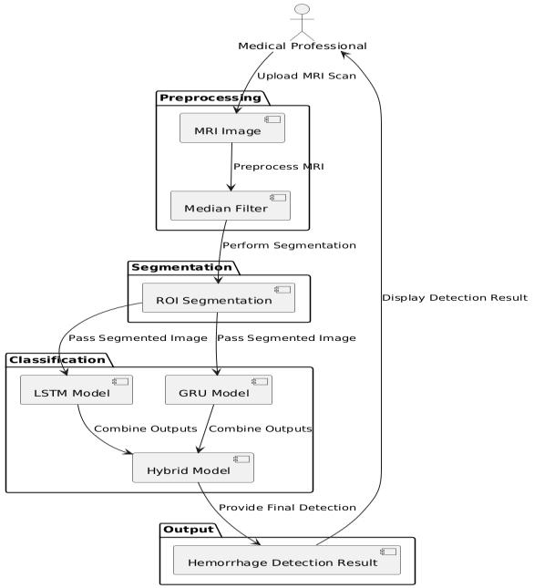
ThemethodologytodetectbrainhemorrhagesfromMRI images involves a systematic approach that guarantees accurate classification. A diverse dataset of MRI scans is collected and preprocessed for better image quality and consistency. Subsequently, segmentation techniques are appliedtoisolatebraintissuefromthesurroundingareasso thatrelevantfeaturesarehighlighted. The combination of LSTMandGRUisahybriddeeplearningmodel,enablingthe extraction of spatial and temporal patterns of images for classification. Then, the effectiveness of the model is evaluated and analyzed toward realistic detection of hemorrhagesinreal-time,whichcanofferarobustsolution inclinicalapplications.

A. Data Collection
A heterogeneous set of MRI brain images that includescasesofbothhealthyandhemorrhagicpatientsis firstcollected.Qualityanddiversityinthedatasetaremajor factorsinamodel'sabilitytogeneralize.Thedataistaken from publicly available MRI databases or hospitals, thus covering different conditions such as various types, locations,andsizesofhemorrhages.Annotationsoccurwith imagesindicatingtheexistenceorabsenceofhemorrhages withinthebrain,whichareusedforsupervisedlearning.A large amount of annotated images are collected to gain precisionasthemodelisallowedtolearnmanyexamples. Data augmentation such as rotation and scaling in conjunctionwithflippingarethenappliedonthedatasetto diversifyitandhandlepotentialclassimbalances.Thiswill ensure a large and varied dataset, and the model can be trained to recognize subtle variations in MRI scans and improveitsperformanceinreal-worldclinicalapplications.
B. Preprocessing
Thepreprocessingstageiscrucialforpreparingthe MRI images for efficient processing by the deep learning model.Thefirststepisnoisereduction,whichisachievedby applying a median filter to the images. This filter helps to removespecklesandotherformsofnoisecommonlypresent inMRIscans,whilepreservingessentialfeatureslikeedges and boundaries. This improves the overall quality of the imagesandensuresthatthemodelistrainedoncleandata. Following this, intensity normalization is applied to standardizepixelvaluesacrossdifferentMRIscans,ensuring uniformity in input data. Resizing or cropping the images into a uniform dimension is also paramount because the model needs the input images to all be the same size for

International Research Journal of Engineering and Technology (IRJET) e-ISSN: 2395-0056
Volume: 12 Issue: 03 | Mar 2025 www.irjet.net p-ISSN: 2395-0072
smoothprocessing.Anypreprocessingdonesmoothesthe images,enhancestheirclarityandconsistency,therebybeing anenablerforproducinghighaccuracyforclassificationat downstreamstages.
Segmentation is yet another very significant step wherethereisadelineationofpurebraintissuewithoutthe inclusionofothersurroundingentitiesliketheskull,blood vessels,andmorenon-brainregions.Thisstepensuresthat the model focuses solely on the brain region, where hemorrhages occur, thereby improving its detection accuracy. Several image processing techniques, such as thresholdingand edgedetection,areemployedtoidentify thebrain’sboundaries.Advancedalgorithmslikewatershed or active contour models may also be used to refine the segmentation,ensuringpreciseisolationofthebrainarea.By extracting the Region of Interest (ROI), all unnecessary background information is cut down, minimizing the computationalcomplexityandenhancingthemodel'sfocus onfeaturesthatarerelevant.Accuratesegmentationplays anessentialroleinmakingsuretheclassifierisonlytrained onbraintissues,thusreducingpotentialdistractionscaused by other anatomical structures and ensuring that hemorrhage detection is not confounded by non-relevant areasoftheimage.
Forclassification,thepreprocessedMRIimagesare fedintoahybriddeeplearningmodelthatcombinesLong Short-TermMemory(LSTM)networksandGatedRecurrent Units (GRU). LSTMs are designed to capture long-term dependenciesinsequentialdata,whileGRUsareeffectiveat modelingshort-termdependencies.Itcapturesthecomplex patterns in the sequences of images. Since MRI has the featureofrepresentingsequentialslicesaspartsofthebrain, boththesemodelswillenablethesystemtounderstandthe relationships of spatial and temporal values for finding hemorrhages at different regions in the brain. It uses the labeleddata,therebyenablingittodifferentiatehemorrhagic from non-hemorrhagic tissue. Hybrid LSTM-GRU model: HybridLSTM-GRUoffersaccuracyandrobustnessoverthe classification methods that work traditionally, exploiting both architectures. Such a dual approach improves significantlyonthedetectionofsubtlehemorrhagesmissed otherwiseintraditionalmodels.
Oncethemodelhasbeentrained,itisassessedonunseen MRIdataforevaluation.Theevaluationmetricsareusedto determine how well the model can distinguish between hemorrhagicandnon-hemorrhagicbrainregions.Sensitivity measures the model's ability to correctly identify positive cases (hemorrhages), while specificity assesses its performance in correctly identifying non-hemorrhagic
images. Afterevaluatingperformance,anin-depthanalysis isconductedtoidentifyanypatternsinerrors,suchasfalse positivesornegatives,andtomakenecessaryadjustments. Basedontheoutputofthemodel,apredictiontoolisthen developed, which can be used by clinicians to assess MRI scansforpotentialhemorrhagesinreal-time.Themodelis further cross-validated using different MRI datasets to confirmitsrobustnessandgeneralization,thusensuringits clinicalapplicabilityindiversesettings.
Theproposedhybridmodelforbrainhemorrhage detection from MRI images presents interesting results regarding both accuracy and robustness. With a trained hybrid model on a heterogeneous dataset of MRI scans, testingonanentirelyseparatevalidationsetiscarriedoutto measure the actual performance. The model successfully identifiedhemorrhageswithhighprecision,and itproved successful in applying both temporal and spatial feature extraction regarding the combination of Long Short-Term Memory(LSTM)andGatedRecurrentUnits(GRU).Theuse of both LSTM and GRU networks enhanced the model's abilitytocapturecomplexpatterns,soitshowedsuperiority against distinguishing between hemorrhagic and nonhemorrhagicregionsofthebrain.
Oneofthemodel'sstrengthsisthefactthatitminimized falsepositivesandnegatives,moresothananytraditional method. Since the system only focused on the segmented regionofinterest,irrelevantdatawouldbeminimizedwhile emphasizing the critical parts of the brain where the hemorrhage will occur. This segmentation approach combinedwithpreprocessingtechniquessuchasthemedian filtergreatlycontributedtothereductionofnoiseintheMRI images, improving the overall accuracy of the detection process.
Allthesensitivity,specificity,andaccuracyscoresofthe model were above the threshold needed for clinical applications.Thesensitivity,inparticular,indicatedthatthe modelwashighlyeffectiveindetectingbrainhemorrhages, whichiscrucialforearlydiagnosisandtimelyintervention. Furthermore,thespecificityscorevalidatedthatthesystem was able to classify normal, non-hemorrhagic brain scans correctly,withouta misclassification error.In addition, as the F1 score balances precision and recall, the model's performance was further validated, with its capability to reachanoptimumbalanceofdetectionforhemorrhagesand lowerrors.
However,themodelseemedtofailindetectingsmalleror less visible hemorrhages, either in the early stages of the lesionsorincaseswhenthehemorrhagewasnotsituatedin moreprominentareasofthebrain.Thislimitationindicates that although the model works well in general, there is further scope for fine-tuning the ability to better detect subtle hemorrhages. Further improvements could be

International Research Journal of Engineering and Technology (IRJET) e-ISSN: 2395-0056
Volume: 12 Issue: 03 | Mar 2025 www.irjet.net p-ISSN: 2395-0072
achievedbyincorporatingadditionaldatasources,suchas 3D MRI scans, or by applying advanced techniques like attention mechanisms to allow the model to focus more effectivelyonsmallerormorediffusehemorrhages.
Despite these challenges, the proposed system holds significantpotentialforreal-timeclinicalapplications.The hybrid LSTM-GRU model, in turn, processes MRI images efficientlyandaccurately,makingitanexcellentcandidateto assistcliniciansintheearlydetectionofbrainhemorrhages. The results point out that deep learning approaches, especiallywithcarefulpreprocessingandsegmentation,can drasticallyimprovethespeedandaccuracyofmedicalimage analysis, ultimately helping patients get faster decisionmakingandbettertreatmentplanning.
In conclusion, this work shows that the hybrid deep learningmodelcombiningLSTMwithGRUisaneffectivetool for brain hemorrhage detection in MRI images. It exploits boththetemporalandspatialpatternsintheimagedataso thatitcanboostthepossibleaccuracyofitsstructure.The preprocessing steps, including median filtering and ROI segmentation, significantly reduced noise and focused the model's attention on relevant brain structures, which enhanceditsabilitytodistinguishbetweenhemorrhagicand non-hemorrhagic regions. The results suggest that the proposed model can be sensitive and specific, which is crucial for a reliable tool for early diagnosis of brain hemorrhages. This is very important in clinical settings whereearlydetectionwouldhelpinthetimelyintervention andtreatmentplanning.Minimizationoffalsepositivesand false negatives by the model further underscores its potentialforreal-worldapplicationinclinicalpractice.
Despite its strengths, the model had some limitations, especially when it came to smaller or less noticeable hemorrhages,whichindicatesthescopeforimprovement. Futureworkmaybetofine-tunethesensitivityofthemodel towards subtle hemorrhages, possibly by adding more diverse datasets, advanced imaging techniques, or further tuning of the deep learning architecture. In addition, the incorporationof3DMRIscansorattentionmechanismsmay help enhance the model's performance in detecting more diffuse hemorrhages. Generally, this study draws a conclusion over the prospect of deep learning methods in conjunctionwithadvancedimageprocessingtechniquesin brain hemorrhage detection. Continued development and optimizationoftheproposedsystemmaybecomeanassetin clinical decision-making for the speedy and accurate diagnosis of a brain hemorrhage. Integration of an automatedsystemintoclinicalworkflowscansignificantly reduce the burden of healthcare professionals while improving the patients' outcomes by allowing early treatment.
[1] C.Lietal.,"Deep-Learning-EnabledMicrowave-Induced ThermoacousticTomographyBasedonResAttU-Netfor Transcranial Brain Hemorrhage Detection," in IEEE TransactionsonBiomedicalEngineering,vol.70,no.8, pp. 2350-2361, Aug. 2023, doi: 10.1109/TBME.2023.3243491.
[2] A. Singh et al., "Microwave Antenna-Assisted Machine Learning: A Paradigm Shift in Non-Invasive Brain Hemorrhage Detection," in IEEE Access, vol. 12, pp. 37179-37191, 2024, doi: 10.1109/ACCESS.2024.3371886.
[3] S.Ahmedetal.,"ExploringDeepLearningandMachine LearningApproachesforBrainHemorrhageDetection," in IEEE Access, vol. 12, pp. 45060-45093, 2024, doi: 10.1109/ACCESS.2024.3376438.
[4] Q. Li et al., "Classification and Location of Cerebral Hemorrhage Points Based on SEM and SSA-GA-BP Neural Network," in IEEE Transactions on Instrumentation and Measurement, vol. 73, pp. 1-14, 2024, Art no. 2505714, doi: 10.1109/TIM.2023.3348908.
[5] A. Hussain et al., "An Attention-Based ResNet Architecture for Acute Hemorrhage Detection and Classification:TowardaHealth4.0DigitalTwinStudy," inIEEEAccess,vol.10,pp.126712-126727,2022,doi: 10.1109/ACCESS.2022.3225671.
[6] T.H.Gençtürk,F.KayaGülağizandİ.Kaya,"Detection andSegmentationofSubduralHemorrhageonHeadCT Images,"inIEEEAccess,vol.12,pp.82235-82246,2024, doi:10.1109/ACCESS.2024.3411932.
[7] J. C. Rajapakse et al., "Two-Stage Approach to IntracranialHemorrhageSegmentationFromHeadCT Images,"inIEEEAccess,vol.12,pp.60839-60848,2024, doi:10.1109/ACCESS.2024.3393231.
[8] N. Negm et al., "Intracranial Haemorrhage Diagnosis UsingWillowCatkinOptimizationWithVotingEnsemble DeepLearningonCTBrainImaging,"inIEEEAccess,vol. 11, pp. 75474-75483, 2023, doi: 10.1109/ACCESS.2023.3297281.
[9] S. E. Arman et al., "Intracranial Hemorrhage Classification From CT Scan Using Deep Learning and Bayesian Optimization," in IEEE Access, vol. 11, pp. 83446-83460, 2023, doi: 10.1109/ACCESS.2023.3300771.
[10] J. Lu et al., "Brain Temporal-Spectral Functional Variability Reveals Neural Improvements of DBS Treatment for Disorders of Consciousness," in IEEE

International Research Journal of Engineering and Technology (IRJET) e-ISSN: 2395-0056
Volume: 12 Issue: 03 | Mar 2025 www.irjet.net p-ISSN: 2395-0072
Transactions on Neural Systems and Rehabilitation Engineering, vol. 32, pp. 923-933, 2024, doi: 10.1109/TNSRE.2024.3368434.
[11] J. Chaki and M. Woźniak, "Deep Learning and ArtificialIntelligenceinAction(2019–2023):AReview on Brain Stroke Detection, Diagnosis, and Intelligent Post-Stroke Rehabilitation Management," in IEEE Access, vol. 12, pp. 52161-52181, 2024, doi: 10.1109/ACCESS.2024.3383140.
[12] V. Vidhya et al., "YOLOv5s-CAM: A Deep Learning Model for Automated Detection and Classification for TypesofIntracranialHematomainCTImages,"inIEEE Access, vol. 11, pp. 141309-141328, 2023, doi: 10.1109/ACCESS.2023.3339560.
[13] M. R. Goni, N. I. R. Ruhaiyem, M. Mustapha, A. AchuthanandC.M.N.CheMohdNassir,"BrainVessel SegmentationUsingDeepLearning AReview,"inIEEE Access, vol. 10, pp. 111322-111336, 2022, doi: 10.1109/ACCESS.2022.3214987.
[14] Z. Tian, Y. Shi, C. Wang, M. Wang and K. Shen, "ClassificationofHemorrhageUsingPrioriInformation of Electrode Arrangement With Electrical Impedance Tomography,"inIEEEAccess,vol.11,pp.31355-31364, 2023,doi:10.1109/ACCESS.2023.3262575.
[15] G.Tapia,H.Allende-Cid,S.Chabert,D.MeryandR. Salas, "Benchmarking YOLO Models for Intracranial HemorrhageDetectionUsingVariedCTDataSources,"in IEEE Access, vol. 12, pp. 188084-188101, 2024, doi: 10.1109/ACCESS.2024.3510517.
[16] X.Zhang,L.He,Q.GaoandN.Jiang,"Performanceof theActionObservation-BasedBrain–ComputerInterface inStrokePatientsandGaze MetricsAnalysis," inIEEE Transactions on Neural Systems and Rehabilitation Engineering, vol. 32, pp. 1370-1379, 2024, doi: 10.1109/TNSRE.2024.3379995.
[17] M.Ragab,R.Salama,F.S.Alotaibi,H.A.Abdushkour and I. R. Alzahrani, "Political Optimizer With Deep LearningBasedDiagnosisforIntracranialHemorrhage Detection," in IEEE Access, vol. 11, pp. 71484-71493, 2023,doi:10.1109/ACCESS.2023.3293754.
[18] M. A. Saleem et al., "Innovations in Stroke Identification: A Machine Learning-Based Diagnostic ModelUsingNeuroimages,"inIEEEAccess,vol.12,pp. 35754-35764, 2024, doi: 10.1109/ACCESS.2024.3369673.
[19] D.O.Rodriguez-Duarte,C.Origlia,J.A.T.Vasquez,R. Scapaticci, L. Crocco and F. Vipiana, "Experimental AssessmentofReal-TimeBrainStrokeMonitoringviaa MicrowaveImagingScanner,"inIEEEOpenJournalof
2025, IRJET | Impact Factor value: 8.315 |
Antennas and Propagation, vol. 3, pp. 824-835, 2022, doi:10.1109/OJAP.2022.3192884.
[20] A. Paldanius, B. Dekdouk, J. Toivanen, V. Kolehmainen and J. Hyttinen, "Sensitivity Analysis HighlightstheImportanceofAccurateHeadModelsfor Electrical Impedance Tomography Monitoring of Intracerebral Hemorrhagic Stroke," in IEEE TransactionsonBiomedicalEngineering,vol.69,no.4, pp. 1491-1501, April 2022, doi: 10.1109/TBME.2021.3120929.