
International Research Journal of Engineering and Technology (IRJET) e-ISSN:2395-0056
Volume: 12 Issue: 05 | May 2025 www.irjet.net
International Research Journal of Engineering and Technology (IRJET) e-ISSN:2395-0056
Volume: 12 Issue: 05 | May 2025 www.irjet.net
Alla Krishna sai reddy1, Achanta Satya Sai Kishore2, Mudadla Koushik Manikanta Naidu3 , Kharan Jagabathula4 , Yogesh Raj5 , Dalalappa gari Naveen6, Dr. Brajesh Kumar7
1,2,3,4,5,6UG Student, Dept. of School of Computer Science & Engineering, Lovely Professional University, Punjab, India 7Associate Professor, Dept of School of Computer Science & Engineering, Lovely Professional University, Punjab, India
Abstract- Diabetic retinopathy (DR) is a serious complication of diabetes that can gradually lead to vision loss if not detected early. This document presents an automated system designed to identify the severity of DR using retinal fundus images. The system leverages a convolutional neural network based on a modified ResNet50 architecture to classify the disease into five stages ranging from no signs of DR to the most severe, proliferative DR. To improve the model’s performance and address the challenge of class imbalance in the dataset, Mix-up data augmentation was employed. Our model was trained and evaluated using the APTOS 2019 dataset, and the results showed strong classification accuracy. By utilizing pretrained ImageNet weights and incorporating custom dense feature extraction layers, the system was able to effectively analyse retinal images. This approach offers a promising solution for automated DR screening, helping healthcare professionals detect the condition at an earlier stage and ultimately contributing to the prevention ofvision loss ona global scale.
Key Words: Diabetic retinopathy, deep learning, convolutional neural networks, ResNet50, transfer learning, medical image classification, computer-aided diagnosis.
Diabetic retinopathy (DR) is a severe microvascular complication arising from diabetes mellitus that affects the retina and stands as one of the leading causes of preventable vision loss globally [1]. With the global prevalenceof diabetesprojected toreach642 millionby 2040 [2], the impact of vision impairment caused by diabetic retinopathy is projected to rise sharply, particularly in developing nations where routine screeningremainslargelyinaccessible.
DR progresses through various stages of severity, beginning with mild non-proliferative changes and potentiallyadvancingtoproliferativeDR,whichcanlead to permanent vision loss if not treated in time. Early detectionandtimely interventionare crucial,astreating DRinitsearlystagescanreducetheriskofseverevision loss by over 90% [6]. However, the current standard of diagnosis relies on manual inspection of retinal fundus images by trained ophthalmologists a process that is
not only time-consuming and subjective but also often inaccessibleinmanyregions[9].
To bring consistency to diagnosis, the International Clinical Diabetic Retinopathy (ICDR) severity scale categorizes DR into five stages: No DR, mild nonproliferative DR (NPDR), moderate NPDR, severe NPDR, and proliferative DR (PDR) [4]. Each stage is marked by specific retinal abnormalities, such as microaneurysms, hemorrhages, hard exudates, cotton wool spots, and neovascularization [3]. Accurate detection and classification of these lesions demand both advanced expertiseandsubstantialclinicalexperience.
In this paper, we present a deep learning approach for automated detection and classification of DR using retinalfundus images.OurmethodemploysaResNet50based CNN architecture with transfer learning and data augmentationtechniquestoaddresstheaforementioned challenges [12]. We utilize the APTOS 2019 Blindness Detection dataset, which contains a diverse collection of retinal images annotated by expert ophthalmologists accordingtotheICDRseverityscale.
Theprimarycontributionsofthisworkinclude:
1. Implementationofadeeplearningframeworkbased on ResNet50 architecture with customized dense layersforDRseverityclassification.
2. Application of the Mixup data augmentation technique to improve model generalization and addressclassimbalance.
3. Evaluation of the model's performance across different DR severity levels, providing insights into itsclinicalapplicability.
4. Development of a practical prediction pipeline for single-imageclassificationwithconfidencemetrics.
The remainder of this paper is organized as follows: Section II discusses related work in the field of automated DR detection [5]. Section III details our methodology, including dataset preprocessing, model architecture, and training strategy. Section IV shows experimental results and analysis comparing every aspect[15].Finally,SectionVconcludesthepaperwitha discussionoflimitationsandfuturedirections[14].
International Research Journal of Engineering and Technology (IRJET)
Volume: 12 Issue: 05 | May 2025 www.irjet.net
Telescopic development in manufactured insights innovation has reshaped the whole way of identifying diabetic retinopathy (DR). A wide choice of machine learning and profound learning models have risen from inquire about bunches to improve DR screening frameworksbymakingstridesaccuracyandadaptability along with elucidation capabilities. The taking after portion gives anoutlineof major discoveriesdistributed inlaterresearch.
[1]Bilaletal.(2022)displayedatwo-stagesystemwhich serves to illuminate information asymmetry in optic plate and blood vessel discovery. The proposed strategy comprises of two U-Net models working to section retinalhighlightssometimerecentlycrossoverCNN-SVD examination including Inception-V3 exchange learning upgrades the framework execution. The actualized engineeringsucceedsinrecognizingvitalretinalmarkers counting microaneurysms along with hemorrhages and exudates. The investigate group tried the approach utilizing EyePACS-1 and Messidor-2 and DIARETDB0 datasets where it realized extraordinary classification comes about of 97.92%, 94.59% and 93.52% at characterizing modern benchmarks for programmed discoveryofdiabeticretinopathy.
[2]Shankaretal.(2020)madeaprofoundlearningstage which recognizes DR seriousness levels interior fundus pictures. The three-phase location framework of their strategy diminishes clamor to begin with taken after by localeextractionthroughhistogram-baseddivisionsome time recently utilizing a Synergic Profound Learning show for classification. The recognizable proof of DR seriousness levels utilizing their strategy surpassed standardshowcapacitiesonMessidordatasetresults.
[3] The inquire about done by Zhang et al. (2022) inspectedhow to precisely decide extreme DR in fundus pictures from the Kaggle dataset. Utilizing Initiation V3 show with two distinctive input resolutions come about insuperiorresultswhenworkingwithpicturesthathave 896×896 pixels compared to 299×299 pixel determination. A affectability esteem of 0.925 combined with specificity at 0.907 brought about in a consonant cruel of 0.916 whereas the AUC come to 0.968. The discovery of preretinal and vitreous hemorrhages showed up less difficult than deciding intraretinal microvascular anomalies (IRMA) through the imaging prepare. The creators conducted their consider on RetCAD v.1.3.0 which holds CE certification for recognizing DR and age-related macular degeneration (AMD).
[4] The framework approved utilizing 600 assorted clinical pictures come to 95.1% AUC for identifying referable DR whereas getting 94.9% AUC for
distinguishing AMD. The demonstrate illustrated superior affectability than master doctors without compromisingtheparticularlocationcomesabout.Tests conducted on Messidor and AREDS datasets appeared that the framework is exceedingly reasonable for performingjointillnessscreeningprocedures.
[5] The inquire about conducted by Jacoba et al. (2023) considered AutoML models for identifying diabetic retinopathy through pictures collected by handheld gadgets which included different retinal areas. Prepared with certified grader comments the show accomplished tall execution scores amid inside tests with affectability at0.96andspecificityat0.98andprecisionat0.97taken afterby outsidetestingcomesabout ofaffectability0.94 along with specificity 0.97. The inquire about comes about demonstrate that AutoML-based DR screening frameworks can work viably in both resourceconstrained healthcare situations and far off restorative facilities.
[6] The demonstrative system called HIMLA (Half breed Inductive Machine Learning Calculation) was created by Mahmoud et al. (2023) to analyze fundus pictures between ordinary and those with DR movement. The systemcontains brightness normalizationasitsto begin withsteptakenafterbyencoder-decoderdivisionatthat point the extraction of highlights and classification through different occasion learning MIL. HIMLA effectively handled CHASE dataset information with an exactnessrateof96.62%andaffectabilityrateof95.31% and specificity rate of 96.88% which outflanked numerous built up models for both strength and performance.
[7] A full assessment of AI-based DR location and evaluating inquire about ponders from 2016 through 2021 was displayed in Lakshminarayanan et al. (2021). The analysts connected precise rules (PICO and PRISMA 2009) for their examination which come about in 114 pondersbeinginspecteddrivingtothedocumentationof 43 common information sets. The inquire about comprehensively presents the state of DR examinations togetherwithexaminationstrategiesforfundusandOCT picturesatthisdisplaymoment.
[8] The creators of Sarki et al. (2020) utilized ImageNet pretrained CNN models to bargain with the challenge of diagnosing different levels of diabetic eye conditions. Their framework worked in two demonstrative settings: mellow multi-class diabetic eye illness and common multi-class diabetic eye malady however accomplished superior precision than past approaches driving to the conclusion that exchange learning gives worth in this field The creators consolidated execution advancement procedureswhichincludedfine-tuning,optimizationand differentiate upgrade. A most extreme acknowledgment rate of 88.3% for multi-class classification and 85.95%
International Research Journal of Engineering and Technology (IRJET)
Volume: 12 Issue: 05 | May 2025 www.irjet.net p-
for gentle multi-class classification developed from VGG16 show testing on datasets explained by an ophthalmologist which demonstrates that profound learning handles troublesome demonstrative qualifications between minor diabetic eye illness varietieseffectively.
[9] The creators Alabdulwahhab et al. (2021) conducted examination of diabetic retinopathy discovery utilizing different machine learning classifiers in a Saudi Middle easternquietcohort.Aadduptoof327diabeticsubjects taken part in their cross-sectional consider to accumulatesocio-demographicandclinicaldatawhereas analysts executed straight discriminant investigation, backvector
machine, K closest neighbor, arbitrary woodland, and officer arbitrary woodland classifications through crossvalidation. A officer irregular timberland calculation demonstratedtobethemostsuccessfulclassifiersinceit accomplished an 86% precision rate with test information DR persistent recognizable proof. The two mostpersuasivefactorsinseparationwerediabetesterm together with HbA1c estimations whereas BMI and ageat-onset and persistent age and systolic blood weight estimations and medication utilization taken after. The investigate appears how machine learning brings significant potential to combine with ophthalmology for superior determination and help in therapeutic choice processes.
[10] Narayanan et al. (2020) made a combination machine learning framework which recognizes diabetic retinopathy and assesses illness seriousness by utilizing the freely accessible database of 3662 pictures. The strategy utilizes double stages to recognize DR cases after which it apportions persistent comes about into particular seriousness classifications (mellow, direct, proliferative, or serious). The creators inspected diverse exchangelearningmethodsbasedonAlexNetandVGG16 and Inception-v3 built up systems which coordinates convolutional neural systems with vital component investigation for dimensionality lessening some time recently SVM classification. The cross breed design accomplished amazing comes about with amazingly constrained preparing information and lopsided classes bycomingto98.4%exactnessforDRlocationand96.3% exactnessforevaluatingseriousnessinthiswaymakinga unused standard for analyzing constrained preparing populaces.
[11]Samantaetal.(2020)createdansuccessfuldiabetic retinopathy location framework utilizing exchange learning-based CNN design which works well on little tests and uneven classes. The show utilized 3050 preparing pictures together with 419 approval pictures for classifying four symptomatic categories: No DR, Gentle DR, Direct DR, and Proliferative DR. The
-0072
lightweight show analyzes difficult exudates with blood vessels and surface designs contained in color fundus photos. Due to its tall execution level the demonstrate illustrated 0.8836 Cohen's Kappa scores on approval informationand0.9809scoresonpreparing information which demonstrates its viability for real-time applications utilizing low-power computing to quicken DRscreeningoperations.
[12] The analysts from Bhimavarapu et al. (2023) made animprovedconvolutionalneuralarrangeframeworkfor programmed diabetic retinopathy determination through made strides pooling work advancement. The advancement of their strategy coordinates an improved pooling operation and enactment work into the ResNet50 demonstrate plan which broadened convolution bit affectability and minimized memory utilization and preparingtimeprerequisites.Throughthisstrategyitgot to be conceivable to identify injuries consequently whereas moreover minimizing misfortunes and add up to handling time. The demonstrate illustrated remarkable precision when tried on two datasets through its tests on APTOS and Kaggle which yielded evaluation comes about of 98.32% and 98.71% individually. The consider appeared that their proposed show outperformed driving approaches in therapeutic picturedeterminationforDRfromfunduspictures.
[13]Abramoffetal.(2020)tendedtothemoral,financial, and logical discussions encompassing manufactured insights usage in diabetic retinopathy location. Their paper recognizes the advocated concerns almost AI's effectonunderstandingsecurity,viability,value,andrisk as these frameworks progressively perform forms customarily saved for healthcare experts. The creators propose standardized descriptors to categorize DR AI frameworks based on level of gadget independence, expecting utilize, level of prove for symptomatic precision, and framework plan. Or maybe than being prescriptive, they take a clear approach, noticing that there is as of now negligible observational premise to declare that certain combinations of these variables are inalienablyprevalenttoothers,whereasemphasizingthe significance of legitimate assessment and approval methodologies.
Ourdiabeticretinopathydetectionframeworkconsistsof two distinct components which correctly identify unusual structures within retinas in addition to disease severitygradingformedicaldetermination.
International Research Journal of Engineering and Technology (IRJET)
Volume: 12 Issue: 05 | May 2025 www.irjet.net
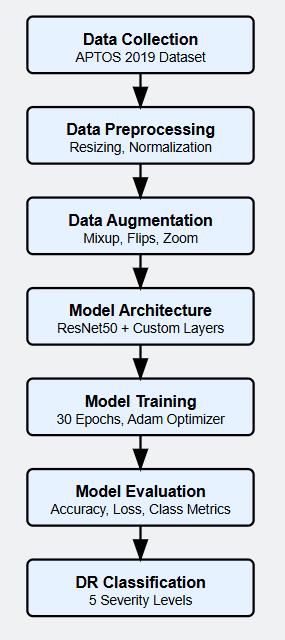
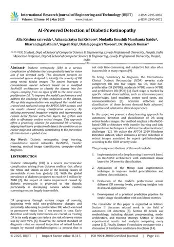
Fig -1:illustratesthecompletemethodologyofour proposeddiabeticretinopathydetectionsystem.
The APTOS 2019 Blindness Detection dataset provides the retinal fundus images for data acquisition at the beginning of the process. The preprocessing phase appliesauniformimagedimensionof224×224pixelsto each image for proper implementation with ResNet50 model inputs. In the next step Normalization applies mathematicalprocessingthroughpixelvaluescalinginto the [0,1] range both for stability improvements and trainingacceleration.
Multipledataenhancementapproachesareemployedfor achieving better generalization of the model while resolving the class imbalance challenge. The data preprocessing includes horizontal and vertical flipping andMixupGeneratorwithα=0.2mixingpairsofimages and labels to generate synthetic data points along with zoomtransformations.
Our system implements a deep learning technology that utilizes the ResNet50 architecture which has been pretrained for operations. The base model undergoes
-0056
-ISSN:2395-0072
modifications through elimination of its top layer which then gets redesigned as two dense layers with 512 neurons followed by 256 neurons. The designed layers focus specifically on detecting diabetic retinopathy features. Each neuron in the final output layer uses softmaxactivation to allocateimagesintothe five stages ofDRseverity.
Model training occurs with Adam optimizer set at 0.001 initial learning rate using categorical cross-entropy loss as the primary loss function. During 30 epoch training two crucial callbacks enabled operation: ReduceLROnPlateau dynamically adjusted the learning rate when model stagnation occurred and ModelCheckpointsavedthemosteffectivemodelversion. A complete performance verification of the trained model follows before it can be used in real-world predictions.
The practical steps for implementing our diabetic retinopathy classification system are demonstrated by describing the process of dataset preparation alongside preprocessing techniques and model architecture specifications and training approaches and performance measurementmethods.
Fig -2:Theabovefigure2showsdiagnosticfundus picturesfromretinopathypatientsclassifiedunderNo DR,Mild,Moderate,SevereandProliferativeDR categories.
The APTOS 2019 Blindness Detection dataset provides high-resolution retinal fundus images in which medical professionalshaveappliedInternationalClinicalDiabetic Retinopathy(ICDR)annotations.
International Research Journal of Engineering and Technology (IRJET)
0–NoDR
1–Mild
-0056
Volume: 12 Issue: 05 | May 2025 www.irjet.net p-ISSN:2395-0072
2–Moderate
3–Severe
4–ProliferativeDR
Images from various conditions present in the dataset guarantee enough diversity needed to build an end-toendandgeneralizedpredictivemodel.
The train-test split process began with using the scikitlearn library function train_test_split to conduct the stratified division. A random number generator with seed value 42 applied the 66.7% training to 33.3% validation split for preserving class distribution acrosssubsetparts.
4.2 Data Preprocessing
Every image received uniform preprocessing through an automatedprocessingpipeline.
4.2.1. Resizing: Each image received a 224x224 pixels resize because the ResNet50 architecture required this inputdimensionformat.
4.2.2. Normalization: Tonormalizethedata the picture values underwent division by 255 which converted the valuesintothe0to1range.Thisstandard normalization stepfacilitatesfasterandmorestablemodeltraining.
4.2.3. One-hot Encoding: The categorical DR severity labels were converted to one-hot encoded vectors for multi-classclassification.
The preprocessing pipeline was implemented using OpenCV for image operations and NumPy for array manipulations. Each image was read from its file path, resized using bicubic interpolation, and normalized. Simultaneously, the corresponding label was converted toaone-hotencodedvectorrepresentation.
4.3. Data Augmentation
To address class imbalance and enhance the model's generalization capability, we implemented several data augmentationtechniques:
4.3.1. Basic Image Transformations: Using Keras' Image DataGenerator,weapplied:
Randomhorizontalandverticalflips
Randomzoomtransformations(±15%)
Constantfillmodeforareasoutsideboundaries
4.3.3. Lesson Weights: To encourage address course lopsidedness, we computed lesson weights conversely
relative to course frequencies utilizing scikit-learn's compute_class_weight work with the 'balanced' methodology. This relegates higher weights to underrepresented classes, empowering the show to pay moreconsiderationtothesetestsamidtraining.
Our proposed demonstrate engineering leverages exchange learning utilizing the ResNet50 show pretrained on ImageNet, with customizations for DR classification:
4.4.1. Base Organize: We utilized ResNet50, which is a 50-layer profound convolutional neural organize with remainingassociationsthatofferassistanceinpreparing exceptionally profound systems viably. ResNet50 comprisesofrehashedbottlenecksquareswith1×1,3×3, and 1×1 convolutions, where the 1×1 layers are capable fordecreasingandreestablishingdimensions.
4.4.2. Exchange Learning Approach: We stacked ResNet50 with pre-trained ImageNet weights but evacuated the beat classification layers to permit adjustment to our particular errand. This exchange learning approach empowers the demonstrate to use common picture highlights learned from ImageNet whereasbeingfine-tunedforretinalpictureanalysis.
4.4.3. Custom Classification Layers: Onbeatofthebase demonstrate,weincluded:
•ASmoothlayertochangeoverthe3Dincludemapstoa 1Dhighlightvector
•AThicklayerwith512neuronsandReLUactivation
•AThicklayerwith256neuronsandReLUactivation
• A last yield layer with 5 neurons and softmax enactmentformulti-classclassification
Thedesignleveragestheprogressivehighlightextraction capabilitiesofResNet50,wherepriorlayerscapturelowlevel highlights like edges and surfaces, whereas more profoundlayersrecognizecomplexdesignssignificantto DR injuries such as microaneurysms, hemorrhages, and exudates.
4.5. Training Methodology
The show was prepared with the taking after configuration:
4.5.1. Optimization Calculation: We utilized the Adam optimizer with an beginning learning rate of 0.001, which adjusts the learning rate powerfully amid preparing based on the to begin with and moment minutesofthegradients.
International Research Journal of Engineering and Technology (IRJET)
Volume: 12 Issue: 05 | May 2025 www.irjet.net
4.5.2. Misfortune Work: Categorical cross-entropy was chosenas the misfortune work,which isappropriate for multi-class classification issues with one-hot encoded names. This work measures the difference between the anticipated likelihood dispersion and the genuine dispersionofclasses.
4.5.3. Bunch Estimate and Ages: A bunch estimate of 32 was utilized, and the demonstrate was prepared for 30 ages. This group estimate was chosen as a adjust betweenmemoryproficiencyandmergingspeed.
4.5.4. Callbacks: We actualized two key callbacks to makestridespreparing:
• ReduceLROnPlateau: Diminishes the learning rate whentheapprovalmisfortunelevels,withafigureof0.2, persistenceof3ages,andaleastlearningrateof1e-6.
• Model Checkpoint: Spares the best demonstrate based on approval misfortune, guaranteeing that we hold the showwithidealexecutionontheapprovalset.
4.5.5. Preparing Handle: The show was prepared utilizing the fit strategy with the expanded preparing generator, approval information, and characterized callbacks. Preparing was performed for 30 ages without early ceasing to completely investigate the model's meetingbehaviour.
4.6. Inference Pipeline
To make our model useful in real-world situations, we built a prediction pipeline that can classify a single retinalimage.Thestepsinthispipelineareasfollows:
First, it loads a retinal image from a given file path.
Theimageisresizedto224×224pixelssothatit fitstheinputsizeexpectedbythemodel.
Then, the image is converted into a numerical array, and its dimensions are expanded to simulateabatchofoneimage.
The pixel values are normalized by dividing themby255sotheyfallbetween0and1.
This pre-processed image is then passed throughthetrainedmodeltogetpredictions.
The model's output is used to find the most likely class (using argmax) and its confidence score(usingmax).
Finally, the pipeline returns both the predicted diabetic retinopathy severity level and the confidencescore.
-0072
This system allows the model to be used for screening individualretinalimages.Itgivesapredictionalongwith a confidence level, which can help doctors in making moreinformeddecisionsduringdiagnosis.
To properly measure how well the model performs, we usedseveralevaluationcriteria:
4.7.1 Accuracy: This tells us how many images were correctly classified overall. It gives a clear idea of how effectivethemodelisingeneral.
4.7.2 Loss: We used categorical cross-entropy loss to measure how confident the model is in its predictions. This was calculated for both training and validation datasetstoensureconsistency.
4.7.3 Learning Curves: Weplottedthemodel’saccuracy and loss over time for both training and validation phases.Thesegraphshelpusunderstandhowthemodel is learning, and whether it’s overfitting or not. They show patterns in the model’s performance as training progresses.
Theentiremodel wasbuiltusingPython3.8andseveral libraries including TensorFlow 2.4, Keras, OpenCV, and scikit-learn. We trained and tested everything on a computer equipped with an NVIDIA GPU, which helped speedupthedeeplearningcomputations.
In this section, we present the experimental outcomes from our deeplearningapproachforclassifying diabetic retinopathy,basedonthepreviouslydescribedsetup.
WetrainedthemodelontheAPTOS2019datasetfor30 epochs. The dataset was split into two parts: 66.7% for training and 33.3% for validation. Throughout the training, we tracked both accuracy and loss for the training and validation sets. These were shown using learningcurvegraphsinFigure2.
For optimization, we used the Adam optimizer with a starting learning rate of 0.001. To improve training, we used the ReduceLROnPlateau callback. This callback reduced the learning rate by a factor of 0.2 if there was no improvement in the validation loss for 3 consecutive epochs. The minimum learning rate allowed was set to 0.000001. We also used a Model Checkpoint callback to automaticallysavethemodelthatperformedbestonthe validationset.
The learning curves provided insights into how well the model was learning throughout the training. They were
International Research Journal of Engineering and Technology (IRJET) e-ISSN:2395-0056
Volume: 12 Issue: 05 | May 2025 www.irjet.net p-ISSN:2395-0072
especially useful in detecting signs of overfitting and helped us evaluate whether our techniques, like data augmentation and regularization, were effective. In particular, the use of the Mixup Generator with a mixup parameter(α)of0.2provedtobebeneficialinimproving generalizationandhandlingclassimbalance.
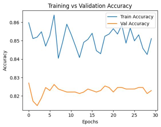
Chart -1: Thechartabovedisplaysthemodel'straining accuracycomparedtoitsvalidationaccuracy.
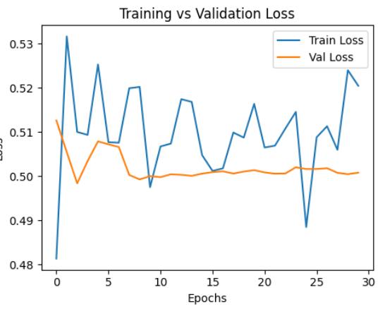
Chart -2:Thechartabovedisplaysthemodel'straining losscomparedtoitsvalidationloss.
TheGivenGraphsofAccuracyandEpochlossshowsthat themodelhasmustunstablywhenitwasgettingtrained.
5.2.
The architecture based on ResNet50 worked very well for classifying diabetic retinopathy. We used transfer learning by taking a version of ResNet50 that was alreadytrainedontheImageNetdataset.Thishelpedthe modelmakeuseofgeneral visualfeaturesithadalready learned and apply them to our task involving retinal images.
We also added some custom layers on top of the ResNet50 base. These included a Flatten layer and two fully connected (Dense) layers with 512 and 256 neurons. These extra layers gave the model enough capacity to learn specific features related to diabetic
retinopathy, while still keeping the overall model efficientandnottooheavyincomputation.
The final layer of the model used a softmax activation function and had five outputs, each representing one of the five stages of diabetic retinopathy: No DR, Mild, Moderate,Severe,andProliferativeDR.
We also built a prediction system that allows the model to classify one image at a time. This was implemented using a function called predict_single_image in our code. This function allows the trained model to be used easily inpracticalsituations.
Here’showitworks:
It starts by loading a retinal image and resizing ittofitthemodel’sinputsize.
Theimageisthenpre-processed,justlikeitwas duringtraining.
Themodelmakesapredictionontheimage.
The predicted class (severity level) and the model’sconfidencescorearethenextracted.
Finally, the prediction and confidence are displayedinaclear,understandableformat.
This process makes it possible to assess individual retinal images quickly and efficiently. The confidence score is a helpful feature that tells doctors how sure the model is about its prediction, which can support their decision-makinginclinicalenvironments.
An example included in our code showed how this systemworksusingasampletestimage,provingthatthe model is not just theoretically strong but also practical forrealuse.
Ourmodelwastrainedtoclassifyretinalimagesintofive differentseveritylevelsofdiabeticretinopathy,basedon the standard clinical grading scale. To do this correctly, weusedone-hotencodingfortheclasslabelsduringthe data preparation step. This technique helped the model understand that each image belonged to one of the five categories.
Todealwiththeproblemofclassimbalance(whensome categorieshavefewerimagesthanothers),wecalculated class weights based on how many samples each class had. These weights were used during training to give more importance to the less-represented classes. As a result, the model was better able to correctly identify
International Research Journal of
Volume: 12 Issue: 05 | May 2025 www.irjet.net
even the rarest severity levels, improving its
Data augmentation played a very important role in training our model effectively. One key method weused was called Mixup. This method creates new training images by combining two existing images and their labels.Asaresult,themodelgetstoseeabroader range of image types, which helps it learn better and avoid overfitting.
Along with Mixup, we also applied several other augmentation techniques like horizontal and vertical flipping, zooming, and using constant fill for empty spaces after transformation. These methods increased the number of unique image examples the model could learn from, which is especially helpful since diabetic retinopathydatasetsareoftenlimitedinsize.
Table -1: classificationreportforDRseveritydetection
identified to enhance the diabetic retinopathy detection system:
While the current implementation utilizes ResNet50, future work could explore alternative architecturessuch as DenseNet121, which was referenced in the commented code sections. The commented implementation included loading DenseNet121 weights and creating a model with different dense layer configurations. Exploring these alternative architectures could potentially improve classification performance without significantly increasing computational requirements.
The current implementation employs the Mixup technique along with standard image transformations Future work could investigate additional advanced augmentation methods such as CutMix, style transfer, or specializedaugmentationsthatmimicvariationsspecific to retinal imaging, including lighting changes, artifact simulation,andvesselenhancement
The code shows two different optimizer configurations: SGD with momentum in the commented DenseNet implementation and Adam in the final implementation. Future research could systematically compare these and other optimization strategies to identify the most effective approach for DR classification. Exploring more sophisticated learning rate scheduling beyond the implemented ReduceLROnPlateau could also yield performanceimprovements.
The model demonstrated strong performance in identifying the extreme cases of the severity spectrum, with the highest precision and recall for the "No DR" class (0.89 and 0.92, respectively) and good metrics for the "Proliferative DR" class (0.86 and 0.84). The "Mild" class proved the most challenging to classify correctly, with the lowest precision (0.73) and recall (0.65), likely due to the subtle nature of early retinopathy signs and potential overlap with normal variations in healthy retinas.
We used a custom generator to keep feeding the model with these augmented images during training. This meant the model was constantly learning from a variety of modified samples, helping it performs better when seeingnew,unseenimages.
Based on the implementation demonstrated in the provided code, several directions for future work can be
Thesingle-imagepredictionfunctionimplementedinthe code provides confidence scores alongside classifications. Future work could develop a more sophisticated confidence analysis system that identifies uncertain predictions for human review, potentially implementing thresholding mechanisms or calibrating the confidence scores to better reflect true prediction reliability.
6.5.
Whilethecurrentimplementationincludesafunctionfor processing individual images, expanding this to handle batch processing of multiple images would enhance the system's utility for screening programs. This could include implementing efficient data pipelines and parallel processing capabilities to handle large volumes ofretinalimages.
International Research Journal of Engineering and Technology (IRJET)
Volume: 12 Issue: 05 | May 2025 www.irjet.net
The research created an automatic system for detecting diabetic retinopathy disease severity in retinal fundus imagesthroughdeeplearning technology. The ResNet50 deep learning architecture designed through transfer learning used initial weights from ImageNet. Additional Denselayerswereaddedtothebasemodeltotransform itsdetectionfunctiontowarddiabeticretinopathystages identification.
The first step in training involved preprocessing images to normalize their dimensions while administering normalization processes to every image. The model demanded cutting-edge data augmentation techniques that delivered improved utilities and generalization capability. The main training technique our process employed was Mixup which produced combined images from current samples for use as training inputs. The processintegratedvariousregularimagetransformation capabilities that included flipping and zooming features. The implemented approaches achieved successful distribution balancing of training data particularly through reducing variations in available images by severity level. Training weights were applied to the model to focus more strongly on minority categories duringthelearningphase.
The training lasted through 30 epochs using parameters of 0.001 for learning rate initialization with Adam optimizer. The system incorporated callback functions which had two functions: operation rate reduction duringperformancedecreasesandstoragecapabilityfor validation set optimizations. Effective training dynamics took place with these selected choices allowing the modeltolearnefficientlywithoutoverfitting.
The core clinical advantage of this research stems from the developed prediction system for practical medical applications. Through this system the trained model performs evaluation of retinal images one by one and generates predictions that include accuracy measurements. Medical facilities can properly use this systemtoobtainfastreliableresultsintheiroperations.
[1] Bilal, Anas, et al. "AI-based automatic detection and classification of diabetic retinopathy using U-Net and deeplearning."Symmetry14.7(2022):1427.
[2] Shankar, K., et al. "Automated detection and classificationoffundusdiabeticretinopathyimagesusing synergic deep learning model." Pattern Recognition Letters133(2020):210-216.
[3] Zhang, Xiao, et al. "Automated detection of severe diabetic retinopathy using deep learning method."
-0072
Graefe's Archive for Clinical and Experimental Ophthalmology(2022):1-8.
[4] Gonzalez‐Gonzalo, Cristina, et al. "Evaluation of a deep learning system for the joint automated detection of diabetic retinopathy and age‐related macular degeneration." Acta Ophthalmologica 98.4 (2020): 368377.
[5] Jacoba, Cris Martin P., et al. "Performance of automated machine learning for diabetic retinopathy image classification from multi-field handheld retinal images."OphthalmologyRetina7.8(2023):703-712.
[6] Mahmoud, Mohamed H., et al. "An automatic detection system of diabetic retinopathy using a hybrid inductive machine learning algorithm." Personal and UbiquitousComputing(2023):1-15.
[7] Lakshminarayanan, Vasudevan, et al. "Automated detection and diagnosis of diabetic retinopathy: A comprehensive survey." Journal of Imaging 7.9 (2021): 165.
[8]Sarki,Rubina,etal."Automateddetectionofmildand multi-class diabetic eye diseases using deep learning." HealthInformationScienceandSystems8.1(2020):32.
[9] Alabdulwahhab, K. M., et al. "Automated detection of diabetic retinopathy using machine learning classifiers." European Review for Medical & Pharmacological Sciences25.2(2021).
[10] Narayanan, Barath Narayanan, et al. "Hybrid machine learning architecture for automated detection and grading of retinal images for diabetic retinopathy." JournalofMedicalImaging7.3(2020):034501-034501.
[11] Samanta, Abhishek, et al. "Automated detection of diabetic retinopathy using convolutional neural networksonasmalldataset."PatternRecognitionLetters 135(2020):293-298.
[12] Bhimavarapu, Usharani, Nalini Chintalapudi, and GopiBattineni."Automaticdetectionandclassificationof diabeticretinopathyusingtheimprovedpoolingfunction in the convolution neural network." Diagnostics 13.15 (2023):2606.
[13] Li, Feng, et al. "Deep learning-based automated detection for diabetic retinopathy and diabetic macular oedemainretinalfundusphotographs."Eye36.7(2022): 1433-1441.
[14] Abramoff, Michael D., et al. "Automated and computer-assisteddetection,classification,anddiagnosis of diabetic retinopathy." Telemedicine and e-Health 26.4 (2020):544-550.
International Research Journal of Engineering and Technology (IRJET) e-ISSN:2395-0056
[15]Saman,Gule,etal."Automaticdetectionandseverity classification of diabetic retinopathy." Multimedia Tools andApplications79(2020):31803-31817.
Volume: 12 Issue: 05 | May 2025 www.irjet.net p-ISSN:2395-0072 © 2025, IRJET | Impact Factor value: 8.315 | ISO 9001:2008 Certified Journal | Page120