Imaging Research Highlights
Imaging Research Highlights
Watch drug delivery in real time Paola Decuzzi, Ph.D. is engineering discoidal polymeric nanoconstructs (DPNs) that can be visualized by MRI and PET as they move through the body to target a disease site (like a tumor, arthritic knee, atherosclerotic plaque, damaged heart
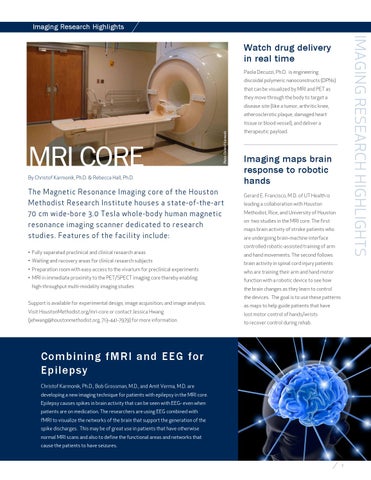
MRI core
Photo: Christof Karmonik
tissue or blood vessel), and deliver a
By Christof Karmonik, Ph.D. & Rebecca Hall, Ph.D.
The Magnetic Resonance Imaging core of the Houston Methodist Research Institute houses a state-of-the-art 70 cm wide-bore 3.0 Tesla whole-body human magnetic resonance imaging scanner dedicated to research studies. Features of the facility include: • Fully separated preclinical and clinical research areas • Waiting and recovery areas for clinical research subjects • Preparation room with easy access to the vivarium for preclinical experiments • MRI in immediate proximity to the PET/SPECT imaging core thereby enabling high-throughput multi-modality imaging studies Support is available for experimental design, image acquisition, and image analysis. Visit HoustonMethodist.org/mri-core or contact Jessica Hwang (jehwang@houstonmethodist.org, 713-441-7979) for more information.
therapeutic payload.
Imaging maps brain response to robotic hands Gerard E. Francisco, M.D. of UT Health is leading a collaboration with Houston Methodist, Rice, and University of Houston on two studies in the MRI core. The first maps brain activity of stroke patients who are undergoing brain-machine interface controlled robotic-assisted training of arm and hand movements. The second follows brain activity in spinal cord injury patients who are training their arm and hand motor function with a robotic device to see how the brain changes as they learn to control the devices. The goal is to use these patterns as maps to help guide patients that have lost motor control of hands/wrists to recover control during rehab.
Combining fMRI and EEG for Epilepsy Christof Karmonik, Ph.D., Bob Grossman, M.D., and Amit Verma, M.D. are developing a new imaging technique for patients with epilepsy in the MRI core. Epilepsy causes spikes in brain activity that can be seen with EEG- even when patients are on medication. The researchers are using EEG combined with fMRI to visualize the networks of the brain that support the generation of the spike discharges. This may be of great use in patients that have otherwise normal MRI scans and also to define the functional areas and networks that cause the patients to have seizures.
7