Houston Methodist Academic Institute


Houston Methodist Academic Institute

It is with great pleasure to share our 2025 Annual Report highlighting notable achievements of the Houston Methodist Academic Institute.
Philanthropist John F. Bookout, Jr. presented a vision—“Houston Methodist will be for the southern United States what is now found on the two coasts and in the Midwest—a leading academic medical center.”
Twenty years later, this vision is realized. The Houston Methodist Academic Institute continues its trajectory of success that followed investing in the visionary ideas of our Centers of Excellence. They delivered by increasing grant funding from $10M annually to more than $100M annually over 10 years.
This success momentum is even stronger. We accepted the challenge at the confluence of uncertainty and probability in science, which is part of our dedicated mission through a global collaborative network that transitions proof-of-concept medical research into clinical impact and educates healthcare professionals on the latest medical breakthroughs in the service of health.
We know Houston Methodist excels in clinical quality and safety outcomes today, but we exist in a constantly changing landscape that requires innovation with science-based programs. One example is the Sepsis Early Recognition and Response Initiative (SERRI), which improves the early identification and treatment of sepsis, a life-threatening condition caused by infection. SERRI put Houston Methodist on the map as the academic medical center that helped 15 health systems save $50 million in medical costs and 2,500 lives.
Houston Methodist is also home to the first coronary artery bypass graft and the first multi-organ transplant. Its innovative education programs, such as PhD programs with Weill Cornell Graduate School of Medical Sciences in Houston and the Texas A&M University School of Engineering Medicine, and extended reality simulation education technology being developed at the Bookout Center.
As we take a moment to celebrate the last 20 years of innovation with this Annual Report, we are simultaneously leaping into the next 20. In 2024, we launched a Quality and Patient Safety Innovation Institute and a Digital Health Institute. We are also investing in AI-powered imaging and bioengineered protein therapeutics to deliver high-quality, cost-efficient care that improves health.
It is an honor to be on this journey for what lies ahead for Houston Methodist’s future of medicine and human health.
Jenny C. Chang, MB.ChirB., MD, MCHM
Ernest Cockrell, Jr. Presidential Distinguished
Chair
Executive Vice President and Chief Academic
Officer
Houston Methodist President and CEO
Houston Methodist Academic Institute

From its conception, the Houston Methodist Academic Institute intended to be different than other academic medical centers. Its driving objective is to develop treatments that can be readily adopted into patient care—using its physical connections to the hospital to source ideas for innovations. Our expertise in FDA approval pathways and collaboration with clinical care teams, positions us to reality test inventions and streamline translating laboratory research into clinical impact.
Houston Methodist met the challenge of translation early in the design of the Research Institute building, housing essential services and technology that support the full cycle of development to efficiently and effectively deliver innovations to the clinic.
The first iteration of the Cycle of Translation was introduced more than 10 years ago to represent our vision of translating laboratory innovations into effective and efficient tangible solutions that benefit patient care and outcomes. In 2024, we invited HMAI faculty to provide feedback on the effectiveness of the cycle in conveying our vision. The overall response was that 95% favored broadening the cycle to capture everything we do today—advancing innovations that improve human health and clinical care into practice.
• Insight from patient care and outcomes defines clinical needs.
• Medically inspired team science performs exploratory research
• Their inventions have real-world value and protectable intellectual property.
• Inventions are further developed through applied research
• These inventions progress through an FDA regulated product development and preclinical validation process, which includes design, production and controlled GLP studies.
• If shown to be effective in clinical trials and studies, inventions can be implemented through piloting, education and implementation, evaluated for quality and safety, and scaled for rollout to patient care
The process is catalyzed by a translational infrastructure that includes intellectual property and regulatory expertise, external partnerships, and funding mechanisms designed to transform patient care.

The Cycle begins and ends with the care of patients in our hospitals and clinics. Clinicians in our hospitals care for more than 2.2 million patients annually, enabling them to identify the most pressing challenges in medicine and provide excellent care.
Clinicians and researchers form interdisciplinary teams to address their needs and clinical challenges.
Our teams of clinicians, researchers and collaborators from around the world have access to a full suite of technology to enable the discovery process.
Inventions that solve a problem have real world value and protectable intellectual property.
Applied research involves testing, evaluating and refining new materials, devices and systems or methods into a final lead experimental product or process with the intent to move into the product development stage.
Product development involves the design and piloting of a test product or process within a cGMP or other controlled environment.
Research teams have access to dedicated Translational Research Initiative bridge funds to take the most promising discoveries into preclinical and early phase clinical development.
Teams have access to early phase trial support, pharmacokinetics and pharmacodynamics support and outpatient clinical care and study management services, including research, nursing, regulatory submissions and budget management support for all phases of clinical trials.
Interprofessional education programs employ the latest advances in simulation and implementation science to facilitate the safe and efficient adoption of new technologies as they are approved for clinical use.
As part of the systemwide goals for quality improvement and patient safety, we develop our education programs to incorporate high reliability organization principles and use team-based training approaches to maximize patient outcomes.
Ushering medical breakthroughs from the lab to the clinic takes many years and millions of dollars. The most promising discoveries are lost in the phase of translation known as the “Valley of Death.”
To help bridge the Valley of Death and to provide our most promising innovations a lifeline, Houston Methodist is drawing on the transformative power of philanthropy.

The John M. O’Quinn Foundation made a $10-million philanthropic commitment to support research, education and patient services across a full spectrum of neurodegenerative disorders. The gift established the John M. O’Quinn Foundation Neurodegenerative Disorders Laboratory at Houston Methodist where physicians and research scientists in the Stanley H. Appel Department of Neurology conduct innovative research and clinical trials to halt the progressive loss of axon integrity, which underlies peripheral nerve disorders, neurological disorders such as Alzheimer’s disease, peripheral neuropathies, Parkinson’s disease, multiple sclerosis, stroke and amyotrophic lateral sclerosis. This gift also included the creation of two endowed chairs and a fellowship for neurodegenerative disorders.

Jun Li, MD, PhD, FANA, FAAN, leads the newly established laboratory where his team of physicianscientists are exploring the best possible combinations of preventive interventions and targeted treatments in neurodegenerative diseases. His research in peripheral nerve diseases and myelin biology has been funded by the National Institutes of Health for nearly two decades. Li’s vision is to provide unparalleled care, clinical trial opportunities and subspecialty programs in Greater Houston to support those battling neurodegenerative disorders is strengthened by this gift. “This generous gift fuels our determination to do even more and to help find therapies for these neurological diseases and supports an interdisciplinary team of neurologists and neuroscientists to further explore treatment options.”, said Li, Chair, Stanley H. Appel Department of Neurology at the Houston Neurological Institute.
Kenneth Podell, PhD, FACPN, Director of the Concussion Center and Neuropsychology Section and Associate Professor of Clinical Neurology, co-leads the first-ever study, “Gut Microbiome
Markers of Sport-related Brain Concussion”, with fellow colleague Sonia Villapol, PhD, Assistant Professor of Neurosurgery, to explore the gut-brain connection in contact-sports student athletes and identify biomarkers for concussions with the athlete’s gut microbiomes.


Atransformational gift from longtime supporters Ann and John F. Bookout III established the Bookout Center in the Houston Methodist Academic Institute.
Houston Methodist’s modern interdisciplinary GME and educational programs have access to advanced resources. The Bookout Center, which launched in 2024, is an advanced surgical training and emerging technology facility for the research, development and integration of robotic, imaging and digital surgery platforms toward more precise, efficient and personalized surgical interventions. The 35,000-square-foot research and training space is dedicated to developing

novel technologies to enable less invasive medical care, demonstrate cutting-edge techniques via live audio and video conferencing within all Houston Methodist procedure areas, and influencing local, regional and national health care professionals and providers.
The Bookout Center is partnering with Rice University scientists as part of its mission to bring surgical innovation around the world. More than 25 faculty members from Houston Methodist and Rice’s Education and Research Initiatives for Collaborative Health are exploring potential Bookout Center partnerships.
We’re excited about the possibilities this collaboration brings. By working together, we can accelerate discoveries that will improve patient experience and outcomes. “ ”
– Stuart J. Corr PhD Associate Research Professor, Bioengineering in Cardiovascular Surgery Director, Innovation Engineering, Bookout Center
Houston Methodist
Three elite Houston Methodist researchers, funded through the Jerold B. Katz Foundation, have been named Jerold B. Katz Investigators.


Jerold B. Katz Investigator
Professor, Pathology and Genomic Medicine
As a leader in synthetic biology and protein engineering, Gollihar’s work focuses on designing and optimizing therapeutic proteins, vaccines and antibodies to tackle urgent public health challenges. His research integrates protein engineering, structural biology and machine learning to address complex issues in immunogen design, protein stability and manufacturability.
Jerold B. Katz Investigator
Associate Professor, Pathology and Genomic Medicine
An expert in neurodegenerative diseases, Cykowski is working to better understand how the distribution and pathologic burden of misfolded proteins in postmortem brain tissue samples from people who had neurodegenerative diseases, including dementias and amyotrophic lateral sclerosis. His team uses data to determine which pathologic markers best correlate with patient symptoms and long-term outcomes with the goal of helping to identify biomarkers in living patients.

Jerold B. Katz Investigator
Associate Professor, Medicine
Kindi, who serves as a preventive and imaging cardiologist at the DeBakey Heart and Vascular Center, is integrating multi-dimensional data—including health records, imaging and environmental factors—to predict cardiovascular disease and related outcomes. He aims to create robust prediction models for diseases such as diabetes, cardiovascular events and dementia by leveraging advanced AI techniques and imaging.

The Houston Methodist Academic Institute recognizes the distinctive and critical role that physician-scientists play in advancing its core missions as an academic medical center. The Clinical Scholars program meets that need by helping to grow our core of rising physician-scientists.


Akshjot Puri, MD
Assistant Professor, Medicine Department of Medicine

Namrata Vasquez, PhD
Assistant Professor, Psychology in Clinical Psychiatry and Behavioral Health
Department of Clinical Psychiatry and Behavioral Health
Paul M. Schoder, PhD, MD
Assistant Professor, Surgery Department of Surgery
The Houston Methodist Endowed Positions and Awards Committee announces the new endowed chairs.


Arica A. Brandford, PhD, JD
Dorothy and Mickey Ables Community Outreach and Engagement Distinguished New Century Chair
Assistant Research Professor, Community Engagement in Medicine Department of Medicine
Sunil Dacha, MD, MBBS
Nina and Michael Zilkha Centennial Chair in Gastrointestinal Health
Associate Professor, Clinical Medicine Department of Medicine
Cindy M. Martin, MD
Lois E. and Carl A. Davis Chair in Heart Failure Professor, Cardiology Department of Cardiology
Kumar Pichumani, PhD
Dagmar Dunn Pickens Gipe Distinguished Professorship in Brain Tumor Research
Associate Research Professor, Neurosurgery Department of Neurosurgery

Houston Methodist brings new scientific discoveries to patients as rapidly as possible. Interdisciplinary research teams identify promising treatments and tools and make diagnostic discoveries through the Cycle of Translation. These findings are accelerated through the development process, positioning them to make the leap from idea to clinic.
The focus is to achieve both market adoption and compliance with these discoveries to provide patients quicker access to safe and effective therapies. Research aims is to help patients maintain optimal physical and cognitive health by developing new treatment technologies and health tools.

As globalization, urbanization and climate change continue, experts agree that future outbreaks of dangerous novel viruses (Disease X) are inevitable. In November 2022, the Coalition for Epidemic Preparedness Innovations (CEPI), published a landmark report, “What Will It Take,” which outlined the paradigm shift needed to speed up vaccine development, and the crucial scientific and technological innovations— including the creation of a vaccine library—that will enable the world to develop new vaccines against future pandemic threats in just 100 days.
Last summer, CEPI and Houston Methodist announced a partnership—and funding of a consortium led by Houston Methodist—to combine cutting-edge artificial intelligence (AI) technology with established laboratory techniques to further the rapid development of future vaccines against Disease X. The Houston Methodist group was awarded $4.98 million to advance the application of AI to analyze the structures of priority viruses from which the next Disease X is likely to emerge. Earlier this year, the award was increased to $34 million over five years.

Led by Jimmy D. Gollihar, PhD, Professor of Pathology and Genomic Medicine and Head of the Antibody Discovery & Accelerated Protein Therapeutics Laboratory, the Houston Methodist team joins experts from Argonne National Laboratory (University of Chicago), J. Craig Venter Institute, La Jolla Institute, The University of Texas Medical Branch and The University of Texas at Austin. Initially, the team will focus efforts on paramyxoviruses and arenaviruses, as well as viral families, which include Nipah virus and Lassa virus, respectively.
“We are delighted to have the Houston Methodist Academic Institute be a part of this program, serving our community and the world. Leading this consortium is an amazing undertaking and a testament to the work that Dr. Jimmy Gollihar, his team in pathology and genomic medicine, and many others in our academic institute are doing to help defeat the next pandemic,” said Dirk Sostman, MD, FACR, Emeritus Professor of Radiology and Distinguished Member of Houston Methodist Academic and Research Institute.
A critical enabler of the 100 Days Mission is the establishment of a global “vaccine library”—an accessible store of scientific knowledge, data and prototype rapid-response vaccine candidates against selected viruses from the 25 priority virus families.
CEPI’s aim is to store AI-generated, lab-tested and verified antigen designs, developed by the Houston Methodist consortium, in the vaccine library to be quickly used to develop vaccine candidates in the event of an outbreak of a novel pathogenic threat. In this scenario, after sequencing the offending virus, these cataloged antigen designs can be inserted into an appropriate rapid-response vaccine platform to start the production of vaccines for clinical testing.
Gollihar’s group is leading immunogen design, but their work encompasses much more. The team has been developing therapeutic monoclonal antibodies for SARS-CoV-2. As variants of concern emerged, they wanted to determine what those mutations were doing to their monoclonals. So, they developed a mammalian display of viral glycoproteins—called a spike display— to allow researchers to study virus mutations and source code in real time.
“We started using spike display to dissect escape mechanisms and realized it was also an engineering tool. We played around with rational design-based approaches with individual variants and then moved to library approaches where we could use millions of variants in human cells and mammalian cell lines and sort, seek and find out what's binding what. The confluence of AI and directed evolution is outright, but protein engineering is really what allows us to do this rapidly,” said Gollihar.
AI experts from Houston Methodist, The University of Texas at Austin, La Jolla Institute and Argonne National Laboratory will use machine-learning approaches to optimize the design of potential epitopes. The University of Texas Medical Branch will validate their immunogenicity in established preclinical models.
When a new pathogen emerges, vaccine developers could quickly respond by selecting AI-identified epitopes that would have already been validated in preclinical tests, enabling vaccine candidates to be moved quickly into clinical testing. This would provide a significant strategic advantage when battling the next pandemic threat, but Gollihar is looking even further into the future.
“The next generation of models for protein engineering will require data sets that don’t yet exist, so we’re really interested in deep mutational scanning,” noted Gollihar.
“Because of our engineering platform, we can now take every amino acid and put it into every single position of a protein and ask: what does that mean to expression, antibody binding or host receptor binding? We’ll learn things we couldn’t otherwise learn. This is the next frontier and we're leading it.”
Health care systems have historically focused on diagnosing and treating people who have developed troubling symptoms and Houston Methodist excels at this. The DeBakey Heart and Vascular Center is the highest nationally ranked hospital in Texas and the Gulf Coast for heart care offering advanced cardiovascular surgery and restorative therapies for unavoidable events. However, Houston Methodist is heavily invested in the next health frontier—lifelong wellness and cardiovascular health—and is leveraging advanced research and experience with caring for patients of all ages to lengthen the time that people spend in good health. The DeBakey faculty recently published a series of papers that advanced our understanding to maintain, protect and enhance cardiovascular health.

While diet plans come and go, low-carbohydrate, high-fat, ketogenic “keto” diets have retained their staying power for more than 20 years. Does following a diet centered on consuming foods high in fats such as meats, eggs, and oils raise heart disease risk?
Cholesterol levels are an index of heart disease risk, as excess cholesterol can contribute to plaque buildup in coronary arteries. A recent meta-analysis of 41 human trials investigating the physiological effects of a keto diet revealed surprising information about the diet’s impact on ‘bad’ low-density lipoprotein (LDL) cholesterol levels. Overweight people following the keto diet for weight loss had no change in their LDL over time. However, this diet is also popular among lean people seeking a high-protein diet to build muscle. When following a keto diet, these individuals LDL cholesterol levels often rise to an unhealthy level, giving rise to the novel “lean mass hyper-responder” (LMHR) phenotype.

Khurram Nasir, MD, William A. Zoghbi, MD
Centennial Chair in Cardiovascular Health and Professor of Cardiology, and his team sought to determine whether the increased cholesterol observed in LMHR keto dieters is accumulating as plaque in their coronary arteries, raising their chances of suffering a heart attack or stroke.
By analyzing coronary computed tomography angiography and coronary artery calcium scan data from large-scale keto dietfocused clinical studies, Nasir’s team evaluated coronary artery plaque levels within LMHR keto dieters compared with matched controls not on a keto diet for an average of 4.7 years.
The team found that for those in the LMHR group, several years of carbohydrate restriction-induced elevations in LDL cholesterol did not increase their coronary plaque burden, relative to controls as detailed in JACC: Advances
Nasir noted, “LMHR is an emerging phenotype of growing research interest with little known about mechanisms and risks. Clinicians’ awareness of the unique aspects of LMHR may facilitate more personalized patient management.”

Viruses are among the more curious denizens of the natural world. Since they aren’t composed of cells, they need an unsuspecting host to help them replicate— infecting and often causing disease in the host as they grow in numbers.
Many different viruses, including those responsible for the flu and COVID-19, can trigger viral myocarditis. This inflammatory cardiac disease can reduce the heart’s ability to pump effectively and is a leading cause of death in young adults. Once a virus infects the heart, there are no effective treatments for myocarditis since the exact physiological mechanisms driving the disease have remained elusive … until now.

Junji Xing, PhD, Assistant Research Professor of Immunology in Surgery, Department of Cardiovascular Sciences, and his team, are helping to develop effective viral myocarditis treatments that protect the heart by revealing the molecular pathways involved of this disease.
“Our curiosity was piqued by the potential role of TRIM29, a protein known for its role in various cellular
processes but not clearly understood in the context of myocarditis,” noted Xing. “We were especially interested in how TRIM29 influenced the protein kinase RNA-like endoplasmic reticulum kinase (PERK)-mediated endoplasmic reticulum (ER) stress response—a pathway impacting cellular survival and stress reactions.”
Xing’s team discovered that cardiotropic viruses highly induced TRIM29 and promoted PERK-mediated ER stress, apoptosis and reactive oxygen species responses that support viral replication in cardiomyocytes in vitro. These findings were published in Nature Communications.
TRIM29 deletion or PERK inhibition with GSK2656157 protected mice from viral myocarditis by disrupting the TRIM29-PERK connection, which bolstered cardiac function, enhancing cardiac antiviral responses and curbing inflammation and immunosuppressive monocytic myeloid-derived suppressor cells in vivo.
“We’re pleased to learn that TRIM29 deficiency and pharmacological targeting of the TRIM29-PERK axis had a significant protective effect against viral myocarditis, reducing viral replication and symptoms,” Xing said. “Our findings make PERK inhibitor a promising drug to move forward to clinical trials for treating patients with viral myocarditis.”


Health care providers have long advised their patients to adopt healthy diets, regular exercise and good sleep habits to reduce cardiometabolic risks. However, an equally important factor is the environment in which patients live. The level of air pollution in their community and the availability of safe, accessible places for physical activity can significantly impact health outcomes.
Sadeer Al-Kindi, MD, Jerold B. Katz Investigator and Associate Professor of Medicine, is pioneering research to identify which neighborhoods correlate with higher risks of heart disease, aiming to tailor medical care more personally to each patient’s living conditions.

Exposure to particulate matter air pollution of less than 2.5 microns (PM2.5) is a major determinant of cardiometabolic disease. The Global Burden of Disease assessment estimates that 20% of global Type 2 diabetes cases are related to chronic exposure to PM2.5. This exposure is also linked with insulin resistance, blood pressure elevation and childhood obesity. Al-Kindi’s detailed analysis on this topic was published in Lancet Diabetes & Endocrinology. With 99% of the global population, including some residents in the Greater Houston area, living in environments with unsafe air pollution levels; Al-Kindi is working to understand the connection between air
pollution and other environmental factors with cardiovascular disease, aiming to develop mitigation strategies.
Even in areas without excessive air pollution, the human-made structures and spaces where we live, work and play are closely linked with cardiovascular health. Previous studies often relied on broad population data or single metrics, neglecting the complex interplay of neighborhood characteristics.
Al-Kindi’s team analyzed data from 1.07 million patients in the Houston Methodist Learning Health System Outpatient Registry. Each patient was assigned a NatureScore™, which measures the quality and quantity of nature in their area, and a WalkScore, assessing neighborhood walkability. The study, published in JACC: Advances, revealed that living in highly walkable neighborhoods with ample nature exposure significantly reduced the odds of cardiovascular risk factors.
Further, Al-Kindi employed deep learning to analyze Google satellite imagery across seven U.S. cities, linking built environment features to neighborhood-level heart disease rates, as published in the European Heart Journal and JAMA Cardiology. These machine vision-derived features better predict heart disease prevalence than traditional demographic and socioeconomic data alone.
“The implications of this work are profound, offering scalable tools to assess environmental impacts on heart health, potentially guiding interventions to tackle health disparities and improve cardiovascular outcomes across U.S. communities,” said Al-Kindi.

Houston Methodist researchers have discovered that certain components of so-called “good” HDL cholesterol may be associated with an increased risk of cardiovascular disease. Led by Henry J. Pownall, PhD, Professor of Biochemistry in Medicine and Khurram Nasir, MD, the research team is investigating the role of certain properties of HDL-C in heart health.

“During routine checkups, adults have their cholesterol levels tested, which includes both “bad” LDL cholesterol and “good” HDL cholesterol,” said Pownall, who is the lead author on a paper recently published in Journal of Lipid Research. “Not all cholesterol, however, is born the same. What is not commonly recognized is that each type of cholesterol has two forms—free cholesterol, which is active and involved in cellular functions, and esterified or bound cholesterol, which is more stable and ready to be stored in the body. Too much free cholesterol, even if it is in HDL, could contribute to heart disease.”
In pre-clinical studies, the team discovered that HDL with a high content of free cholesterol is likely dysfunctional. To validate their findings, they are currently at the halfway point of the NIH-funded Houston Heart Study with 400 patients with a range of plasma HDL concentrations.
“The most surprising finding from our study, thus far, is that there is a strong link between the amount of free cholesterol in HDL and how much of it accumulates in white blood cells called macrophages, which can contribute to heart disease,” emphasized Pownall.
While it was previously thought that the transfer of free cholesterol to HDL was beneficial for heart health by removing excess cholesterol from tissues, the data shows that in the context of high plasma HDL concentrations, the reverse is true—free cholesterol transfer from HDL to the white blood cells in blood and tissues could actually raise one’s risk for cardiovascular disease.
Once they reach their immediate goal of showing that excess free cholesterol in HDL is associated with excess cardiovascular disease, the research team plans to develop new diagnostics and treatments for managing heart disease, as well as use HDL-free cholesterol as a biomarker to identify patients requiring HDL-lowering therapies.

In the spirit of collaboration, Houston Methodist and Rice University joined forces with the opening of the Center for Neural Systems Restoration (CSNR), which is aimed at discovering how the human brain processes perception, cognition and behavior to find novel solutions for brain and spinal cord diseases and injuries. The Center serves as an incubator for advancing the frontiers of translational neuroscience— understanding the function of the human brain and developing next-generation technologies, neural prosthetics and rehabilitation regimens for the treatment of neurological conditions.
People who take short naps during the day—typically associated with non-rapid eye movement (NREM) sleep—demonstrate the benefits of even short periods of sleep with improved learning, memory, and perceptual performance. This idea that sleep improves cognitive function has been around for a century, but the underlying neural mechanisms have remained elusive.

To further interrogate the underlying neural mechanisms of the cognitive benefits of sleep, Valentin Dragoi, PhD, Rosemary and Daniel J. Harrison III Presidential Distinguished Chair in Neuroprosthetics, and team used multiple-electrode recordings in areas of the cortex involved in task-related activity to examine the dynamics and information coding in neural populations before, during and after sleep, and their impact on behavioral performance.
In a study published in Science, Dragoi’s team used multi-electrode intracranial arrays positioned on the head to examine the spiking activity of 4,422 neurons recorded across three cortical areas. To define epochs of NREM sleep,

the team utilized automated sleep recognition software incorporating all components of polysomnography, including electroencephalogram, electrooculography and electromyography.
Evidence shows that brief naps can consolidate memory and improve behavioral and perceptual performance. The team also focused on studying the effects of short sleep periods (30 minutes of rest). They learned increased delta power (2-4 Hz) during slow wave sleep was accompanied by increased synchronized firing in neural population activity in each brain area recorded. Surprisingly, delta-band synchrony in population activity during sleep further caused a desynchronization of neural responses and improvement in perceptual performance during subsequent cognitive tasks.

Hypothesizing that the beneficial effects of sleep could be reproduced in their model by electrically stimulating neural populations in the delta-frequency band, they conducted blocks of stimulation for 20 to 30 minutes by generating synchronous electrical pulses in the delta frequency band on eight channels of the electrode array. Remarkably, electrical stimulation of the visual cortex during quiet wakefulness emulated the restorative effects of sleep in the absence of sleep.
This could be a mechanism by which the brain maintains stability of population activity after sleep-induced synchronized neural responses. Our results provide proof of concept for stimulation procedures to improve perceptual performance in the absence of sleep and may set the stage for future neuromodulation in humans.
”
– Valentin Dragoi, PhD
Rosemary and Daniel J. Harrison III Presidential Distinguished Chair in Neuroprosthetics
Scientific Director, Center for Neural Systems Restoration
Houston Methodist
Professor, Electrical and Computer Engineering
Rice University
This study is one of many research areas in collaboration with CSNR to nurture functional interactions between Rice engineers and researchers, and Houston Methodist neurosurgeons, neuroscientists and regeneration biologists to design and test novel prosthetics, interventions and diagnostics on patients. The Center is led by Houston Methodist’s Gavin Britz, MD, MBCCH, MPH, MBA, FAANS, Candy and Tom Knudson Centennial Chair in Neurosurgery and Rice University’s Behnaam Aazhang, PhD, Jr., Director, Electrical and Computer Engineering, Rice Neuroengineering Initiative.
Pedro T. Ramirez, MD, FACOG, is dedicated to questioning the gynecological cancer treatment status quo. As Chair of the Department of Obstetrics & Gynecology, he has spent more than 25 years treating gynecologic cancer through both open and minimally invasive surgery (MIS). While open to new surgical technologies, including MIS, Ramirez is a trailblazer in conducting randomized clinical trials that challenge the efficacy of these medical advancements to ensure the best patient outcomes. His studies have influenced international standards of care and have helped shape how these surgical technologies are evaluated during development, comparative study and clinical monitoring.

Robotic control systems and artificial intelligence (AI) programs are quickly advancing in the surgical technology landscape.
In fact, Ramirez, believes that “surgical robots could perform entire procedures” in the near future. However, surgical technology, including robots and AI, are historically under-evaluated before implementation.
For most of the early 2000s, minimally invasive surgery was considered the gold-standard approach to treating cervical cancer. Then, a landmark study in 2018 led by Ramirez suggested otherwise. The study, published in the New England Journal of Medicine, revealed that minimally invasive surgery using robotics and laparoscopy to treat early-stage cervical cancer was associated with worse health outcomes when compared to the open surgery approach. These results changed the standard of care for treating cervical cancer.
Since then, Ramirez has become more involved in ensuring surgical robots are used in the safest, most effective capacity. Accomplishing this goal requires a keen focus on the continuous evaluation of surgical technology once it becomes available on the market.


“We’re moving toward a technology where the surgeon can potentially be on autopilot or standby,” Ramirez said. “These types of advancements come with ramifications if the technology is not evaluated and used properly, so, I joined the IDEAL Collaboration to help implement a robotics colloquium”
Founded at the University of Oxford, the IDEAL Collaboration is comprised of a group of research methodologists and surgeons with a common goal of improving surgical innovation research. While the group has already established an IDEAL Framework to evaluate surgical innovation and devices, the nature of surgical robots and AI pose new questions and concerns that go beyond the IDEAL Framework and the boundaries of classical evidence-based medicine.
We felt that robotics needed its own evaluation framework because it is a technology that is growing so quickly and being integrated so fast. We need to become more critical of robotic technology as it is introduced to the field, especially when AI is involved and there’s potential for increasing device autonomy.
”
– Pedro T. Ramirez, MD, FACOG Chair, Department of Obstetrics & Gynecology
Dr. Mary and Ron Neal Cancer Center Houston Methodist
Taking these concerns into account, in 2024 Ramirez and his team published the IDEAL Robotics Colloquium in Nature Medicine. The new framework outlines recommendations for surgical robot evaluation during development, comparative study and clinical monitoring. This framework also provides practical recommendations for developers, clinicians, patients and health-care systems while considering multiple perspectives, including economics, surgical training, human factors, ethics, patient perspectives and sustainability.
There’s no question that medical innovations have saved countless lives by increasing healthcare efficiency and efficacy, not all innovations benefit patient health outcomes compared to traditional treatment methods.


That’s why Pedro T. Ramirez, MD, FACOG, Chair of the Department of Obstetrics & Gynecology, prescribes a healthy dose of scrutiny toward such innovations.
Well-known for trailblazing the first prospective randomized clinical trial (RCT) that challenged minimally invasive surgery (MIS) as the standard of care for treating cervical cancer, Ramirez recently evaluated the feasibility of a phase three, multi-center RCT comparing the efficacy of MIS with that of the traditional open method approach (laparotomy) in treating epithelial ovarian cancer.
Published in JAMA Network Open, Ramirez explained what the feasibility study results mean for future evaluations of gynecological cancer treatment. “Currently, the open approach is considered the standard of care. If MIS shows inferior outcomes compared to the open approach, then we will have level-one evidence that MIS should not be offered as an option when performing interval cytoreductive surgery in patients with advanced ovarian cancer,” he said.
Why are health-care providers scrutinizing MIS as a treatment?
Despite its wide use, there is a concerning absence of high-quality evidence supporting this surgery’s safety and effectiveness. We need to confirm that we’re not putting patients at a disadvantage by using MIS, especially given the results of my cervical cancer treatment study. As physicians and surgeons, we have a responsibility to provide treatment based on evidence-based research. I believe that the way forward in medicine is to provide a level of scrutiny that ensures the best outcomes for patients. “ ”
– Pedro T. Ramirez, MD, FACOG Chair, Department of Obstetrics & Gynecology

Final analysis of the first prospective randomized clinical trial comparing overall survival between open and minimally invasive radical hysterectomy for treating early-stage cervical cancer reveals that an open approach should be the standard of care.
The clinical trial update, published in the Journal of Clinical Oncology, reaffirms conclusions drawn from the initial 2018 publication that analyzed results at 84% lower. At the time of the initial publication, both laparotomy and minimally invasive techniques were acceptable radical hysterectomy approaches, despite limited retrospective study data evaluating survival rates of the robot-assisted surgery.
While researchers hypothesized that minimally invasive radical hysterectomy would not be inferior to laparotomy, results suggested otherwise. Of the 289 patients undergoing minimally invasive surgery and 274 undergoing open surgery, disease-free survival was 85% in the minimally invasive group and 96% in the open group at follow up (4.5 years).
Overall survival was also higher in the open surgery group (96.2% vs. 90.6%), revealing a 2.5 times higher risk of death from cervical cancer in the minimally invasive group. Further, rates of recurrence of carcinomatosis were 9% in the open group versus 23% in the minimally invasive group.
“Given the higher recurrence rate and worse overall survival with minimally invasive surgery, an open approach should be the standard of care for patients undergoing radical hysterectomy for early-stage cervical cancer,” said Ramirez, study PI.
While the results were surprising to the field, there’s been significant international shifts in how cervical cancer is surgically treated.
“New technologies are exciting and may provide some benefits; it’s important to remember that evidence-based medicine should be our highest priority,” Ramirez said.
Every transplant is a unique, lifesaving event that can powerfully affect organ recipients, donors and their families. The J.C. Walter Jr. Transplant Center program continues to make indelible impact on the lives of countless patients and families. In 2024, the Center completed 738 transplants across four organs— 60 hearts, 90 Lungs, 293 livers, 287 kidneys—and generated 134 living donor transplants and implanted 35 LVADs, bringing the total to 773 end-organ interventions and 134 living donor procedures.

Every day, 17 people die waiting for organ transplants, but getting a new organ doesn’t guarantee survival. Despite immunosuppressive medications, rejection of transplanted organs happens in up to 50% of patients.

Houston Methodist researchers, led by Wenhao Chen, PhD, Associate Professor of Transplant Immunology in Surgery, are advancing the field with the identification of a troublesome subset of CD4+ T cells that may be a more effective therapeutic target for preventing transplant rejection in patients.
Transplanted tissues and organs are recognized as “foreign” by our immune systems, thereby triggering an immune response that leads to the physiological rejection of the tissue. To prevent this, physicians must modulate immunity for the foreign object to be tolerated and adjust immune responses. While the molecular pathways leading to T cell activation have been extensively studied, the mechanisms that modulate subsequent CD4+ T cell effector programs resulting in transplant rejection are not fully understood.
To gain deeper insight into the role of CD4+ T cells in transplant rejection, Chen’s study utilized single-cell RNA sequencing to analyze the CD4+ T cell response in transplantation scenarios. The results, published in Nature Immunology, indicate the presence of stemlike CD4+ T cells in transplant recipients, as well as the stem-like program directing the CD4+ T cell response in models of transplantation.
More specifically, they found stem-like CD4+ T cells which recognize transplant antigens that can differentiate into effector cells that attack transplanted organs. However, the effector cells lose their ability to proliferate and fail to reject allografts upon adoptive transfer into new recipients.
“T cells play a central role in fighting infections and cancer, but they are also the key players in mediating autoimmune diseases and transplant rejection,” Chen said. “Our study demonstrated that IRF4 is a master regulator


of T cell function; a discovery that will allow development of innovative therapies for patients with chronic infections, cancers, autoimmune diseases and transplanted organs.”
These results provide robust evidence for a stem-like program that controls the self-renewal and effector
differentiation abilities of CD4+ TEP cells—stem-like CD4+ T cells continually replenish the effector cell pool, they may be the more effective therapeutic target for preventing transplant rejection and in other T cell-related immunotherapies.
“ The identification of these stem-like CD4+ T cells as a key driver of transplant rejection opens a promising new frontier in clinical transplant care. By targeting these cells specifically, we could enhance our ability to prevent rejection, improve long-term outcomes, and ultimately offer patients a better chance at a healthy life with their new organs. This work brings us closer to personalized, precision therapies that could one day redefine transplantation success. ”
– R. Mark Ghobrial, MD, PhD
J.C. Walter Jr. Presidential Distinguished Chair Director, J.C. Walter Jr. Transplant Center

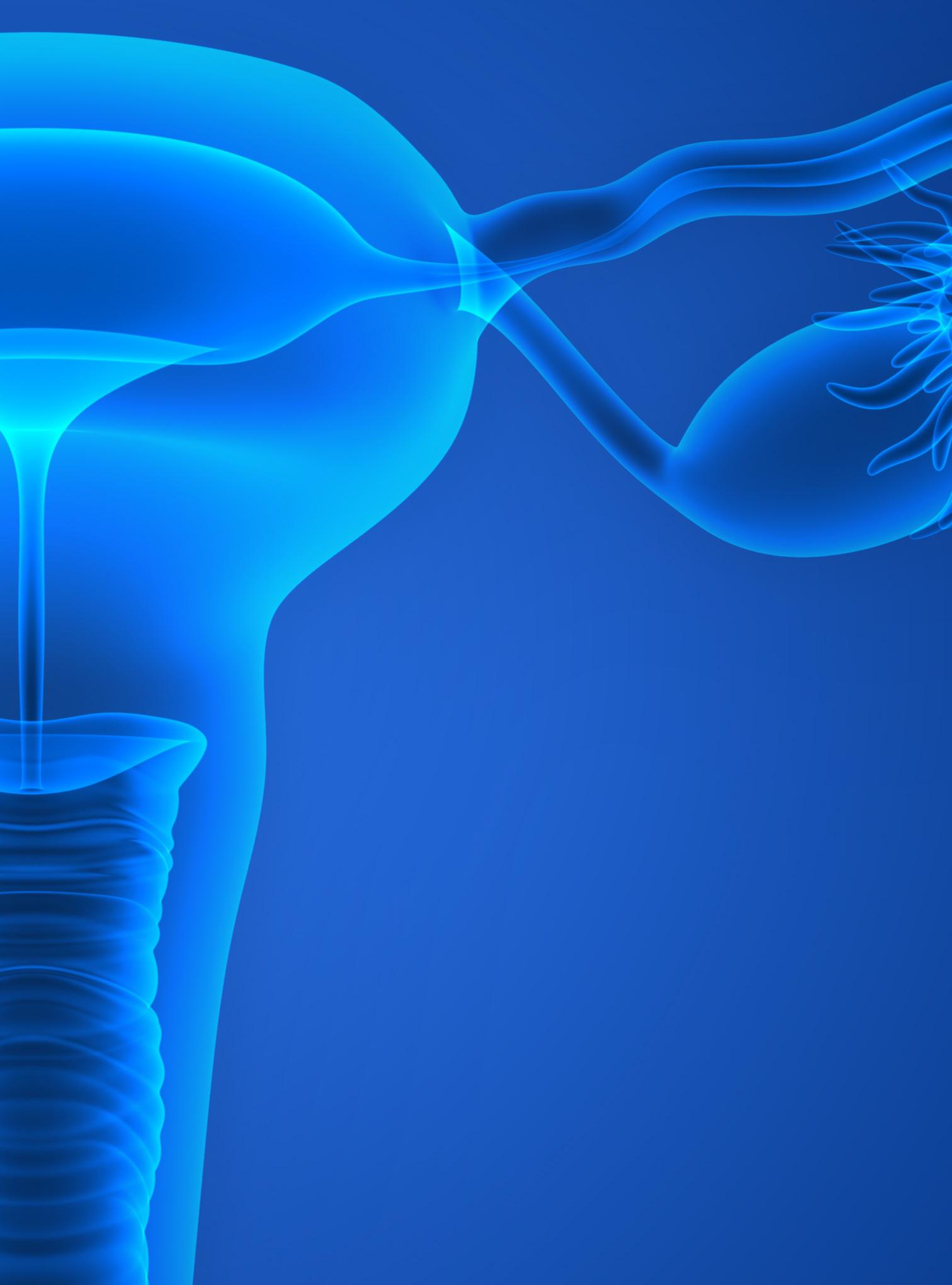
Meet the lymph node. These small, bean-shaped organs found at the convergence of major blood vessels in the body filter substances in a person’s lymph fluid. An adult has approximately 600-800 nodes located in the neck, armpit, chest, behind the ear, abdomen and groin.
Houston Methodist researchers are now in a Phase 2a Clinical Trial—Safety, Tolerability, and Efficacy of Hepatocyte Transplantation into Periduodenal Lymph Nodes Among Subjects With End-Stage Liver Disease.


Under the leadership of Constance Mobley, MD, PhD, FACS, researchers have successfully performed a first-inhuman miniature liver hepatocyte transplant to change a patient’s lymph nodes into ectopic miniature livers. Houston Methodist is only one of two sites partnering with LyGenesis, Inc.
More than 10,000 liver transplants were performed in 2023; however, over 1,700 patients die annually while on a waitlist per the United Network for Organ Sharing.
The team dosed four patients currently on a transplant waitlist, using their lymph nodes as living bioreactors to regenerate an ectopic organ. Selected patients must have a Model for End-Stage Liver Disease score greater than 10, but less than 25 at the time they are enrolled in the study. The goal is to have a total of 12 patients for the study, which began in 2022.
Mobley’s colleague, Sunil Dacha, MD, MBBS, performs an endoscopic ultrasound to locate the lymph nodes in the periportal area. Once Dacha locates a lymph node in that area, he endoscopically injects that lymph node with the liver cells.
“The procedure is simple; however, the difficulty lies in identifying the nodes around the liver due to their size— one centimeter or less. They are not easy to find, but with meticulous examination, we’ve successfully performed these procedures,” said Dacha, Associate Professor of Clinical Medicine.
The first stage of this study is to meet the FDA requirements of safety, tolerance and efficacy.
What’s so exciting about this study is that these types of studies have never been performed in patients. We are the first hospital in the world to successfully inject these cells into a human. We always hear that Houston Methodist is leading medicine; in this instance, we actually are the first in the world to accomplish this goal.
”

“With our initial patient cohort, we want to ensure that their lymph nodes are functioning like livers,” said Mobley. “Our primary endpoint is safety and tolerability.” Mobley’s team monitors their patients for a year to note any changes in their baseline liver function with the hope that their liver will become healthier.
Houston Methodist is the first hospital in the world to inject these cells into a human successfully.
If this first-in-class allogeneic regenerative cell therapy obtains FDA approval, it would enable one donated liver to treat dozens of end-stage liver disease patients, which could vastly affect the organ supply and demand imbalance for patients suffering from liver failure.
“There’s such a great need for the success of this study because there aren’t enough organs for the number of patients who are in need,” Mobley emphasized. “Once we are successful in this study, the number of patients who we will be able to treat and save from liver disease will be immeasurable.”
Mobley also noted how this can be a game changer when treating patients with liver failure who may have additional underlying conditions that can be a barrier to receiving a liver transplant.
“Ultimately, my hope is that using liver cells will allow us to rescue patients who are extremely sick and can’t tolerate surgery,” Mobley said. “Theoretically, we could get so good at this that maybe patients wouldn’t need liver transplants which would be incredible.”
Update: Data and Safety Monitoring Board Approves LyGenesis’ Phase 2a Clinical Trial to Continue Dose Escalate In Novel Trial Organ Regeneration.
–
Constance M. Mobley, MD, PhD, FACS
Associate Director of Liver Transplantation
J.C. Walther Jr. Transplant Center
Houston Methodist Center for Critical Care
Associate Professor, Clinical Surgery
Department of Surgery
Sherrie and Alan Conover Center for Liver Disease & Transplantation
Houston Methodist
Chronic rejection of a transplanted kidney refers to degeneration in kidney function over time as the body’s immune system attacks the transplanted kidney. Around 50% of transplanted kidneys may experience chronic rejection to some degree within five years of transplant surgery.

Research shows that chronic rejection entails processes regulated by the actin cytoskeleton and the RhoA/Rock pathway. To preserve kidney function and prevent kidney transplant failure, Houston Methodist is leading two Phase 2 clinical trials, investigating the safety and efficacy of two different pharmaceuticals impacting the RhoA pathway in transplant patients.
Interstitial fibrosis (IF) is an abnormal scar tissue between the kidney tubules and tubular atrophy (TA) is a degeneration of the tubules. Both usually occur together, signifying significant and often irreversible kidney damage and are present in approximately 45% of kidney biopsies by one-year post-transplant. Even though IF/TA plays a significant role in renal graft and kidney dysfunction after transplantation, efforts at prevention—or slowing the progression— of allograft fibrosis have been unsuccessful.
Fingolimod for the Abrogation of Interstitial Fibrosis and Tubular Atrophy Following Kidney Transplantation
The first clinical trial is ongoing and investigates whether the RhoA inhibitor fingolimod (brand name Gilenya®), safely abrogates the development of IF/TA in kidney transplant recipients compared to placebo. Fingolimod binds to sphingosine 1-phosphate (S1P) receptors on immune cells, causing internalization of the receptor and decreasing downstream RhoA activation. This fingolimod-induced disruption of immune cell trafficking also indirectly impacts activation of the Akt/mTOR pathway, leading to decreased mammalian target of rapamycin (mTOR) signaling. Preliminary data strongly suggest that fingolimod inhibits chronic rejection of transplanted hearts in a murine model by inhibiting RhoA and downregulating mTOR.

The second clinical trial, recently initiated, investigates whether belumosudil (brand name- REZUROCK®) safely abrogrates the development of IF/TA in kidney transplant recipients compared to placebo. Belumosudil works by blocking the activity of Rho-associated coiled-coil kinase 2 (ROCK2), which contributes to the development of immune and fibrotic diseases. Evidence shows that belumosudil was effective in preventing fibrosis and chronic rejection in a murine cardiac transplantation model.
Each clinical trial is a randomized, double-blind, placebocontrolled trial in de novo kidney transplant patients to determine if the addition of either drug—on the background of standard immunosuppression—will prevent fibrosis in the kidney transplant.
“ We hypothesize that mitigating the fibrogenic effects of RhoA, with either belumosudil or fingolimod, will reduce structural damage in transplanted kidneys and possible subsequent transplant failure. ”

– Ahmed Osama Gaber, MD, FACS, FAST John F. Jr. and Carolyn Bookout Presidential Distinguished Chair in Surgery Department of Surgery Chair, Department of Surgery
J.C. Walter Jr. Transplant Center Houston Methodist
The Cancer Prevention and Research Institute of Texas (CPRIT) offers various funding opportunities for cancer research, product development and prevention programs. Two Houston Methodist researchers received $3.4 million in grants and awards from CPRIT in 2024.

Qing Yi, MD, PhD, Ralph O’Connor Centennial Chair and Professor of Cancer Biology, received a research award of $1.9M for individual investigator translational research, “Combination Therapy Using ATRA and Carfilzomib to Treat Proteasome Inhibitor Refractory Multiple Myeloma.”
Yi’s research will focus on multiple myeloma—a bone cancer characterized by the accumulation of tumor cells in the bone marrow. Myeloma remains incurable despite the many chemotherapy drugs available. Patients often become resistant to myeloma treatments, including traditional chemotherapeutics and novel agents. Additionally, some patients exhibit initial resistance, not responding to chemotherapy at all. Those who do respond often experience relapses after treatment and succumb to the disease.

“Proteasome inhibitors, such as bortezomib and carfilzomib, have good therapeutic efficacy for multiple myeloma, yet their initial response rate in myeloma patients is only 27-48%,” said Yi.
To overcome resistance and advance the development of more effective therapies, Yi’s team performed a high-throughput screening of 1,855 FDA-approved drugs and found that a treatment used for patients with acute promyelocytic leukemia—all-trans retinoic acid (ATRA)—can overcome human multiple myeloma cell resistance to the current standard-of-care drugs. ATRA alone does not kill myeloma cells.
Yi believes that ATRA may be used to treat patients with myeloma to restore their response to these drugs. The CPRIT grant will be used to conduct a first-in-human phase IB/II clinical trial to determine the safety, tolerability, efficacy and recommended phase II dosing of ATRA/carfilzomib combination therapy to treat proteasome inhibitor resistance in multiple myeloma patients.


Nestor F. Esnaola, MD, MPH, Diane Modesett Chair and Interim Director, Dr. Mary and Ron Neal Cancer Center, received $1.4M for a prevention grant for cancer screening and early detection, “Cancer Prevention and Outreach for Individuals Disproportionately Affected by Cancer in Medically Underserved Regions (C-CUR).”

The proposed program leverages well-established partnerships and incorporates dissemination and implementation of evidence-based cancer education, screening and early detection, education and patient navigation.
Esnaola’s research will focus on improving cancer screening and prevention education services in urban medically underserved areas (MUAs) within health-disparate populations in Texas, which is a focus of significant and ongoing concern. Low-income individuals residing in Texas MUAs suffer from significantly higher cancer incidence and mortality.
Federally Qualified Health Centers (FQHCs) help address these issues and are critical to the health care safety net. FHQCs reduce the cancer burden within their populations through cancer education, screening and early detection. With a community-led approach to improve access to cancer-related preventive services through Legacy Community Health, an FHQC, in collaboration with the Cancer Center and the Texas A&M Health Science Center, Esnaola and collaborators hope to make substantial contributions to screening, early detection and reduction of mortality rates for certain cancers, as well as for hepatitis C.
“The CPRIT project will create a link between the community and clinical providers to make cancer-related preventive services available via three Legacy Community Health FHQCs to patients in Greater Fifth Ward Lyons, Santa Clara and San Jacinto/Baytown over three years,” said Esnaola.


Collaboration, innovation and education go hand-in-hand and is critically essential in training the next generation of physicians, researchers, caregivers, medical engineers and learners through emerging technologies that will pave the way for better health.
From high-realism mannequins to standardized patients and virtual reality, simulation training has helped health care professionals hone their skills for decades.

These critical training tools improve health care effectiveness and efficiency while fostering open conversations about human error and how to improve situational responses. While simulation training has historically been siloed— nurses training nurses and doctors training doctors—simulation collaboration among various health care professionals has grown in the last 20 years.
Take maternal and neonatal care, for example. According to a 2023 March of Dimes report, U.S. maternal deaths have nearly doubled since 2018, and infant mortality remains alarmingly high, especially among Black birthing people.
Now, a national team of anesthesiologists and surgeons are specifically calling for more collaboration between simulation and health care quality professionals to improve patient safety and quality of care. This call to action was published in Simulation in Healthcare and The Joint Commission Journal of Quality and Patient Safety
“Quality of care and patient safety are two areas where simulation is underutilized,” explained Randolph H. Steadman,
MD, MS, Carole Walter Looke Centennial Chair in Anesthesia and Critical Care and co-author of the call-to-action paper.
“The reason is that quality and simulation professionals rarely cross paths to communicate and collaborate, resulting in missed opportunities for institutions to improve in quality of care and patient safety.”
Last year, Steadman led several Maternal and Infant Simulation Instructor Courses at MITIE in the Bookout Center. These trainings brought maternal and neonatal professionals together from all Houston Methodist campuses. In addition to learning how to work better as a team and how to simulate their own training scenarios, participants identified ways to standardize emergency response protocols systemwide. This way, health care professionals who work at multiple Houston Methodist campuses will not have to worry about code calls or emergency supply carts varying by hospital, which can increase efficiency and patient safety in critical situations.
“Data shows that we’ve since quickened our response times to treat delivery-associated hemorrhaging and pre-eclampsia in maternal patients, resulting in improved patient safety and quality of care,” Steadman said.

Education is a never-ending path. Fostering key affiliations furthers Houston Methodist’s mission to advance, through our Cycle of Translation, promising innovations into effective treatment that provides clinical benefits and impact for our patients.
One such affiliation is with the Weill Cornell Graduate School of Medical Sciences (WCGS), which has expanded its PhD program to the Houston Methodist Academic Institute’s Education Institute. This new enhancement builds upon the 16-year academic affiliation between Weill Cornell Medicine (WCM) and Houston Methodist to train the next generation of physicians and scientists.
The Weill Cornell Graduate School of Medical Sciences, based in New York City, offers two Weill Cornell PhD programs at Houston Methodist in Physiology, Biophysics, and Structural Biology, and Neurosciences. These programs provide deep engagement in translational predoctoral research by collaboratively working on projects with
academic medicine teams that include clinicians at the Houston Methodist Research Institute and physician-scientists at Houston Methodist Hospital.
Houston Methodist students in the program undertake the same curriculum as students who complete the programs in NYC and complete their research studies in Houston— jointly mentored by faculty located at both sites. New York
A partnership with the Sloan Kettering Institute


WCGS faculty provide the didactic curriculum and thesis research is performed at the Houston Methodist Academic Institute (HMAI), under the direction of WCGS faculty located at both campuses. In turn, graduate students and faculty based in New York City can benefit from Houston-based faculty expertise and state-of-the-art facilities and technology at HMAI. This collaboration enhances the experience of students and faculty at both locations, promotes scientific interactions and enriches our student body.
These Houston-based students receive Cornell’s Doctor of Philosophy degrees— a first for Weill Cornell Medicine awarding the degree outside of New York.

In addition to the affiliation with Weill Cornell Medicine, the Houston Methodist Education Institute and Texas A&M University launched the Engineering Medicine Program at the Texas A&M University School of Engineering Medicine. The Institute also oversees continuing medical education and graduate medical education and supports more than 758 students and postgraduate trainees for medical, nursing, allied health and research education programs.
The Office of Graduate Medical Education hosted its annual Match Day event marking the moment when graduating students discover where they will train for residency and in what specialty. The National Resident Matching Program releases the results simultaneously to all applicants and programs across the United States.

On Match Day, 75 medical students from around the world officially became incoming residents at Houston Methodist for the 2024-2025 academic year. These incoming Houston Methodist residents joined the 421 graduate medical education (GME) residents and fellows who work with inpatient teams and one-on-one with subspecialists during rotations within 76 GME programs.
Annually, Houston Methodist trains more than 41,000 students, researchers and clinicians with a team medicine and science approach that offers insight into the workings of a high performing academic medical center relentlessly focused on quality improvement, innovation and biodesign of next-generation technologies.
Snapshot of Matches 2024-2025 Incoming Residents
• 60 U.S. Graduates (80%)
• 8 from Texas (50%)
• 22 from other U.S. medical schools
• 15 International Graduates (20%)
• 3 Urology positions in the American Urological Association Match (EnMed; University of Minnesota School of Medicine; University of South Carolina School of Medicine)
Aga Khan Medical College
Aristotle University of Thessoloniki
C.M.H. Medical College
Jinnah Sindh Medical University
King Edward Medical College
Mu’Tah University
Tanta University
Tec de Monterrey SOM and Health Sci.
University Sidi Mohammed Ben Abdellah
Universidad Americana
University of Alexandria
University of Jordan

“Physcianeers”
Thirty-four students matched in the Texas A&M University’s Engineering Medicine (EnMed) program, 59% matched in residency programs in Texas. These students were among the hundreds of medical students in the Houston area matched to resident training programs across the U.S. The EnMed program, founded in 2019, is a partnership with Houston Methodist Texas A&M University to integrate medical education with research focused on innovation and entrepreneurship, empowering a new generation of “physicianeers” with the clinical skills to diagnose and treat patients along with the engineering perspective to problem-solve, invent new technologies, and rapidly advance their innovations to transform patient care.

Dedicated to the development of next-generation human performance devices, technologies and training regimes, the Center for Human Performance, a collaborative partnership with Houston Methodist and Rice University, brings together academic researchers and university students working side by side with student-athletes, trainers and coaches to advance research and education in human performance and protecting student-athletes.


For the last several years, advanced technologies and equipment at the Center of Human Performance offer Houston Methodist the opportunity to test and enhance human physical performance. The information gathered is applicable to athletes and patients alike—to aid rehabilitation and prevent injuries.

Bradley Lambert, PhD, Assistant Research Professor of Orthopedic Surgery and Manager of the Center of Human Performance, is engaged in various lines of research including novel rehabilitation methods, strategies to minimize post-surgery opioid use, biomechanics following total joint replacement and blood flow restriction (BFR) therapy— all of which have various applications in sports medicine, performance research as well as improving clinical and patient care.
Bradley S. Lambert, PhD
As the name suggests, BFR therapy is a methodology to restrict blood flow. External pressure is applied using a specialized automated pressure cuff in the upper arm or thigh which restricts vascular flow via direct compression
when activated. The automated pressure cuff can also be outfitted with a Doppler probe to determine the degree of vascular occlusion.
It uses partial occlusion of blood flow to mimic the stress response of high-intensity exercise in a way that helps preserve muscle and bone and can be performed at exceptionally low intensities, which is optimal for a post-surgery patient. Modified from a form of resistance training called “KAATSU Training,” BFR therapy has been shown to stimulate muscle anabolism and increase muscle mass and strength when combined with low-intensity exercise in clinical trials.
BFR therapy has garnered considerable attention from scientific communities and the media in recent years due to its many applications and potential benefits in muscular hypertrophy, strength and endurance. Combined with low-load resistance exercise, BFR may be used in various sports training to increase skeletal muscle mass. In healthy populations, BFR is an applied methodology to decrease stress on the joints during strength training and is particularly useful in populations that cannot lift heavy weights.


“Blood flow restriction therapy was still under investigation and not commonly utilized as a standard of care for rehabilitation before we started looking at it in 2017. We’ve performed extensive research on this topic, and it has now become a standard of care for all of our sports medicine physicians,” noted Lambert. “Presently, we’ve branched BFR into older patient populations for total joint reconstruction.”
Applications of BFR is also being explored in patients following wrist fractures, and those with Achilles tendon
rupture repair. More remains to be explored about the mechanisms of action at the cellular level in response to BFR.
Lambert noted, “We’ve now looked at blood flow restriction therapy in collegiate baseball players, and some of the protocols that we’ve come up with are now standardized for the Houston Astros and several major league baseball teams, particularly for the shoulder and elbow. There is potential for this therapy to be used to combat unloading muscle loss associated with disuse in spaceflight and other numerous applications.”
BFR therapy is opening new frontiers in both athletic training and patient care. By harnessing the body’s natural responses to enhance muscle strength and recovery with minimal stress, we’re helping athletes optimize their performance and revolutionizing rehabilitation for patients to offer safer, more effective paths to recovery and redefine how we support human performance across all stages of life.

For the next two years, 120 Division I Rice University athletes will participate in a concussion study with Houston Methodist researchers to identify reliable and novel concussion biomarkers in their gut microbiomes in ways that standard brain imaging cannot.
Sponsored by the NFL Players Association, this project will advance a 2022 study that explored the gut-brain connection in contact-sports student-athletes and identified biomarkers for concussions within the athletes’ gut microbiomes. Although the study, initially involving 33 football players, was cut short by the COVID-19 pandemic, it was the first study to show a measurable drop in certain bacteria found in the stools of athletes without concussions compared to those who had suffered sports-related concussions. It also found a connection between inflammatory proteins found in the blood and the ratio of good and bad gut bacteria of athletes who suffered concussions.

“This larger study will give us a clearer understanding and measurement of concussions and their association with gut microbiota functions, and whether these are gender-dependent,” said Sonia Villapol, PhD, Assistant Professor, Neurosurgery, who leads the study with Kenneth Podell, PhD, John M. O’Quinn Centennial Chair in Concussion Research and Care. “Our ultimate goal is to better protect and maintain athletes’ health and also develop treatments to improve their safety and well-being.”
The study is part of the Center for Human Performance, a Houston Methodist and Rice University partnership, to advance research and education to prevent injuries, improve rehabilitation, enhance performance and speed athletes’ return to life, work and sports.
Dubbed “Gut Microbiome Markers of Sport-related Brain Concussion,” the study will include 80 Rice male and female athletes from collision/contact sports (football and soccer) and 40 athletes from non-contact sports, including swimming, track and field, and tennis. Researchers will collect blood and stool samples throughout the two-year study and use machine learning and advanced microbiome sequencing to analyze the data.
Researchers hope that analyzing the gut microbiomes of these college athletes can lead to the development of personalized treatments for concussions and a system that can be used on the field to detect early signs of mild traumatic brain injury through markers in the microbiome.
The Houston Methodist Academic Institute oversees the Education Institute and Research Institute, including 842 faculty and 41,100 learners. The Academic Institute aligns our research and education initiatives in service to the clinical mission, providing solutions that answer the call for new technologies and skills our clinicians need for patient care.
The Houston Methodist Education Institute coordinates our primary academic affiliation with Weill Cornell Medicine and other joint programs, including the Engineering Medicine Program at Texas A&M University School of Engineering Medicine. The Education Institute also oversees continuing medical education and graduate medical education, and supports more than 758 students and postgraduate trainees for medical, nursing, allied health and research education programs.
The Houston Methodist Research Institute supports research programs and infrastructure that enable faculty across the system to bring new scientific discoveries to patients as rapidly as possible through the full Cycle of Translation from conceptual bench research to prototyping and development to clinical trials and FDA approval. The Research Institute supports more than 1,900 clinical studies and trials and $126 million in extramurally funded translational research programs.
Houston Methodist Academic Institute
Judge Ewing Werlein, Jr., Senior Chair
W. Benjamin Moreland, Chair
Edward R. Allen, III
Steven Birdwell
John F. Bookout, III
Marc L. Boom, MD
P. Embry Canterbury
Jenny C. Chang, MD
David Chao
Martin Craighead
Leslie Doggett
Elaine Finger
Antonio Gotto, MD, D.Phil
Robert A. Harrington, MD
Edward A. Jones
Evan H. Katz
Sippi K. Khurana, MD
Steven Looke
Ransom Lummis
David A. Modesett
James Muschalik
Gregory Nelson
Joe Bob Perkins
Ward Sheffield
Jeffrey F. Simmons
Suzanne (Sue) Smith
Christopher G. Stavros
Steven D. Stephens
Spencer A. Tillman
David M. Underwood, Jr.
Amy Waer, MD
Martha S. Walton
Donna Sims Wilson
