IMPrESS Perio Implant Center | Dr. Noroozi, Certified Periodontist | Burnaby, BC
From Our Office to Yours
Dear Colleagues,
Thank you for taking time in your busy schedule to look into our newest newsletter. For our referring doctors, friends, and patients, we provide these newsletters fi lled with the latest periodontal health news and implant reconstructive surgical procedures. I would like to sincerely thank our loyal referring colleagues for their continued support during the past decade. We are looking forward to collaborating with your of fi ce for the best comprehensive patient care possible. Should you have any questions and comments, please do not hesitate to contact me.
All materials and contents are courtesy of Dr. Mehdi Noroozi Inc and patients treated in his practice and subject to copyright. Not to be copied or duplicated for any purpose.
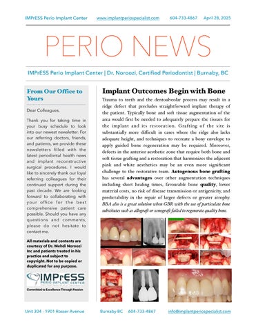
Implant Outcomes Begin with Bone
Trauma to teeth and the dentoalveolar process may result in a ridge defect that precludes straightforward implant therapy of the patient. Typically bone and soft tissue augmentation of the area would first be needed to adequately prepare the tissues for the implant and its restoration. Grafting of the site is substantially more difficult in cases where the ridge also lacks adequate height, and techniques to recreate a bony envelope to apply guided bone regeneration may be required. Moreover, defects in the anterior aesthetic zone that require both bone and soft tissue grafting and a restoration that harmonizes the adjacent pink and white aesthetics may be an even more signi fi cant challenge to the restorative team. Autogenous bone grafting has several advantages over other augmentation techniques including short healing times, favourable bone quality, lower material costs, no risk of disease transmission or antigenicity, and predictability in the repair of larger defects or greater atrophy. BBA also is a great solution when GBR with the use of particulate bone substitutes such as alograf or xenograf failed to regenerate quality bone.
Here is a successful implant reconstruction of a complex partial edentulism in the anterior mandible. A 50 year old male presented with grade II mobility of the existing failing dental bridge with hopeless prognosis for the retainer teeth. Significant soft tissue deficiency along with extremely thin alveolar bone (knife edge) and saddle shape ridge make esthetic implant therapy impossible. Soft tissue augmentation, 3D bone reconstruction with allograft shell block technique, CBCT guided restoratively driven implant placement and custom made abutments were applied to deliver a prosthesis that resembles natural teeth.
Significant bone resorption following tooth extraction. Autogenous bone from mandibular ext. oblique ridge was used to reconstruct the horizontal and vertical bone for implant therapy. 4 months re-entry shows significant bone gain and successfully integrated implant in the posterior mandible.
Here is a referral for peri-implant bone loss shortly after implant insertion in a different office. The patient received two bone-level implants in the molar areas with horizontal bone loss extending to 3-4 mm coronal aspect of implants. The implant threads were exposed even before insertion of the crowns. With the patient's consent, implants were removed. Guided bone regeneration with 50-50 Auto-Xeno and Ti-reinforced Cytoplast membrane was completed to gain some vertical bone height. 8 months later, Tissue level Straumann SP implants were inserted in excellent bone quality and quantity.
Here is a case of missing central maxillary incisor with significant bone and soft tissue deficiency due to a dental trauma. A young male 25 years old has been wearing a maryland bridge due to loss of his front tooth at very early age. 3D bone augmentation with autogenous block graft and particulate bone according to the Khoury technique along with VIP-CTG for soft tissue enhancement. Guided implant surgery 4 months following bone grafting. Restoration was done by his referring dentist.
Keys for Successful Esthetic Zone Single Implants
To achieve a successful esthetic result and good patient satisfaction, implant placement in the esthetic zone demands a thorough understanding of anatomic, biologic, surgical, and prosthetic principles. The ability to achieve harmonious, indistinguishable prosthesis from adjacent natural teeth in the esthetic zone is sometimes challenging. Placement of dental implants in the esthetic zone is a technique- sensitive procedure with little room for error. Atraumatic extraction, 3D guided immediate implantation, bone and soft tissue grafting along with immediate screw retained provisional restoration are some of the most documented strategies to achieve aesthetic outcome.
Soft Tissue Management Around Implants
Here is a case of an implant in the area of the missing right lateral incisor with bone and soft tissue deficiency. This implant was placed 7 years ago by a different dentist. No soft tissue grafting was done at the time of implant placement. Due to the large implant size, buccal positioning, and thin tissue phenotype, this young patient in her 30s experienced a significant esthetic dilemma. The existing implant crown was removed, and connective tissue grafting was done followed by a new screw retained zceram crown.
The amount of keratinized tissue you have might play a critical role in the implant's appearance, implant placement, and ability to keep the implant clean. This case presents the soft tissue augmentation around severely compromised implants with absence of attached mucosa. gingival tissue was harvested from patient's palatal area. The postoperative image shows establishment of adequate attached tissue and more optimal plaque control plus some implant surface coverage with newly formed soft tissue attachment.
Implant Complications Management
Here is a referral case regarding a dental implant with the presence of infection, bone loss, pus, and deep probing. The implant was placed in a different office a few years ago. Radiographically, bone loss is evident and has continuously got worse over time. The existing crown was removed, fl ap surgery was performed and the implant surface and the surrounding bone were decontaminated and detoxified with YSGG ER CR laser. Upon surgical entry, an infrabony defect was noted. Guided bone regeneration with the application of autogenous bone and xenograft was applied to reconstruct the lost bone. Once the healing period was over, a new crown was inserted by the referring dentist. No more evidence of deep probing or suppuration was found. Radiographic bone gain was evident in two year follow up.
Here is a case of a referral for a young patient who received implants by another dentist in the area of congenitally missing maxillary lateral incisors. Unfortunately, one of the implants did not integrate, and the other implant showed significant amount of bone loss, even before restoration. Upon clinical and radiographic examination, I found out that the implants were not surrounded by sufficient bone and soft tissue. Therefore the implants were removed and sites were developed with autogenous bone and soft tissue. Four months later adequate bone and peri-implant soft tissue was established, two new Straumann implants were placed surgically according to 3D restoratively driven protocols with excellent bone quality and primary stability. Final implant restorations were placed with optimal aesthetics.
Aesthetic Crown Lengthening & Lip Repositioning -
Gummy Smile Solutions
Gummy Smile Treatment- Altered Passive Eruption. Do you feel your patient’s teeth look too short and their smile is too gummy or do gingiva cover too much of some teeth while leaving the others the right length? If so, dental crown lengthening might be the solution for your patient. During this procedure, the excess of gingival and bone tissue is removed to expose more of the crown of the tooth. Then the gingival margin is sculpted to give your patient new smile just the right look. Although teeth appear short, they may actually be the proper length. The teeth may be covered with too much gum tissue. We can correct this by performing the periodontal plastic surgery procedure, crown lengthening. During this procedure, excess gingival and bone tissue are reshaped to expose more of the natural tooth. This can be done to more than one tooth, to even the gum line, and to create a beautiful smile.
Optimizing Implantology: Pterygoid Implants as an Alternative to Cantilever and Sinus Lift Procedures
Pterygoid implants are an innovative solution in dental implantology, especially when traditional options like cantilever bridges or sinus lifts aren’t ideal. They offer several advantages, particularly for patients with significant bone loss in the posterior maxilla (upper jaw). Here’s why pterygoid implants can be a game-changer:
• Avoiding Sinus Lift Surgery
• No Need for Cantilever Bridges
• Minimizing Bone Grafting
• High Success Rate and Immediate Functionality
• Less Invasive, Shorter Recovery
• Ideal for Atrophic Maxilla
• Preservation of Natural Bone Structure
All On X Implant and Prosthetic Rehabilitation
The fixed implant supported hybrid prosthesis also known as All on 4 has become an efficient and reasonable opportunity for patients to achieve a fresh new start with their dentition. However, for the dental provider, it can also be considered as the final frontier, the last definitive opportunity for patients with terminal dentition. Success or failure is in our hands. It is incredibly important for us to properly treatment plan these cases to mitigate potential risk and complications. Certainly the greatest risk is not providing enough prosthetic space. It is paramount the surgical team create enough vertical height from the implant level to the incisal edge of the prosthesis. There has to be enough vertical height to stack components: abutments, Ti bar, the acrylic wrap and denture teeth. It is commonly reported in the literature that a minimum vertical height requirement is 15 to 16 mm from the fixture to the incisal edge. Cases below demonstrate full arch fully guided bone reduction and implant placement in preparation for implant supported hybrid prosthesis. The bone needed to be removed to gain the essential space for prosthetic components. This bone was invaluable source of autogenous bone to augment the thin bone on buccal aspect of anterior implants. Surgical and prosthetics completed by IMPrESS multispecialty team. While All on 4 implants are an amazing tool for many people, restoring an entire arch of teeth on 4 implants isn’t the “go to” full arch implants option. Instead it’s a fairly specific procedure that’s really only suitable in a small fraction of cases. Instead it’s often better to get what are known as either a “Fixed Zirconia Bridge” or “Fixed Hybrid Bridge” restoration. These procedures use 6-8 traditional implants to support a bridge, on which the dental prosthetic (full set of upper or lower teeth) is then placed.
A variety of periodontal plastic surgical techniques have been proposed to obtain root coverage of gingival recession defects. All of the available root coverage procedures are able to provide significant root coverage for Miller Class I and II recession - type defects. However, only the subepithelial connective tissue graft in conjunction with a coronally advanced flap appears consistently effective across all clinical parameters, and is therefore currently considered the gold standard for gingival recession therapy. The major shortcomings of connective tissue graft procedures include patient morbidity associated with the second surgical site and limited availability of palatal donor tissue. Connective tissue gingival graft with the apical access point and tunnel technique to manage the extreme generalized gingival recessions are routinely performed in our office.
Mucogingival Surgery, Palatal Connective Tissue for Lingual Recessions
The goal of this post is to demonstrate the practicality and results of increasing the zone of keratinized tissue on the lingual surface of mandibular anterior teeth. Calculus is most commonly found on the lingual surface of mandibular teeth, so they are subjected to in fl ammatory elements resulting in tissue deformation and destruction. Significant attention has been paid to grafting the buccal surface, but there is a paucity of information addressing the lingual surface of mandibular anterior teeth. Gingival augmentation procedures are essential before fixed restorations to prevent further recession and facilitate plaque control.
Mucogingival Deformities
-
Role of Keratinized
Attached Gingiva
Mucogingival deformities are a group of conditions that affect a large number of patients. Among the mucogingival deformities, lack of keratinized tissue and gingival recession are the most common. Lack of keratinized tissue is considered a predisposing factor for the development of gingival recessions and inflammation. Gingival recession occurs frequently in adults, has a tendency to increase with age, and occurs in populations with both high and low standards of oral hygiene. Recent surveys revealed that 88% of people aged ≥65 years and 50% of people aged 18 to 64 years have ≥1 site with gingival recession. The presence of recession is esthetically unacceptable for many patients; dentin hypersensitivity may occur; the denuded root surfaces are exposed to the oral environment and may be associated with carious and non-carious cervical lesions (NCCL), such as abrasions or erosions.
Allograft Dermal Matrix with Modified Tunnel Technique for Soft Tissue Augmentation- Root Coverage
Since its introduction to dentistry in 1997, Allograft Regenerative Tissue Matrix has been a widely accepted acellular dermal matrix (ADM) for soft tissue applications. It supports tissue regeneration by allowing rapid revascularization, white cell migration and cell populationultimately being transformed into host tissue for a strong, natural repair. This product has been widely used in my practice over the past several years to substitute for autogenous gingival grafting to treat gingival recessions, to enhance the tissue phenotype and to achieve root coverage. Since the amount of tissue that can be harvested from palate is relatively limited, patients with generalized gingival recessions have to go through multiple operations with long and slow recovery. The application of donor tissue has the following benefits:
•Incision and scalpel free
•Accelerated recovery
•One visit can treat multiple areas of recession
•Less discomfort for the patient after treatment
•No need for scalpels or invasive surgical tools
•No need to take donor tissue from the patient’s palate
“Our goal is to support our referring clinicians in providing exceptional health care for patients through colaboration, peer support and a culture of knowledge sharing. We welcome referrals fom al dentists and specialists and are equipped to support you based on your level of experience. IMPrESS team is dedicated to working closely with your office and we are proud to provide evidence-based periodontal & implant procedures. Your patient’s care and comfort are our priority fom the initial consultation through to future treatment. Our office wil work with you to ensure a positive experience and optimal results for your referrals.”