ANIMAL RESEARCH REPORT 2022–23


CONTENTS 3 FOREWORD Professor Ian Walmsley FRS 5 DISCOVERIES Recent findings from animal research 8 PROVOST’S AWARDS FOR EXCELLENCE IN ANIMAL RESEARCH 2022–23 winners 13 MOVING CLOSER TO A CURE The new techniques helping to find a cure for diabetes 19 HERE COME THE ORGANOIDS Growing mini organs from stem cells to understand diseases 25 UNLOCKING THE SECRETS OF DIGESTION What the digestive tract of mice can tell us about obesity 30 KEEPING IMPERIAL’S ANIMAL FACILITIES RUNNING Keeping conditions clean and safe with the help of robotic machinery 32 LEADERS IN COMMUNICATIONS Why Imperial’s approach to communication earned us a Leader in Openness award 34 THE CONCORDAT ON OPENNESS ON ANIMAL RESEARCH
Some
animals, like frogs, are used in animal research to understand and treat diseases that put their species at risk
FOREWORD Animal Research at Imperial
Back in 2018, Imperial became the first UK university to gain accreditation from the Association for Assessment and Accreditation of Laboratory Animal Care (AAALAC). This recognition of high welfare standards is given for a period of three years. Institutions must take part in the same rigorous inspection process to retain this recognition and I am glad to say that since our last annual report, our AAALAC accreditation has been reconfirmed.
In our last report, we highlighted the role of Imperial’s animal research in tackling COVID-19. We are now building on the knowledge and insights we gained during the pandemic with the creation of a new Institute of Infection that brings together scientists, medics, engineers and business expertise from across Imperial. Animal research will support their work in understanding viruses, bacteria, fungi and parasites, as well as transforming diagnosis, prevention and treatment of infectious disease.
Previously, we also announced our plans to create a 3Rs Hub, to promote research that reduces, refines and replaces animal research. Now fully operational, the Hub is empowering our researchers to share expertise and resources to help Imperial become a world leader in the 3Rs – refinement, reduction and replacement.
Since our last report, the Medical Research Council (MRC) London Institute of Medical Sciences (MRC LMS) has moved into a new £110m building. The building houses up to 40 research teams conducting
behavioural, metabolic and imaging studies on mice. This work will help us address major health challenges such as obesity, diabetes, heart disease and cancer.
At Imperial, we are actively engaged with industry experts to ensure the discoveries we make can be translated into real-world benefits. Our White City Campus is home to a growing community of new and established businesses. As part of our interactions with industry we plan to share our knowledge of animal welfare and the 3Rs for the benefit of the scientific community.
Indeed, Imperial has also been recognised a second time for its willingness to communicate about animal research with a Leader in Openness Award from Understanding Animal Research. The award gives independent affirmation for this publication as well as the animal research news stories, podcasts and live Q&As that Imperial has produced.
Since our last report, there has been a change to the operating model of the Home Office Animals in Science Regulation Unit (ASRU), the body that oversees and licences animal research in the UK. There is now an increased requirement to ensure we demonstrate how we meet the Standard Conditions of Licences and our participation in audits.
Our compliance has always been excellent, and we are embracing these changes. We have established a compliance committee to review
our processes and supporting documentation to implement a risk-based approach to the new regulatory regime, and to ensure we maintain our excellent compliance record. Our commitment to the highest animal welfare standards and the 3Rs is unwavering.
This is my last report as Establishment Licence Holder, as this role is now being taken up by Professor Mary Ryan, Vice Provost (Research and Enterprise). It has been an immense honour to work with the dedicated and talented staff in Central Biomedical Services. Imperial is at the forefront of good practice and I am proud of our commitment to the 3RS, the training we provide and the integrity of our work. My thanks go to Marina Botto, Rob Floyd, Helen Jones, Charlotte Dean and Sian Harding for providing such inspiring and capable leadership, as well as to Mary for accepting this important role.
Professor Ian Walmsley FRS Provost Establishment Licence Holder 2019–2023

Animal Research Report 2022–23 | 3
Animal research remains indispensable to enable better human health, and at Imperial we are committed to maintaining the highest calibre of facilities to support this work where it is absolutely necessary, and to maximise benefit for society through its impact on the UK’s health system.
DISCOVERIES Recent findings from animal research
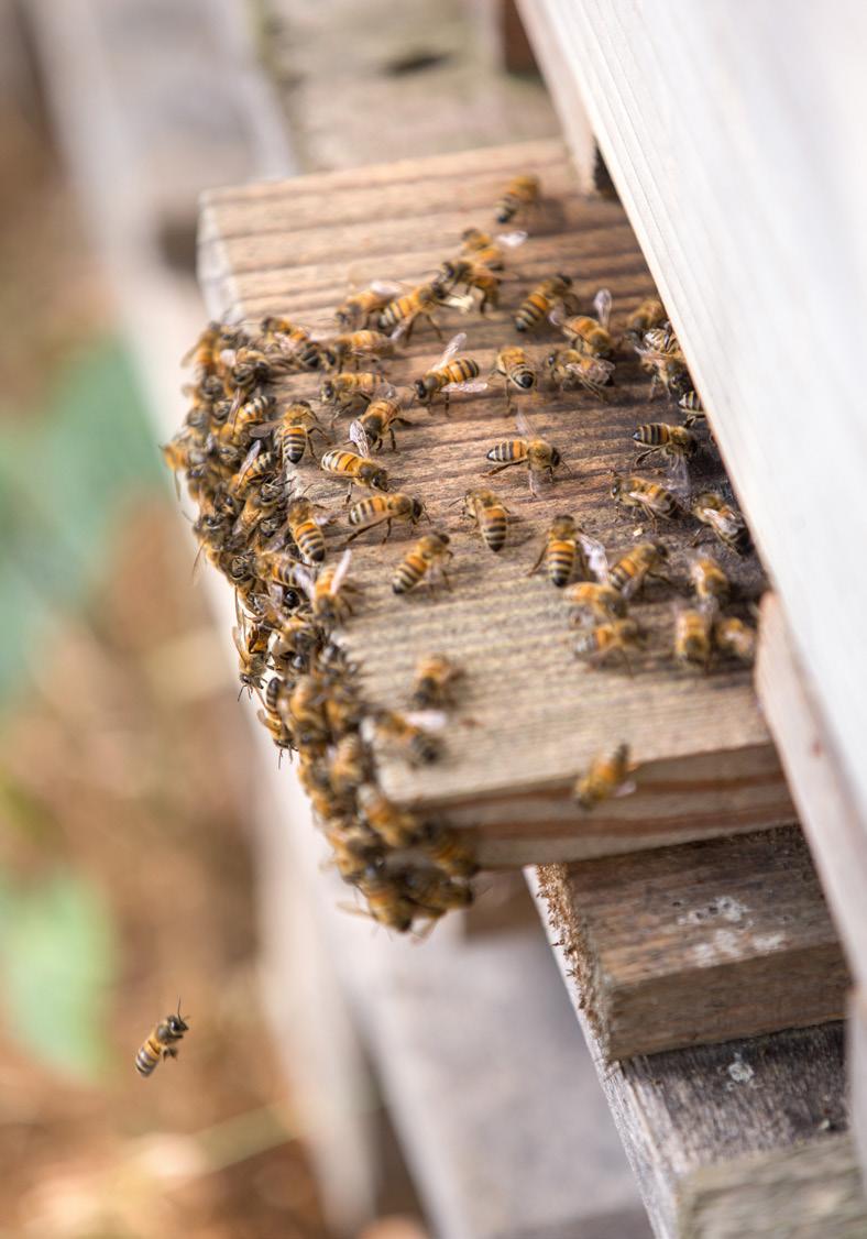
Changing temperatures increase pesticide risk to bees
A team of Imperial researchers have found that temperature has an influence on how badly pesticides affect bees’ behaviour. It is already known that pesticides can have a negative effect on bees and these new findings indicate that future extreme temperature events under climate change could increase the impact of pesticides on bees’ survival and their ability to pollinate crops.
The team studied six bumblebee behaviours at different temperatures, under the influence of two pesticides (the neonicotinoid imidacloprid and the sulfoximine sulfoxaflor). Four of the behaviours – responsiveness, likelihood of movement, walking rate, and food consumption rate – were affected by imidacloprid more strongly at the lower temperature. This suggests cold snaps could increase pesticide toxicity on behaviours that are important for bees’ nest duties. However, a key behaviour – how far the bees could fly – was most strongly affected by imidacloprid at the highest temperature. This relationship showed a strong drop-off, with flight distance measuring the same between 21°c and 27°c, before falling sharply when reaching 30°c.
The findings will help researchers look towards developing a toxicity forecast framework, which allows predictions on how bee populations will respond to climate change.
Scientists engineer mosquitoes that can’t spread malaria
Imperial scientists have engineered mosquitoes that slow the growth of malaria-causing parasites in their gut, preventing transmission of malaria to humans.
The genetic modification causes mosquitoes to produce compounds in their guts that stunt the growth of parasites. This means they are unlikely to reach the mosquitoes’ salivary glands and be passed on in a bite before the insects die. So far, the technique has been shown to dramatically reduce the possibility
of malaria spread in a lab setting, but if proven safe and effective in real-world settings, it could offer a powerful new tool to help eliminate malaria.
The innovation is designed so it can be coupled with existing ‘gene drive’ technology to spread the modification and drastically cut malaria transmission. The team is looking towards field trials but will thoroughly test the safety of the new modification before combining it with a gene drive for real-world tests.
 Left: Beehive, Silwood Park Campus
Right: Scientists have engineered mosquitoes to prevent the spread of malaria
Left: Beehive, Silwood Park Campus
Right: Scientists have engineered mosquitoes to prevent the spread of malaria
Animal Research Report 2022–23 | 5
If proven safe and effective in real-world settings, it could offer a powerful new tool to help eliminate malaria.”
Flexible, steerable device could help treat brain diseases
Imperial scientists successfully placed a bioinspired steerable catheter into the brain of a living animal for the first time in October 2022.
The early-stage research tested the delivery and safety of the new implantable catheter design in two sheep at the University of Milan’s Veterinary Medicine Campus, to determine its potential for use in diagnosing and treating diseases in the brain. The technology includes a soft, flexible catheter to avoid damaging brain tissue while delivering treatment, and an artificial intelligence (AI)-enabled robotic arm to help surgeons navigate through brain tissue.
If proven effective and safe for use in people, the platform could reduce the risks associated with diagnosing and treating brain diseases. It could help surgeons to see deeper into the brain to diagnose disease, deliver treatment like drugs and laser ablation more precisely to tumours, and better deploy electrodes for deep brain stimulation in conditions such as Parkinson’s and epilepsy.
Robber flies combine navigation systems to catch prey
Using high-speed cameras, researchers have revealed how robber flies combine navigation systems to avoid obstacles while chasing down their prey.
The findings show that the flies combine two navigation systems to steer themselves mid-flight. One system is called ‘pure proportional navigation’, which is often used by homing missiles. This works by connecting the robber fly’s eyes to their prey, through their line of sight. Changes in speed and direction are then determined by how their line of sight changes – for example if a target veers to the left or right. The second system is a simple avoidance algorithm to ensure that the fly does not collide with obstacles. If an obstacle is coming towards them, it appears as if it is growing larger in their field of view so the robber fly veers away. The bigger the obstacle grows, the more the robber fly turns away to avoid it.
The researchers believe that their work may one day inspire the future design of miniature aerial drones.
Treatment improves spinal cord regeneration in mice after injury
Weekly epigenetic treatment in mice can support spinal cord regeneration after severe injury, according to Imperial research.
Currently, spinal cord injury does not have any effective treatments and in severe cases, the outcomes are extremely limited by the failure of spinal neurons to regenerate naturally after injury.
An Imperial team found that weekly treatments with an epigenetic activator can aid the regrowth of sensory and motor neurons in the spinal cord, when given to mice 12 weeks after severe injury. They used a small molecule called TTK21 to activate genetic programming that induces nerve fibre regeneration in neurons. The team tested TTK21 treatment in a mouse model of severe spinal cord injury. The mice lived in an enriched environment that gave them opportunities to be physically active, as is encouraged in human patients. Treatment began 12 weeks after severe spinal cord injury and lasted for 10 weeks. Researchers found several improvements after TTK21 treatment compared with control treatment.
6 | Animal Research Report 2022–23
Bioengineers find new way heart valves grow
Imperial bioengineers have discovered a new mechanism driving the growth of heart valves in zebrafish embryos.
The findings shed light on how heart valves grow and find their shapes in embryos. Heart valves are constantly challenged by the mechanical forces generated by heartbeat, and diseases of the heart valves can mean that blood fails to circulate properly. Some people are born with congenital heart valve defects, but exactly how valves grow in embryos is poorly understood.
The research team, from Imperial and institutions in France, used zebrafish to identify the processes at play during the development of the valves found between atria and ventricles, known as atrioventricular valves. They found that alongside mechanisms that were already known to generate heart valve tissue, another mechanism works in parallel to determine the shape and function of the canal from which the atrioventricular valves develop.
Further studying the role of mechanical forces in congenital valve defects could eventually help researchers understand how to prevent and treat them.
Woodcocks have the brightest white feathers ever measured
An Imperial-led team has found that the woodcock, a large wading bird, uses its bright-white tail feathers to communicate in semi-darkness, and its feathers reflect 30 per cent more light than any other measured feather.

Birds that are most active during the day often use colourful plumage to communicate information with each other. Birds that are most active at dawn and dusk, or at night, such as woodcocks, tend to have less showy plumage, as they need to be camouflaged to avoid predators while they sleep during the day.
It was thought that birds active during low-light conditions used sounds or chemicals to communicate. However, many have bright white, reflective patches, which could be used for communication in environments with very little natural light.
The Imperial team used specialised microscopy to image the structure of Eurasian woodcocks’ white tail feathers from a collection of specimens in Switzerland. They were surprised to find that the feathers reflected up to 55% of light.
Intermittent fasting may help heal nerve damage
Researchers have shown how intermittent fasting changes the gut bacteria activity of mice and increases their ability to recover from nerve damage.
In the mice, fasting led to the gut bacteria increasing production of a metabolite known as 3-Indolepropionic acid (IPA), which is needed to regenerate nerve fibres called axons. These thread-like structures at the ends of nerve cells send out electro chemical signals to other cells in the body. The bacteria that produces IPA is found naturally in the guts of humans as well as mice and is also present in humans’ bloodstreams.
The researchers, from the Department of Brain Sciences, assessed nerve regeneration of mice where the sciatic nerve, the longest nerve running from the spine down the leg, was crushed. Half of the mice underwent intermittent fasting while the other half were free to eat with no restrictions at all. The length of the regrown axons was measured and was about 50% greater in mice that had been fasting.
The next stage for this research will be to test this mechanism for spinal cord injuries in mice as well as testing whether administering IPA more frequently would maximise its efficacy.
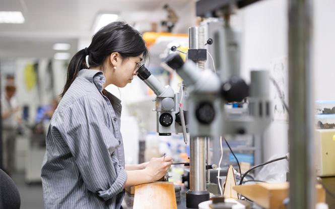
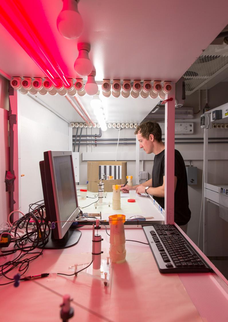
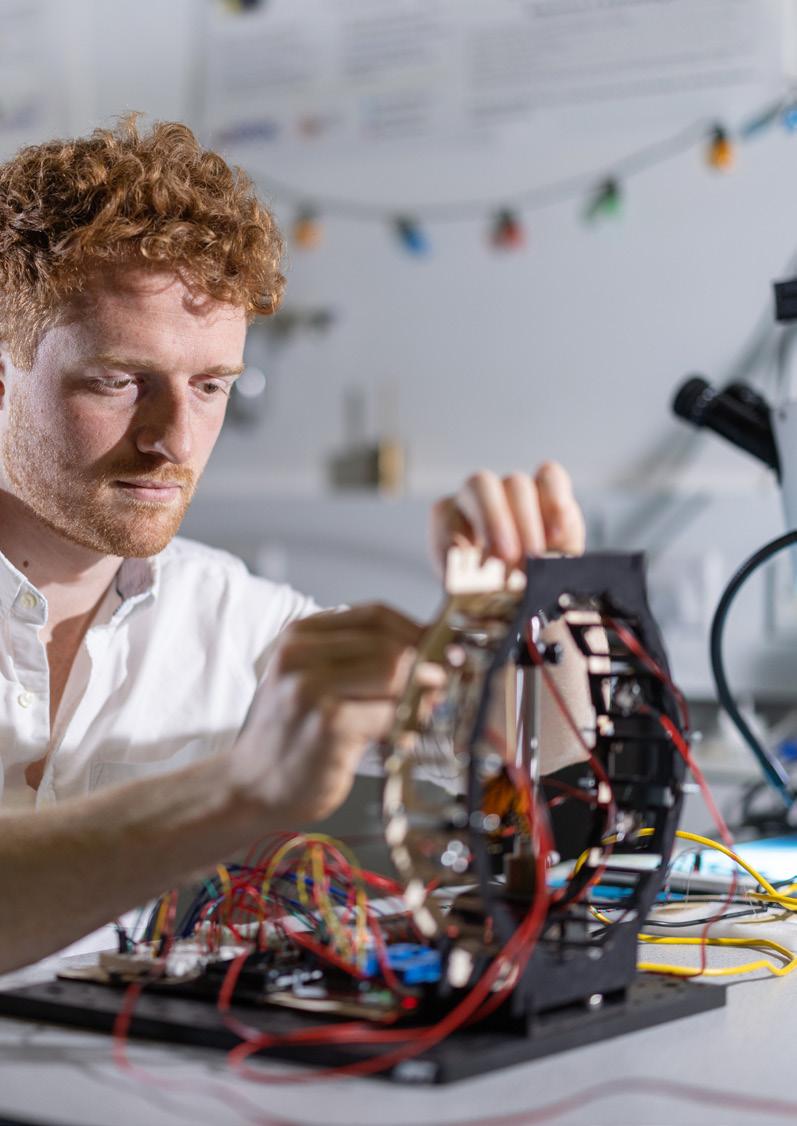
Above left: Biomedical Engineering student building a cable-driven parallel robot to simulate flying insects
Left: Eurasian woodcock (Scolopax rusticola) on the lake
The Imperial research communications team regularly publishes news stories about the impact of the College’s research with animals. Visit www.imperial.ac.uk/news to read the latest stories or search ‘animal research’ on the site.
Animal Research Report 2022–23 | 7
PROVOST’S AWARDS for Excellence in Animal Research
The Provost’s Awards for Excellence in Animal Research recognise best practice and acknowledge staff who have made advances in the 3Rs, shown openness in communicating about animal research, or demonstrated outstanding collaboration between research and Central Biomedical Services (CBS) staff. Winners receive £1,000, which can be used to cover costs associated with the presentation of the awardwinning work to a wider audience.
PROVOST’S AWARD 2023 WINNERS
Application of the 3Rs, Researchers
Dr Jonathan Miles, Research Associate and Dr Michael Wortley, Research Associate, National Heart and Lung Institute (NHLI)
Jonathan and Michael received the Provost’s Award for developing a new technique for the pre-clinical study of chronic cough in guinea pigs. They have created a custom-made system to monitor guinea pigs within their standard housing, allowing the animals to be studied in a less stressful environment than had previously been possible. They also worked to reduce the number of animals needed for this kind of study, including replacing the guinea pigs with dry lab work for part of the project. They believe their novel methods will provide results with increased statistical power that are easier to transfer to the clinic. The development of this cough monitoring technique brings the pre-clinical study of cough in line with state-of-the-art clinical cough research. Their technique should provide more clinically translatable research that will accelerate the discovery of new medicines for cough.
Below: Using dry lab work has reduced the use of animals in a guinea pig cough study
8 | Animal Research Report 2022–23
Application of the 3Rs, CBS staff
Mr Alex Stepney, H2 Facility Manager and Mr Alex Martinez, A-tune manager, Central Biomedical Services
Alex Stepney and Alex Martinez received the Provost’s Award for introducing a novel automatic system to genotype animals within the breeding unit. The new refined system has reduced the number of human or genotyping errors, and improved welfare for the animals. The project first began with the ongoing review of the breeding systems in operation at Imperial facilities, during which Alex Stepney proposed introducing the option of genotyping transgenic mice via a commercial supplier, to increase the accuracy in genotyping and reduce the need for re-sampling. He contacted the supplier and worked with several research groups to test the process. Alex Martinez worked with the supplier to create as streamlined a system as possible and is currently working to integrate the system into A-tune.
Team award
Dr Shane Foo, Senior Scientific Officer, Institute of Cancer Research and Miss Justyna Glegola, Training and Competency Assessor, Central Biomedical Services
This team received the Provost’s Award for working together to ensure excellent training was given on intertumoral injections; a very precise procedure where researchers need specific and rigorous guidance. Shane and Justyna were recognised for their collaborative work in devising and delivering excellent training for the procedure to the Principal Investigator on the project, working across different teams and institutes to improve both animal welfare and science.
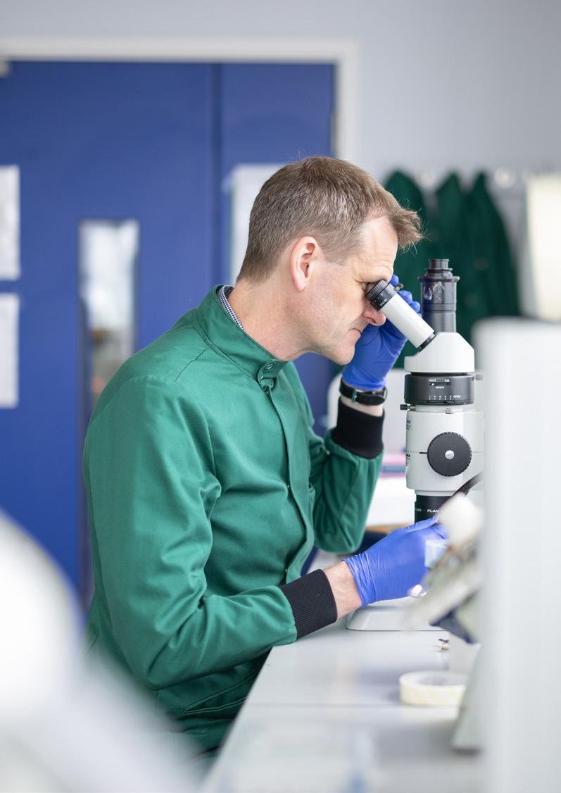
Top: Scientists who work with animals at Imperial are supported by a team of vets and technicians responsible for maintaining high levels of animal welfare
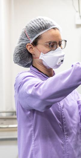
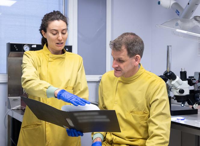
Above: Staff at Imperial work to replace, reduce and refine the use of animals in research
Animal Research Report 2022–23 | 9
Application of the 3Rs, Researchers
Dr Claire Dunican, PhD student and Dr Athina Georgiadou, Research Fellow
Claire and Athina received the Provost’s Award for devising a framework for choosing mouse models of malaria. Many combinations of mouse strains and malaria parasites are used in the malaria research field. Until now, the rationale for selecting one model over another to study disease mechanisms relied on subjective parameters. Using comparative transcriptomics, Claire and Athina defined how closely mouse models mirror the biological processes occurring in different severe malaria syndromes, providing a framework for selecting the most relevant models. Their approach can dramatically reduce the number of mice used in experiments that have limited relevance to human disease.
Public Engagement award
Mrs Rebecca Frise, Laboratory Manager
Rebecca received the Provost’s Award for volunteering as a speaker for the 2021 Institute of Animal Technology (IAT) webinar series to educate the public about the use of animals in research. Rebecca delivered an informative talk and had multiple helpful interactions with members of the public. She also participated in an external event, ‘How animal research has helped unlock COVID-19 research’, answering follow-up questions about the role of animal research in biomedical discovery.
Dr Masanori Asai, NC3Rs Training Fellow
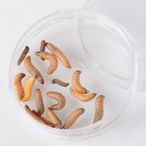
Masanori received the Provost’s Award for his work reducing the number of animals used in tuberculosis (TB) research. He has pioneered a novel mycobacterial infection model using Galleria mellonella (greater wax moth) as an animal model. Masanori completed a proteomic and gene expression analysis of the G. mellonella-TB model, the first of its kind, showing that the infection model recapitulates all the hallmarks of mycobacterial disease. Pre-screening in G. mellonella can reduce animal use in the TB research field by 70 per cent.
Above: Galleria mellonella (greater wax moth larvae)
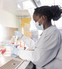
Below: Provost Award winner, Rebecca Frise, working in the laboratory
Right: Silwood Park provides a unique environment setting for studying bees
PROVOST’S AWARD 2022 WINNERS 10 | Animal Research Report 2022–23
MOVING CLOSER TO A CURE

Hardly a day seems to pass without new headlines about the ever-growing costs – in lives, health and funds – of diabetes. Researchers at Imperial have developed a new technique using both mice and human tissue to help them better understand the mechanisms underlying the disease. Now they are using these insights to seek improved treatments for people living with diabetes.
Around one in 10 adults worldwide –more than 500 million people – live with diabetes.1 Prevalence in the UK is rising rapidly, with the latest estimate, published in April, putting case numbers at more than 5 million. The disease directly causes an estimated 2 million deaths per year worldwide, and is a major cause of blindness, kidney failure, heart attacks, stroke and lower limb amputations.2
The key to understanding and potentially finding a cure for diabetes lies in clusters of cells called pancreatic islets. These tiny groups of cells produce hormones including insulin, which is key to keeping blood glucose levels within a normal, safe range. Diabetes occurs when pancreatic islets are either attacked by our immune systems (type 1), or become damaged through overwork and, as a result, fail to make enough insulin (type 2).
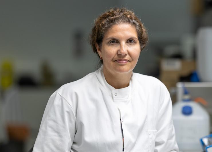
“Diabetes is one of the major noncommunicable diseases affecting humankind today,” says Dr Victoria Salem, Clinical Senior Lecturer in Diabetes and Endocrinology in the Department of Bioengineering. “If we want to find a cure, something we are still some way from doing, we really need to understand pancreatic islet biology.”
Scientists have found it challenging to unpick precisely how these vital structures operate, not least because they measure only around 0.1mm in diameter and are embedded deep within the pancreas. It has long been known that beta cells within the pancreas can both sense glucose and produce insulin. More recently, Victoria and other researchers have shown that rather than operating individually, beta cells act within coordinated networks in which some drive the activity of others.
Until a few years ago, these clues to the complexities of beta cell activity could only be shown in experiments that could not accurately replicate the natural functioning of pancreatic islets. That’s because they were often carried out on islets removed from human cadavers or animals, without the continuous blood supply and neural inputs they get in a living body.
1 www.idf.org/aboutdiabetes/what-is-diabetes/facts-figures.html
2 www.who.int/news-room/fact-sheets/detail/diabetes
Animal Research Report 2022–23 | 13
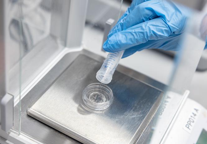
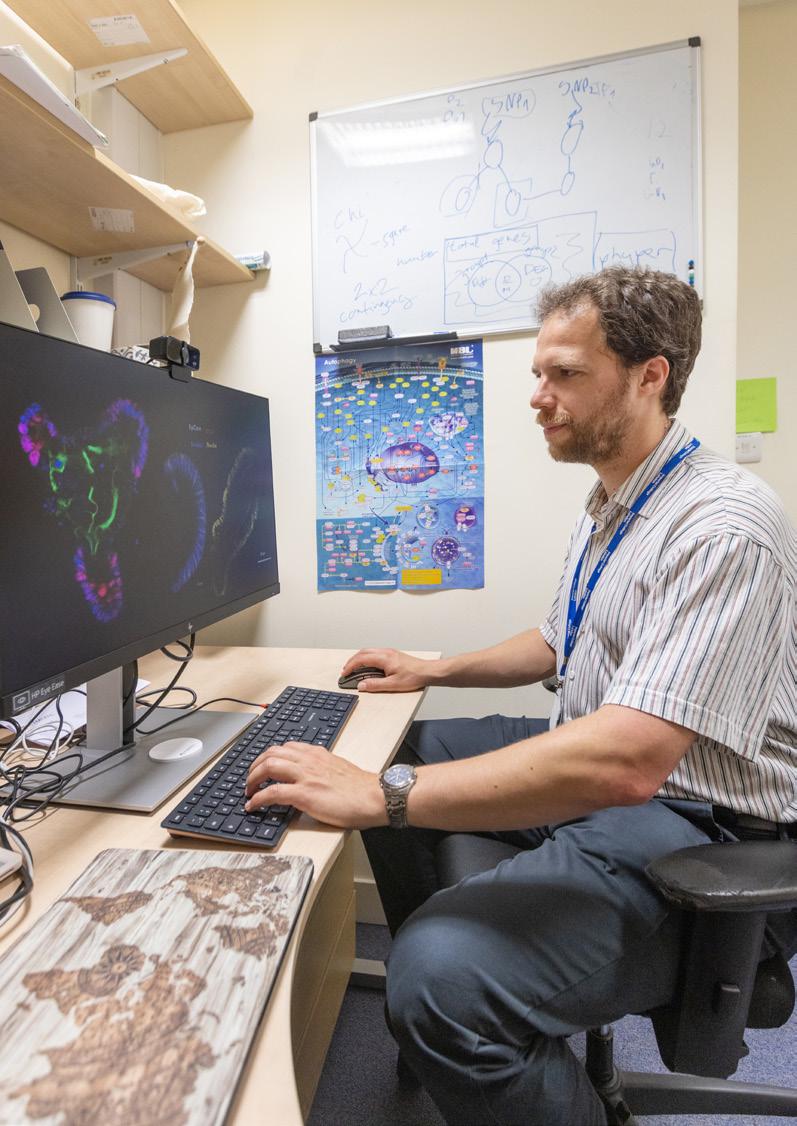
Left: Dr Victoria Salem is studying pancreatic islet biology to better understand diabetes. Her team genetically engineers islets to become fluorescent when they release insulin so that this can be imaged in real time
The insights gained from these types of experiments were useful but limited in how they could be applied clinically.
One way to study pancreatic islets ‘in vivo’ is to repeatedly take blood samples from humans or animals, however this too has its limitations. “We used to infer pancreatic islet function indirectly by taking regular peripheral blood samples, but this involves taking it a long way from where the insulin is released and so it doesn’t accurately reflect what is going on,” says Victoria. “And even the most advanced modern imaging technologies don’t have the resolution to be able to directly image these tiny islets deep within the pancreas.”
Scientists have long used a method in laboratory mice involving transplanting tissue into a space between the cornea and the iris called the anterior chamber of the eye (ACE) as a way to better study it.3 Researchers at the Karolinska Institute in Stockholm, Sweden, successfully did this with murine pancreatic islets, and showed that these developed a blood supply and used the technique to study how they responded to a variety of interventions.4
Victoria and her team went a stage further by genetically engineering mice so that beta cells in their pancreatic islets produce a fluorescent chemical when they release insulin. They also engineered pancreatic islets from human bodies donated for medical research to do the same thing. The group transplanted both the mouse and human islets into the ACE of mice, and observed them developing a blood supply. “Efforts to find a cure for diabetes have been held back by the difficulties involved in studying pancreatic islets in their natural, living environment. Our method is the only way, to date, that allows us to image and interrogate the pancreatic islet and see its changing behaviour over time in response to different medicines or stimuli, in a live animal.”
The new technique, outlined in a paper published in 2019 in the journal Nature Metabolism, allowed Victoria’s group to observe individual beta cells releasing insulin over time and showing how they act in a coordinated manner. Applying statistical methods and network theory, they were able to identify highly connected ‘hub’ beta cells that they say appear to play a coordinating role in directing the activity of the islet.
3 www.frontiersin.org/articles/10.3389/fendo.2021.652853/full (ref 17) 4 www.nature.com/articles/nm1701
If we want to find a cure, something we are still some way from doing, we really need to understand pancreatic islet biology.”
14 | Animal Research Report 2022–23
– Dr Victoria Salem
“We also found evidence that type 2 diabetes is associated with a loss of this beta cell connectivity and of these hub cells,” she says. “Because we can’t directly image pancreatic islets in a living human, the only way to show this was to do it in a living animal.”
The use of the transplantation to the murine ACE technique is not only helping to generate new and important scientific insights. It is also enabling reductions in the number of mice used in research. “Historically, if we wanted to understand what a chronic stimulus did to pancreatic islet function, we would have to expose a group of animals to that stimulus and sacrifice them at maybe one week, two weeks, four weeks, six weeks and six months, and look at the pancreases at each of those different time points,” says Victoria. “Now, we can re-image one group of animals with transplanted islets in their eyes time and time again, which means a big reduction in the number of animals needed to give us longitudinal data.”
Furthermore, laboratory mice that are used in such experiments are able to be anaesthetised while they have the 10-minute procedure, which involves an injection into the front of the eye. “We worked closely with human cataract surgeons and vets to think about the best way to anaesthetise the animals, and to devise preoperative care so that when they wake up, they recover as quickly and painlessly as possible,” she adds.
The group went on to use their technique to allow them to observe the behaviour of individual beta cells over periods of several weeks. In a paper published only as a preprint at the time of writing, they showed that mice genetically engineered to lack the GCGR gene in their beta cells, which is key to maintaining normal beta cell function, had impaired pancreatic islet function when given a sugary diet. In vitro tests confirmed the islet cells in the mice lacking GCGR had impaired ability to produce insulin.5 The researchers also found evidence that the identity of
5 www.biorxiv.org/content/10.1101/2022.09.01.506223v1
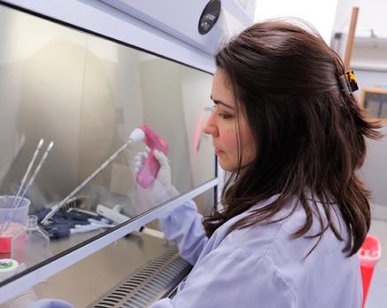
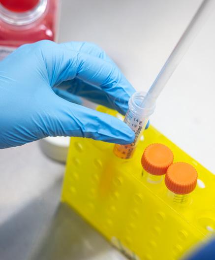
Far left: Diabetes studies use both mice and human tissue to understand the disease
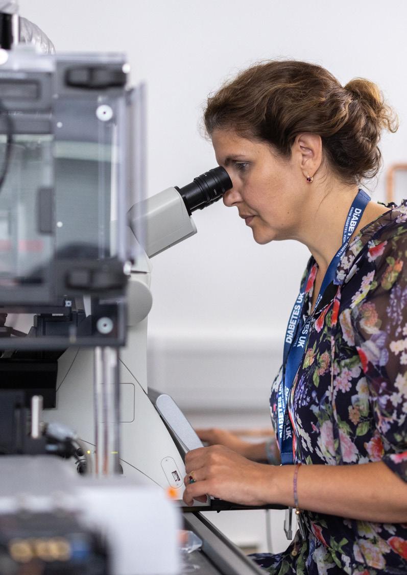
Above: Scientists are imaging the pancreatic islet to see its changing behaviour over time
We worked closely with human cataract surgeons and vets to think about the best way to anaesthetise the animals and devise preoperative care so they recover as quickly and painlessly as possible.”
Animal Research Report 2022–23 | 15
– Dr Victoria Salem
highly connected hub beta cells is fluid rather than fixed. “This finding opens up new questions about the environmental factors that drive a beta cell to become a hub beta cell.”
It’s a question also being addressed by Victoria’s colleague Professor Guy Rutter, of Imperial’s Department of Medicine, Diabetes and Reproduction. Now based primarily at the University of Montreal, Canada, Guy is seeking to identify whether these key hub beta cells exhibit molecular signatures that could be targeted with medical interventions that could protect them as a means to slow the development or progression of type 2 diabetes.
Alongside her work advancing our understanding of islet biology, Victoria also works as a clinician working with patients who have type 1 diabetes. The immense challenges they face in matching their insulin doses with their blood sugar levels, and the mental load that comes with concern about serious
complications, are a strong motivation for her fundamental research.
“When patients ask me about the possibility of a cure for diabetes, they’re not talking about an insulin pump that needs to be programmed, that they have to worry about falling off or stopping working,” she says. “What they usually mean is no longer having to worry about managing their own blood sugar ever again – and that would be a replacement in the form of a transplant of those beta cells that have been destroyed.”
In March, Victoria was awarded a Senior Research Fellowship by the Type 1 Diabetes Grand Challenge, a partnership between the Steve Morgan Foundation, Diabetes UK and diabetes charity JDRF UK. She will lead a team using the £2m grant to try to find a cure for type 1 diabetes based on delivering stem cell-derived beta cells to patients via an implantable device.
16 | Animal Research Report 2022–23
Alongside her work advancing our understanding of islet biology, Victoria also works as a clinician working with patients who have type 1 diabetes.
Victoria and her colleagues are aware they will have to overcome some major hurdles to achieve their ambitious goals. The new beta cells have to be delivered in such a way that they survive transplantation, develop a blood supply and avoid autoimmune attacks. The group is seeking to use a synthetic hydrogel barrier to shield their beta cells from immune system attack, in the hope that this can avoid the tissue scarring seen by other researchers who have used harder Teflon coatings around pancreatic islets in previous experiments.
The group is collaborating with other leading researchers at Imperial to help them tackle these challenges. Molly Stevens, Professor of Biomedical Material and Regenerative Medicine in the Faculty of Engineering, is leading on the use of biomaterials and bioprinting to make the implants. Working with Dr Blerina Ahmetaj-Shala, a Research Fellow in the Faculty of Medicine, the team is using cells taken from patients’
blood to grow new blood vessels in the lab to provide their implants with a blood supply. Their use of microfluidic chips and hydrogels in this work avoids the need for animal experimentation. The researchers are also planning to use computer simulations to carry out high-throughput screening of synthetic materials to use for their implants and to develop immune system response assays.
“We are really pleased that much of the work we are doing in the sphere of regenerative medicine has gone hand in hand with developing new technologies that are helping us replace animal studies,” says Victoria. “This is also a wonderful example of how Imperial can bring together cutting edge technologies and ideas from across disciplines towards really transformative clinical goals.”
Above: Dr Salem’s team is growing beta cells and blood vessels from stem cells on chips to understand islet biology and develop advanced therapeutics to one day transplant back into people with type 1 diabetes
Animal Research Report 2022–23 | 17
HERE COME THE ORGANOIDS
Growing numbers of researchers are using mini organs grown from stem cells to better understand cell biology and the causes of diseases. Organoids can generate deeper scientific insights than traditional cell culture methods and have the potential to significantly reduce the use of laboratory animals. Scientists at Imperial are using organoids to unpick the cellular and molecular causes of inflammatory bowel disease in work that is laying the foundations for improved, more personalised treatment.
Researchers have been growing and observing cells from living organisms for more than 100 years. Since the 1940s, use of animal models and human cell cultures has been standard practice in the life sciences and has generated innumerable insights, from basic cell biology and disease mechanisms, to toxicity testing and drug discovery.
Both approaches are, however, limited in important ways. Critically, cells grown on flat, usually plastic surfaces do not behave in the same way as they would in their natural 3D environments. Animal research can be used to get around this problem, but many biological processes, such as brain development and aspects of metabolism, are unique to humans.6 That could help to explain why around 90% of new drugs fail to make it through clinical trials, according to the U.S. Food and Drugs Administration (FDA).7
Heightened awareness of these limitations, together with a desire to reduce the number of animals used in research, has driven a rapid increase in the use of organoids – self-organising, 3D tissue cultures derived from stem cells – as an alternative.
6 www.nature.com/articles/s41580-020-0259-3
7 www.ncbi.nlm.nih.gov/pmc/articles/PMC9293739
8 www.nature.com/articles/nature07935
In 2009, researchers at the Hubrecht Institute in the Netherlands reported that they had successfully coaxed adult stem cells from mice, in culture, to grow into mini-guts along with all the intestinal cell types present in living animals.8 Since then, scientists have successfully worked out how to create the right environment for stem cells to develop into organoids that resemble the liver, kidney, lung, intestine and stomach.
The number of research papers that include the word “organoid” published annually on PubMed, the life sciences and biomedical research search engine, grew from under 300 in 2015 to more than 3,000 in 2022 – a figure that is still growing. “The use of organoids is really exploding,” says Dr Tamas Korcsmaros, a systems biologist who works on the gut in the Department of Metabolic, Digestion and Reproduction at Imperial. “They were originally mostly used to study the ways cells develop in different tissues, however more recently they have been used increasingly to better model biological functions and disease. Organoids have a lot of potential in the screening of new drug candidates and personalised medicine.”
Tamas has long been interested in new approaches to treating patients with inflammatory bowel disease (IBD) – a group of chronic inflammatory disorders of the gastrointestinal tract including Crohn’s disease and ulcerative colitis. He has, in recent years, used organoids to better understand the mechanisms that maintain health and fight disease in the gut. In particular, he has studied the role of autophagy, a process that recycles cellular components, and Paneth cells, which regulate microbial composition and help regulate immune system defences in the small intestine.
Paneth cell abnormalities are known to be associated with Crohn’s disease. Previous research has also shown that mutations in autophagy-related genes – especially one called ATG16L1 – are linked to Paneth cell abnormalities and poor immune defences against bacterial pathogens. However, the precise impact of autophagy impairment on Paneth cells has been unclear.
To better understand mechanisms underlying Crohn’s disease, Tamas’s group used a mouse model in which autophagy was impaired because cells in the lining of the intestines, called the intestinal epithelium, lacked the
Animal Research Report 2022–23 | 19
Left: Dr Tamas Korcsmaros works with computational and experimental approaches to study signalling networks in the gut
ATG16L1 gene. They used gut tissue samples from these mice and from normal controls to grow organoids, adding drugs to increase their numbers of Paneth cells. The autophagyimpaired organoids generated significantly higher levels of 198 different proteins, compared to control organoids.
“We were able to show that autophagy is not just a cellular recycling system,” says Tamas. “It also specifically targets proteins and degrades them. The Paneth cells our organoids generated had higher levels of proteins that can undermine the health of the gut.” Improved understanding of the specific genes, proteins and mechanisms underlying Crohn’s disease could inform the development of novel treatments for the condition, such as the use of autophagymodulating drugs.
9 www.pubmed.ncbi.nlm.nih.gov/30814064/
The study by Tamas’s group, published in 2019, highlights a key advantage of organoid models.9 In most instances, scientists want to study the behaviour of tissue consisting of cells that are still dividing, so cell cultures are usually grown from cancer cells capable of evading natural cell death processes, or those that can be reprogrammed to become ‘immortalised’. This can make observations based on them biased or less relevant. Additionally, some human cell types are inaccessible, such those in the brain, while others are unavailable in cancerous forms or can’t be immortalised. Paneth cells are among many types of cells that, as a result, cannot be studied in traditional cell cultures. “In organoids you can have non-cancer, non-immortalised human cells, and, with the right type of stem cell, you can grow almost any type of organ,” says Tamas. “Organoids can also be patient-specific.”
Many scientists and doctors are excited about the ability to grow organoids from stem cells taken from patients because of the potential to unlock personalised medicine. This was demonstrated in a study Tamas’s group undertook in collaboration with gastroenterologist Dr Nick Powell, of Imperial’s Department of Metabolism, Digestion and Reproduction, to better understand the roles of immune system proteins called cytokines in IBD, including Crohn’s disease and ulcerative colitis.10 By treating human colon organoids with a range of cytokines, they were able to identify some that play key roles in triggering inflammation and others with antiinflammatory effects.
The researchers, who published their results last year, found the same patterns of cytokine responses when they did the same tests on tissue
10 www.cell.com/cell-reports/fulltext/S2211-1247(22)01280-3?_returnURL=https%3A%2F%2Flinkinghub.elsevier. com%2Fretrieve%2Fpii%2FS2211124722012803%3Fshowall%3Dtrue
20 | Animal Research Report 2022–23
development of novel treatments.
samples from more than 1,000 patients with IBD. There are a range of treatments for IBD but only around 50–60% initially respond to therapies, and, of these, around 25% go into remission11. “Organoids can help explain why only some patients respond to specific drugs based on mechanisms such as cytokine responses,” says Tamas. “I think we will see pharmaceutical companies using them to design clinical trials so they can better target new therapies towards patients in whom they are more likely to work.”
‘good’ and ‘bad’ bacteria are the cause of health and disease rather than the other way around.
A major surge in interest in the microbiome over the last decade, from both academic and commercial groups, has led to a significant increase in the use of mouse models. Yet some microbes that colonise the human gut are not found in mice. And animal models rarely allow systematic, high-throughput and reproducible investigations.
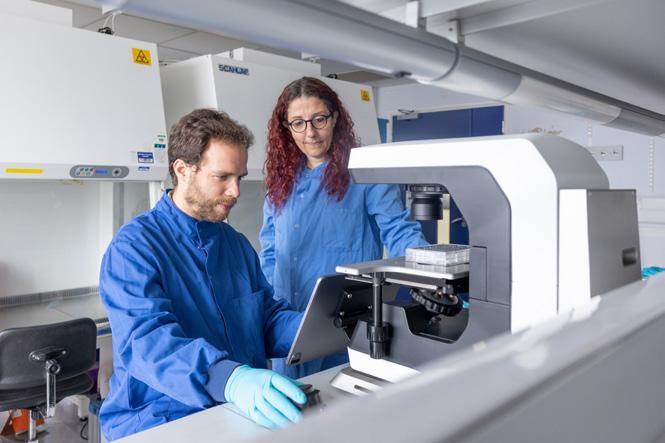
Above: Dr Tamas Korcsmaros co-leads the NIHR Imperial BRC Organoid Facility that has a large research and training laboratory to catalyse organoid research
Organoids also have major potential in research into the roles of microbes in the human gut. The microbiome, as these are often collectively known, is believed to be key to health and disease. Many studies have shown that having certain populations of bacteria in the gut makes it either more or less likely that individuals have a wide range of conditions, from asthma and IBD to type 2 diabetes and obesity. However, the research methods used, such as human cohort studies, have often been unable to find out whether
Microbes colonising the human gut are thought to play a role in modulating autophagy. Detecting autophagy is, however, challenging. Immunohistochemistry, the common method used, involves labelling cell parts with antibodies and requires that samples are fixed at pre-selected time points. It can involve the use of a large number of mice because it is hard to know when it is best to fix samples, and labelling antibodies must be observed separately. Organoids are therefore emerging as an alternative.
11 www.gastroenterologyandhepatology.net/archives/june-2022/approach-to-treatment-failure-ininflammatory-bowel-disease/
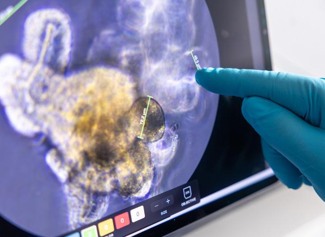
Left: Gut organoids provides an excellent model system to study human cells and intestinal processes
Animal Research Report 2022–23 | 21
Improved understanding of the specific genes, proteins and mechanisms underlying Crohn’s disease could inform the
Tamas’s group has developed a new experimental tool called the human Autophagy Reporter Colon Organoid (hARCO) with the potential to both improve research on gut microbes and autophagy, and reduce animal experimentation. His team has genetically engineered cells in human organoids so that they express proteins that show up as red and green under a microscope. When autophagy occurs, the environment becomes more acidic, which eliminates the acid-sensitive green, enabling researchers to identify when autophagy is taking place. Tamas highlights how, for example, use of the system could have spared 3,315 mice had it been used in all 51 studies PubMed identifies as having used immunohistochemistry to detect autophagy in 2019.
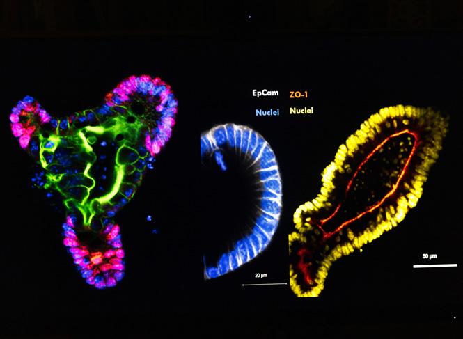
“If we want to understand human diseases, our results will be more relevant and applicable if we use human tissue-based organoids,” says Tamas. “In developing hARCO, we were motivated by both a desire to address the ethical issues of mouse
experiments, and their practical limitations. We hope that, in the near future, systems like hARCO can be used to screen hundreds of microbes or drug molecules.”
This does not, however, mean the end of animal-based research. “Organoids are more complex than traditional cell cultures but they are still simplified models,” says Tamas. “They can help us better understand how the epithelial layer works in the gut, but do not include, for example, the impacts of neighbouring immune cells or neuronal cells, or physical conditions that are equally relevant.” Efforts are underway to add complexity into ‘organs-onchips’ that can include more cell types than simpler organoids and better replicate the physical forces that act on cells in natural environments. Animal experiments will however still be needed for many years to come, not least because many labs do not have the resources to work with organoids, which are still complex and expensive to produce.
These issues help explain the launch in June 2023 of the NIHR Imperial BRC Organoid Facility at Imperial, a research and training hub designed to make it easier to use stem cell and organoid models. The Facility will provide technical expertise, training, laboratory space, arrange industrial partnerships, coordinate the supply of consumables to lower costs and encourage multidisciplinary working across faculties at Imperial. “The Organoid Facility brings together engineers working on developing methodologies with clinical research scientists with ideas about the unmet clinical needs that need to be addressed,” says Tamas, co-lead of the Facility.
“It can act as a catalyst and help organoids to fulfil their potential as a preclinical and precision medicine tool, by supporting the full pipeline of work from patients to publications and clinical output.”
22 | Animal Research Report 2022–23

 Left: Gut organoids can be used to investigate disease relevant cell types
Above: In certain research studies, organoids can replace the use of animals
Right: Dr Tamas Korcsmaros with Dr Isabelle Hautefort, senior gut organoid researcher of the NIHR Imperial BRC Organoid Facility
Left: Gut organoids can be used to investigate disease relevant cell types
Above: In certain research studies, organoids can replace the use of animals
Right: Dr Tamas Korcsmaros with Dr Isabelle Hautefort, senior gut organoid researcher of the NIHR Imperial BRC Organoid Facility
In developing hARCO, we were motivated by both a desire to address the ethical issues of mouse experiments, and their practical limitations.”
– Dr Tamas Korcsmoros
UNLOCKING THE SECRETS OF DIGESTION
A technique that uses ultrasound to examine the microscopic structure of the mouse digestive tract is helping researchers’ understanding of how human bodies respond to food. These discoveries could ultimately help millions of people around the world living with obesity and related diseases such as diabetes and heart disease.
“I’m interested in how the body senses what you’ve eaten, how that regulates your metabolism and what you think you’re going to eat next. For instance, what makes you feel hungry and what makes you feel full,” says Kevin Murphy, Professor of Endocrinology and Metabolism in the Faculty of Medicine.
Professor Murphy joined Imperial in 1998, first as a technician, then a PhD student, and he has remained here ever since, holding a variety of teaching and management roles alongside his research. Through his research he is trying to unpick how the gut detects the nutrients in our food and then signals to the brain and other organs to process the food and coordinate our appetite. Studying these processes is critical to understating the global rise in obesity and related conditions, such as type 2 diabetes.
Professor Murphy explains: “If we understand how you control appetite, and how you control glucose, then we can identify new targets for drugs or dietary approaches that could help manage those diseases.
“We know that obesity is bad, but it’s not being obese that kills you. It’s linked to other diseases like cardiovascular disease, liver disease, diabetes and so on. Many of these problems seem to start with what you eat and gut dysfunction, which then leads on to dysfunction in other organs.
“I’m particularly interested in diet because diet could have a role in either trying to prevent people gaining weight and developing diabetes to start with, or addressing diabetes once people have it. Can you slow progression or even turn it around? If you understand the specific components of food that might do that, then you can give people advice or add those components into foods.
“We’re looking for the molecules produced by digestion that punch above their weight in terms of metabolic control. If we can understand the specific amino acids that are produced by protein breakdown, that are most important in regulating glucose control, then you could load foods with those rather than just adding extra protein.”
Professor Murphy’s recent work has focused on how the gut senses particular forms of protein and fat, and how this regulates glucose control.
“We usually think of carbohydrate intake as the major factor regulating glucose metabolism, and yes, the

Animal Research Report 2022–23 | 25
Left: Professor Kevin Murphy, Professor of Endocrinology and Metabolism in the Faculty of Medicine
pancreas senses and regulates blood sugar. But the other macronutrients you eat have a really big effect as well. So, for example, if you eat protein, the insulin needs to pack that protein away in muscles, or it needs to send it to the liver to be processed. Also, the fat you eat seems to have a real effect on glucose control. But the mechanisms that mediate these processes aren’t really very clear.”
This work is revealing how the gut sends signals along nerves to the brain and then on to the pancreas, where insulin is manufactured, while other signals pass directly from gut to pancreas. The work includes various research techniques, from using organoids and animals in the lab through to studies in humans.
“We need a mixture of all these systems to be able to do the work because some of them can answer questions that others can’t. If you’re looking for very specific molecular mechanisms, things like organoids are really good. But if you’re trying to look
at something like the effects on food intake, you can’t measure food intake in a cell. And if you’re looking at whole body glucose control, which involves the gut, the pancreas, the liver and the brain, there is no way of looking at that very effectively without using a whole animal model.
“Often, we’ll take inspiration from work in humans, where, for example, we can measure what’s going on in the blood when people eat certain foods and use a tube down the throat to pick up what’s going on in the gut. But then to tease apart the mechanisms we switch to the in vitro and the in vivo models.”
This lab work allows Professor Murphy and his team to study the effects of different foods at the level of molecules or cells and to adjust, perhaps with potential drugs, to see how the system responds.
Recently, this work has been boosted by a collaboration with Professor Meng-Xing Tang and his team from the Department of Bioengineering.
The team have developed a noninvasive system that can visualise what is happening in the gut at a microscopic level. The system uses harmless microbubbles to allow ultrasound scanning to pick out tiny details in the guts of living mice. This could be the intricate mesh of blood vessels that serve the gut or even the villi, the tiny finger-like projections that line the small intestine and help absorb the nutrients from food.
The researchers have collaborated on a mouse study of inflammatory bowel disease and possible new treatments. The ultrasound technique can be used to study disease progression and possible treatment over time in the same animal. Using the same animals over a period of time produces more reliable results as well as reducing the number of animals needed.
“The hope is that we can validate the platform to prove that it can pick up small changes in gut health to model disease and see improvement in gut function with new therapies,”
26 | Animal Research Report 2022–23
Right: Research has involved organoids, animal research and studies in humans
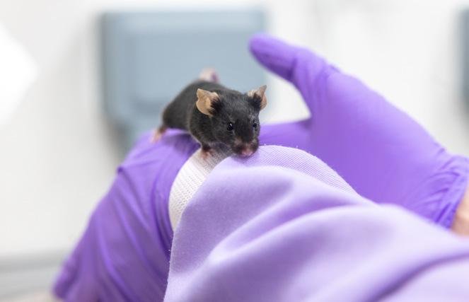
Below right: Animals are anaesthetised for short periods during imaging

says Professor Murphy, “Drug companies could use this as a platform in the future to more rapidly, and more effectively, screen potential drugs for inflammatory bowel disease. We don’t have good drug treatments at the moment, but I think this will accelerate that drug development.”
Professor Murphy has recently been awarded funding from Diabetes UK to use the mouse ultrasound imaging to understand vascular changes hat occur as a result of diabetes. He explains, “Some people can have impaired regulation of glucose but the point when this switches to type 2 diabetes is when the pancreas can’t keep up with the pressure and this is often reflected in changes in vascular health. People get microvascular disease, retinal disease, where they can go blind, neuropathy in the legs that can lead to amputation, and some can experience heart disease as well. The idea is to use the mouse model to longitudinally image vascular health and to see if anything promotes blood flow to specific tissues and whether that can actively help with the diabetes itself and those downstream vascular problems.”
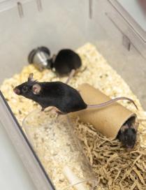
In the past, Professor Murphy has also had funding from the UK National Centre for the Replacement, Refinement and Reduction of Animals in Research (NC3Rs) for non-invasive physiological studies looking at what factors affect food intake in animals. He also contributed to work that proved that giving animals environmental enrichment, which can be as simple a cardboard tube to play in and chew on, does not interfere with food intake. This work means that animals in food studies can now be given enrichment and researchers can be confident that the animals will still eat normally.
Professor Murphy adds, “I think the 3Rs are really important. In the work my lab does we’re always thinking about the 3Rs; whether we can refine an experiment and whether we really need to use animals at all.
“Animal work is important and, at the moment, it can’t be entirely replaced. But you can’t treat animals like test tubes. You also need to weigh up the harms that you’re causing at the same time. I think the 3Rs provide a great framework for looking at that.”
We’re always thinking about the 3Rs: whether we can refine an experiment and whether we really need to use animals at all.”
– Professor Kevin Murphy
Left: Professor Kevin Murphy with Cecilia Dunsterville, PhD student at the Faculty of Medicine
Drug companies could use this as a platform in the future to more rapidly, and more effectively, screen potential drugs for inflammatory bowel disease.”
Animal Research Report 2022–23 | 27
– Professor Kevin Murphy
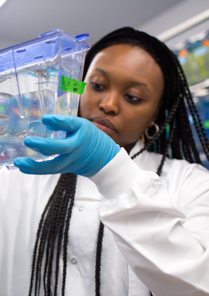
Zebrafish at Imperial are the focus of a study by bioengineers to discover a new mechanism driving the growth of heart valves in their embryos
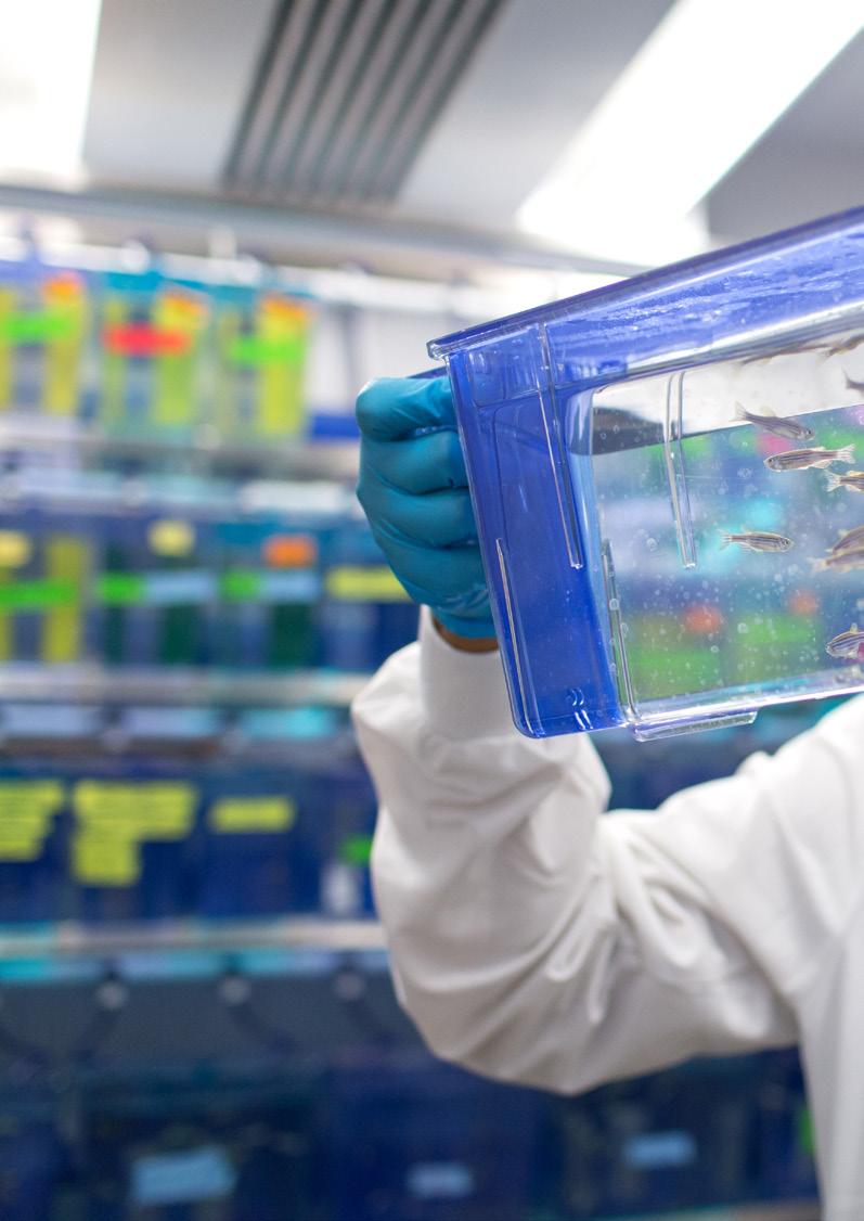
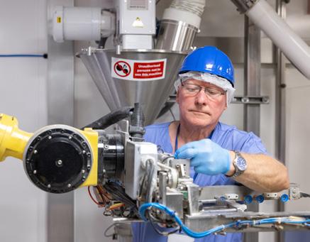
KEEPING IMPERIAL’S ANIMAL FACILITIES RUNNING
Modern animal facilities rely on robotic machinery to keep conditions clean and safe. This is vital for the health of animals and the staff who care for them, as well as the quality of the research being conducted.
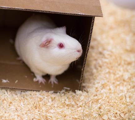
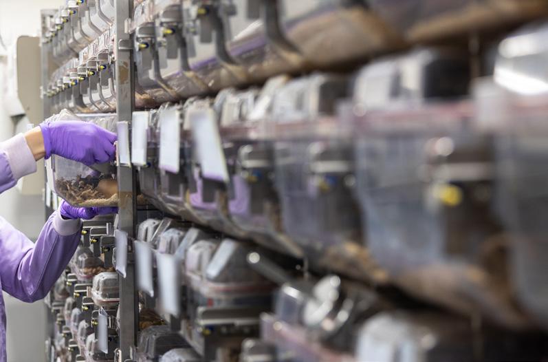
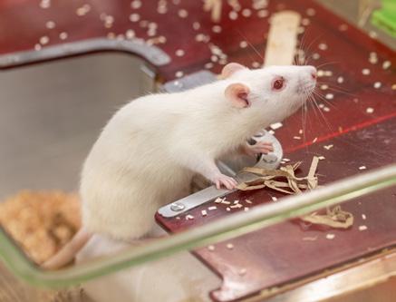
Thousands of research animals, mostly mice, are cared for at Imperial’s animal research facilities. This means that every week, hundreds of cages and water bottles must be cleaned and replaced.
At the mouse breeding facility this process is overseen by Support Service Supervisor Piotr Czapski. Piotr studied in Poland before moving to the UK. He joined Imperial as an animal technician in 2007 and qualified as a Named Animal Care and Welfare Officer (NACWO) before taking on his current role.
The cleaning work itself is increasing done by robots and this machinery is maintained by CBS service engineer Steve Steel across all Imperial’s animal facilities. Steve has worked as an
engineer since the late 1970s and has been in-house at Imperial for the last nine years.
Steve says: “Ten to fifteen years ago, the cage wash operators would have to do everything manually. One person would receive the dirty cages, empty them and manually load them onto a tunnel wash. Then, when it was clean, another operator would have to take it off the machinery, fill it up with fresh bedding and present it ready to go back to animal rooms. Now, in most of our sites, that’s all taken care of by robots.”
Piotr adds: “We can see the difference in the case of a breakdown when this work needs to be done by hand. The amount we need to process is huge. And it’s a constant process, so the robots are essential.
“Like all technology, the machinery involved is continuously improving and a new, more compact bottle washing machine has just been installed at Piotr’s facility. He explains: “Every single cage needs a water bottle, and these must be changed twice weekly. The new bottle wash takes crates of used bottles, places them on a conveyor, de-caps them, washes the bottles and caps, refills them with clean water and replaces the caps so they’re ready to go.
“Using a machine guarantees the bottle are completely clean. It also removes the need for manual handling – some of the crates will weigh around 20kg.”
These mechanised processes are part of a wider system designed to keep the facilities clean for the benefit
30 | Animal Research Report 2022–23
of the animals, the staff who care for them and the research being carried out.
“The technology reduces allergen levels across the unit,” Piotr explains. “For example, when I started work here in 2007, we used to have open cages. This meant allergens like dander were airborne. We wore masks but animal technicians could still easily get asthma. A few years later, we had individually ventilated cages which are sealed to protect the animals from their external environment, and everything is channelled away to protect technicians from allergens.
“Whenever you change an animal’s cage it has to be opened on an animal transfer station where the air is filtered to prevent cross contamination. From a researcher’s points of view that’s very important. There might be different studies going on in the same room and we need to make sure pathogens aren’t transferred from one cage to another.”
The very high standards of hygiene at Imperial’s animal facilities are part of the reason why it was the first UK university to achieve accreditation from the prestigious international animal welfare society the Association for
Assessment and Accreditation of Laboratory Animal Care (AAALAC).
Now, Piotr, Steve and their colleagues must also consider the environmental impact of their work. Steve says: “Manufacturers are already clued up on this and they’re working on a new generation of equipment which will be as efficient in sanitisation but will use less energy and less chemicals. That will be the next step forward.”
In the meantime, Piotr and Steve continue their work with existing machinery and the staff who operate it. “I enjoy fixing things,” says Steve. “I also enjoy training other people and helping them improve their own knowledge.
“Most of the time the equipment works trouble free. But on occasion the operator will have to intervene and sort a problem out, so it’s a higher-level job now than it used to be.”
Piotr adds: “When there’s a problem and you’re able to resolve it, that is something you should be proud of. But sometimes I need another pair of hands so I call on Steve.
“One cannot work without the other, we are dependent on each other’s work so we’re always aiming to work as a team.”
Ten to fifteen years ago, the cage wash operators would have to do everything manually. Now, in most of our sites, that’s all taken care of by robots.”
–
Steve Steel, CBS Service Engineer
Left: Imperial was the first UK university to achieve accreditation from the Association for Assessment and Accreditation of Laboratory Animal Care
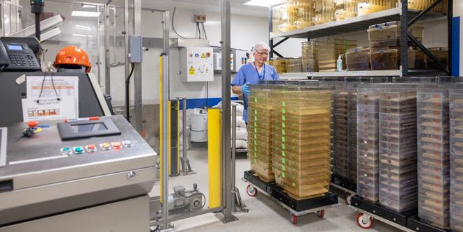
Right: Steve Steel checks equipment at Imperial’s animal facility
Animal Research Report 2022–23 | 31
Left: Cleaning and replacing of cages at the mouse breeding facility is overseen by Support Services
LEADERS IN COMMUNICATIONS
Leaders in Openness on Animal Research
Imperial College London was re-confirmed as a Leader in Openness on Animal Research in May 2022 by the not-for-profit mutual society, Understanding Animal Research.
Leaders in Openness are signatories to the Concordat on Openness in Animal Research who are recognised for committing considerable resource and energy to following best practice, embedding openness within their organisations, and making the aims of the Concordat a reality.
The assessment process involved a public panel, a peer review process, and a public review.
Imperial’s media engagement strategy around animal research was also highlighted as a case study in the 2022 Annual Report on the Concordat on Openness. The Concordat highlighted the work of Imperial’s Communications team and the good practices around openness in Central Biomedical Services.
Talking about animal research on social media
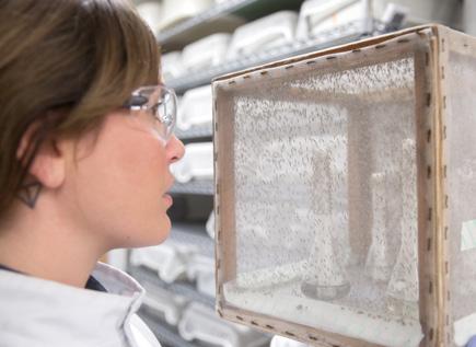
Imperial facilities were featured both in 2022 and 2023 in photos and videos on Understanding Animal Research’s social media channels.
In May 2023, during the annual #MiceInResearch social media event, Imperial took over Understanding Animal Research’s Instagram account. Instagram Takeovers give organisations the opportunity to talk to a different audience by engaging with another organisation’s followers. The content was also promoted by Imperials’ own social media accounts.
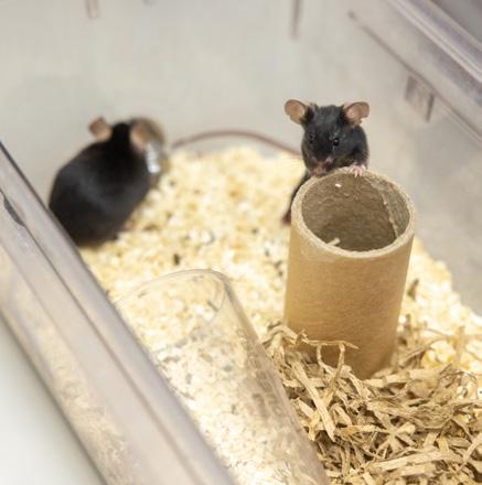
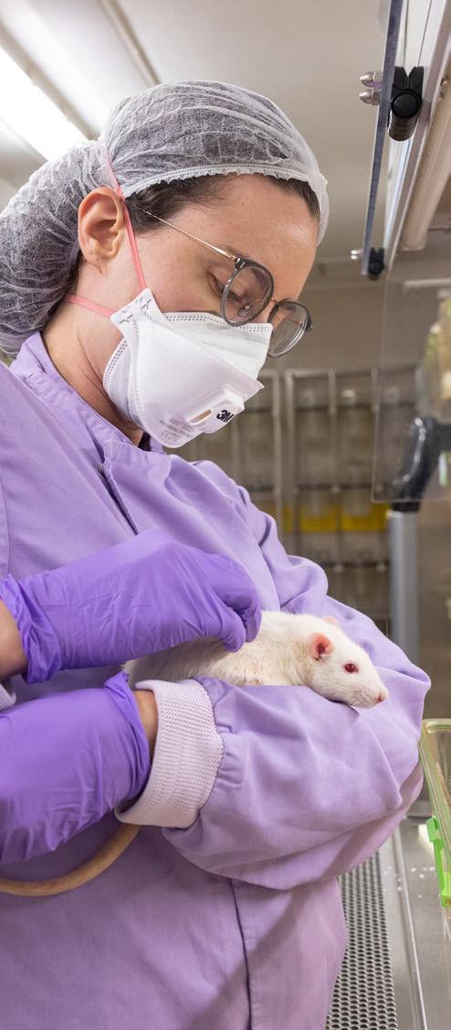
The Central Biomedical Services department, which cares for the animals used by the College for research, opened the doors of Imperial’s Breeding Unit to talk openly about Imperial’s work with mice. The takeover included video and images from within the animal facilities and the London Medical School (LMS-MRC) laboratories and it was run in collaboration with Senior Animal Technician, Stephanie Natario; Senior Researcher, Elaine Irvine; and PhD student, Chiara Pojani.
The trio created content to show the public how we care for mice within our units, why we use mice in research at the College and how animal research is regulated at Imperial.
“I am pleased I was allowed to openly discuss my research on social media” said Chiara, “I am keen on helping people understand how animal research works and why animal models are still crucial nowadays. Both our Comms team and the Understanding Animal Research team were supportive, and the whole process ran smoothly.”
The event reached thousands of followers on the Understanding Animal Research, Imperial and MRC social media channels. Stephanie said, “I thoroughly enjoyed being a part of the Instagram takeover. I had a really good time, and it was interesting to see what types of research are conducted involving the animals I care for.” You can watch the stories on the Understanding Animal Research Instagram Highlights: @understandinganimalresearch.
Imperial also made use of other external social media campaigns to promote its animal research community. Early in 2023, Imperial joined the Institute of Animal Technology’s #TechMonth Campaign. This month-long event drew attention to the critical role of animal technicians and technologists.
In collaboration with several CBS staff members at different career levels, the Social Media team produced a series of threads to share on Twitter, giving voice to Imperial’s animal technologists through quotes describing their experiences and feelings about working in animal research.
32 | Animal Research Report 2022–23
Animal research on Imperial web pages
Since the last animal research report, the animal research web pages have been reviewed to make it easier to navigate and ensure all the content is accessible to a broad public audience. The Digital team have also made the animal research section of the Imperial website easier to find from the College’s homepage and improved accessibility for people with visual impairments.
Spotlighting members of the Imperial community
Imperial has been working to ensure that staff carrying out animal research at Imperial are included in broader communications initiatives. One example of this is the Making Waves project, a recent series of immersive features showcasing the great science and teaching taking place at the National Heart and Lung Institute. The series shone a spotlight on Simone Walker, a Senior Research Technician, who manages and runs a specialist technical facility measuring lung function in mice.
To keep up-to-date on animal research at Imperial, including the latest news on research projects and discoveries, awards and resources for researchers and technicians, visit: www.imperial.ac.uk/research-andinnovation/about-imperial-research/ research-integrity/animal-research/
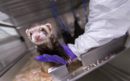
Far left: Ferrets have been used to study influenza as their respiratory system is physiologically similar to humans
Right: An animal technologist holds a guinea pig used to train technologists and researchers how to handle animals
I am keen on helping people understand how animal research works and why animal models are still crucial nowadays.”
– Chiara Pojani, PhD student
The Concordat on Openness on Animal Research
By signing the Concordat on Openness on Animal Research, Imperial has made the following commitments:
Commitment 1
We will be clear about when, how and why we use animals in research
Commitment 2
We will enhance our communications with the media and the public about our research using animals
Commitment 3
We will be proactive in providing opportunities for the public to find out about research using animals
Commitment 4
We will report on progress annually and share our experiences
Design, editorial and photography: Communications Division, Imperial College London
34 | Animal Research Report 2022–23
Right: Moth research into why insects gather at artificial lights
www.imperial.ac.uk/animal-research
