Make amazing artwork with our microscopy reagents
We have spent over twenty years partnering with the scientific community to craft the products needed for breakthroughs in immunology, cancer research, and neuroscience. From established immunohistochemistry and immunofluorescence methods to advanced iterative photobleaching techniques, we are supporting researchers with directly conjugated and fluorescent antibodies, chemical probes, and secondary reagents.
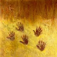
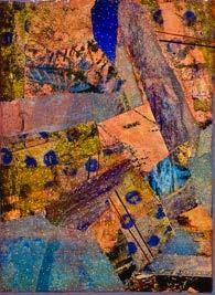
Each of the images below was submitted by researchers for the BioLegend 2025 Image Contest, where at least one BioLegend reagent was used to generate stunning results.
Learn more at biolegend.com/en-us/microscopy
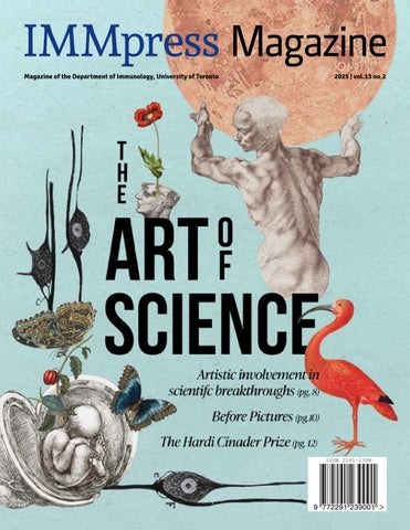
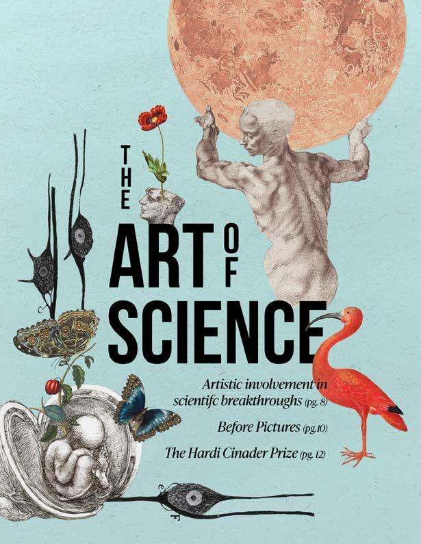
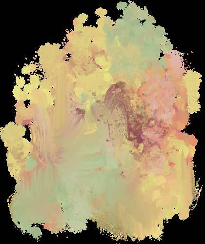
THIS ISSUE’S COVER
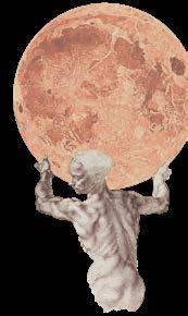
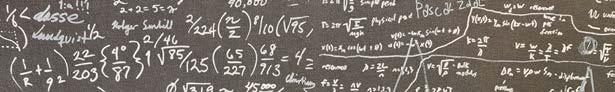
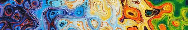
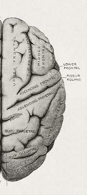
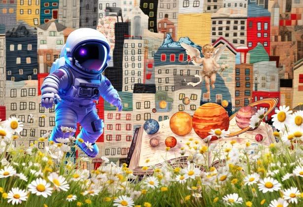
Is a surrealistic digital collage celebrating the works of notable artist-scientists. Pieced together are the masterful creations of – Michelangelo, Leonardo Da Vinci, Santiago Ramón y Cajal, Maria Sibylla Merian, Dorothea Maria Gsell, and John Russell. This cover is an homage to the greats who revealed the beautiful marvels of our world with their scientific curiosity and artistic sensibility.
Read below to learn more about the featured artists.
John Russell (1745–1806) was a British painter and an astronomy enthusiast. Enamored by the moon seen through his telescope, he carefully depicted every patch, crevice, shadow of the alluring celestial object. His dedication to detail can be seen in his stippled (patterned dotting) engraving, Lunar Planisphere, Flat Light (1805).
Michelangelo (1475–1564), an iconic Italian Renaissance artist, deeply appreciated the human anatomy. His love for the human form is shown by the fine details of the man’s torso sketched in his Studies for the Libyan Sibyl (circa 1510-1511).
Maria Sibylla Merian (1647-1717) was a German scientific illustrator. With body color (opaque watercolor now known as gouache), she painted colorful and accurate studies of insects and plants - as exemplified by the Great Blue Butterflies and Red Fruits (circa 1705-71) on the left.
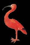
Her daughter, Dorothea Maria Gsell (1678-1743), also painted beautiful botanical and animal illustrations - as shown by the Scarlet Ibis with an Egg (1699-1701) at below right.
Santiago Ramón y Cajal (1852-1934), a Spanish neuroscientist and Nobel laureate, published seminal papers on the central nervous system. His pioneering work featured meticulous and captivating drawings of the brain and spinal cord with their constituting neurons - one of which is shown above.
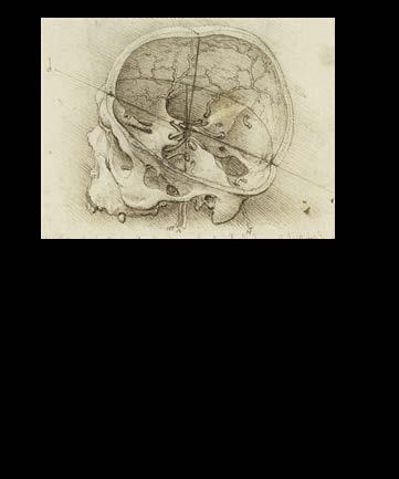
The legendary Italian Renaissance polymath, Leonardo Da Vinci (1452-1519), strove to understand the inner workings of the human body. After performing postmortem dissections, he recorded his observations through exquisite sketches - as shown by the above Studies of the Foetus in the Womb (circa 1510-1513).

Jennifer Ahn Design Director
EDITORS-IN-CHIEF
Meggie Kuypers
Manjula Kamath
DESIGN DIRECTOR
Jennifer Ahn
SOCIAL MEDIA COORDINATOR
Tianning Yu
SENIOR EDITORS
Jennifer Ahn
Daniah Alkassab
Zi Yan Chen
Baweleta Isho
Manjula Kamath
Meggie Kuypers
Annie Pu
Faizah Numa Sayeed
Siu Ling Tai
Boyan Tsankov
Deeva Uthayakumar
Nicolas Wilson
Tianning Yu
Adriana Zutic
DESIGN ASSISTANTS
Jennifer Ahn
Zoeen Carter
Yinhan Dechi Castro
Milea DiPonzio
Baweleta Isho
Manjula Kamath
Vera Lynn
Ana Sofía Mendoza Viruega
Preya Patel
Faizah Numa Sayeed
Victoria Sephton
Sophie Sun
Tianning Yu
WRITERS
Jennifer Ahn
Zi Yan Chen
Milea DiPonzio
Mahdieh Golzari-Sorkheh
Rishabh Johri
Vera Lynn
Ana Sofía Mendoza Viruega
Annie Pu
Faizah Numa Sayeed
Raven Stoddart
Siu Ling Tai
Christopher Ryan Tan
Boyan Tsankov
Deeva Uthayakumar
Tianning Yu
FOUNDING EDITORS
Yuriy Baglaenko
Charles Tran
C ontributors
Copyright © 2013 IMMpress Magazine. All rights reserved. Reproduction without permission is prohibited. IMMpress Magazine is a student-run initiative. Any opinions expressed by the author(s) do not necessarily reflect the opinions, views or policies of the Department of Immunology or the University of Toronto.

Join our mentorship Join our mentorship team team
WE’RE RECRUITING MOTIVATED MENTORS AND MENTEES
THE GRADUATE PEER SUPPORT NE T WORK IS A FACULT Y OF MEDICINE INITI ATIVE TH AT AIMS TO IMPROVE WELLBEING OF GRADUATE STUDENTS.
OUR MENTORSHIP PROGRAM PAIRS TRAINED STUDENTS WITH MENTEES TO PROVIDE SUPPORT WITH CH ALLENGES TH AT STEM BEYOND THE SPHERE OF ACADEMIC AND CAREER GOALS.
SIGN UP USING THE QR CODES BELOW:
MENTOR: MENTEE:


Solving for (X)-istential Patterns Within Nature
The Science of Music
An Immunologist’s Suite: How Dr. Gabriel Victora Composed a Life in B cells
Beyond Left and Right: The Neuroscience of Creativity and Logic
The combined impact of art and science on society & the power of mixing fact with feeling
Painting and Science: What Photography and Impressionism Teach Us About Innovation
An AI-generated picture worth a thousand words
Michelle Letarte: Building Community with Art and Science

Artistic Involvement in Scientific Breakthroughs
Before Pictures
The Hardi Cinader Prize: Celebrating Graduate Students Crafting Scientific Breakthroughs with Artistry and Precision
Bioart: when science and creativity join paths
The inseparable nature of art and science: Does the “Renaissance man” still exist today?
The Science of Culinary Arts Body horror and horrific bodies: what the horror genre can tell us about responsible science
Letarte & El-Maklizi Art Showcase
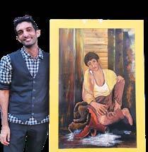
Shaping identity with Science & Art: Mahmoud El-Maklizi


FROM THE
CHAIR L etter
“The most beautiful thing we can experience is the mysterious. It is the source of all art and science.”
- Albert Einstein
Thank you to our talented IMMPress writers for bringing this concept, and this quotation from Albert Einstein, to our attention. Einstein also said, “The greatest scientists are always artists as well”, and while we may not all be avid practitioners of the arts, I would argue there is an artist inside all of us, just waiting to be released. Indeed, in my experience, it can be hard to separate the scientist from the artist. I am thinking about my faculty colleagues who play active roles in the arts, including Drs. Chris Paige (chair of the Tafelmusik board of directors), Rob Rottapel (board member of the Toronto Symphony Orchestra and talented flautist), Michelle Letarte and Eleanor Fish (talented visual artists) among others. Clearly there is a strong vein of artistic talent that runs through our collective Departmental history –after all, the Cinader award, named after one of our founding members, recognizes senior PhD students who have not only pursued scientific excellence, but also a passion for the arts.
I myself find inspiration at my weekly choir practices where I can focus entirely on something that is not science. And that is a big theme of this issue – how

using the creative side of our brains for something unrelated to our science can bring unexpected epiphanies. It’s like trying the crossword in the morning, then returning to it in the evening and realizing you have been unconsciously solving those tough clues while your brain marinates in something else. But art is more than brain diversion – we know that the representation of data in our papers is highly visual, particularly in this age of multidimensional datasets. This can make a big difference to readership when your paper is published, the equivalent to hanging a painting on a gallery wall for the rest of the world to see.
While the visual arts can be a springboard to communicating science, music has the power to teach us about the importance of practice, of repetition and of gaining resilience. The biography of Dr. Gabriel Victora, who began as a concert pianist and is now a renowned B cell biologist, reminds us that a similar discipline drives the scientist and the musician to strive for perfection. As noted in the article on “beyond left and right” the best art and science come from “a blend of focus and creative freedom”. After all, there is only one way to get to Carnegie Hall…
Beyond the production of art (and science) I really liked how Dr. Michelle Letarte described her experience among artists at Propeller Gallery as not just about the art, but about the community. It was that sense of community that also prompted Dr. Mahmoud El-Maklizi to return to art during his PhD in our Department. Is it not the same in science? We are at our creative best when we urge each other to take risks and we can help each other with creative problem solving.
Summing up this issue, I quote from the article on Fact and Feeling: “At their core, artists and scientists often share a common objective: to challenge conventional ways of thinking and uncover new perspectives that allow us to enhance our experience as human beings”. As scientists, that’s our job in a nutshell – and what an amazing job it is.

Jen Gommerman, PhD Canada Research Chair in Tissue Specific Immunity Professor and Chair, Department of Immunology
While art and science are often thought to belong within the opposing realms of creativity and logic, both are at their core characterised by curiosity, exploration, and the spirit of innovation. It cannot be coincidence, after all, that some of the most renowned scientists in history were also great artists — and vice versa. In this issue of IMMpress Magazine, we seek to bring this connection into the light. Science offers precision, methodology, and the pursuit of truth, while art infuses these discoveries with beauty and meaning. By delving into the intersection between these two contrasting yet complementary disciplines, we celebrate the shared drive that fuels discovery and expression; the desire to push beyond our boundaries and see the world not just as it is, but as it could be.
We begin with a historical perspective, looking at how scientists have previously used art as both a form of inspiration (p.8) and documentation (p.10). Science and art exist in tandem within the natural world (p.14-16) and are inextricably linked within the neuroscience of our perception (p.24). It
L etter
FROM THE
EDITORS
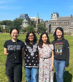
In order of left to right: Tianning Yu (Social Media Coordinator), Manjula Kamath (Co-Editor-in-Chief), Jennifer Ahn (Design Director) & Meggie Kuypers (Co-Editor-in-Chief)
is undeniable that both have dramatically shaped our societies (p.26), especially as technological advancements have pushed our capabilities forward to enable photo-realism (p.28), artificial intelligence-generated art (p.30), and the manipulation of biology itself into an art form (p. 32). With such tools at our fingertips, it is perhaps to be expected that art and science have become largely inseparable within the modern world (p. 34), to the extent that their combined application can be found within mundane practices such as cooking (p.35) and entertainment (p.36).
As a highlight of this issue, we feature two interviews with Professor Emerita Dr. Michelle Letarte (p.20) and alumnus Dr. Mahmoud El-Maklizi (p.22) about the duality of their passion as artist-scientists and how they have managed to incorporate these unique facets of themselves into their everyday lives. A collection of their work is showcased on the back panel (p.38), reprinted with their permission. We also feature a biographic article of Dr. Gabriel Victora (p.18), detailing his remarkable journey as a scientific researcher with a background in music, as well as Dr. Bernhard (Hardi) Cinader
(p.12), whose legacy as an artistscientist continues with the annual awarding of the Hardi Cinader Prize. We are incredibly grateful to these generous individuals who were willing to share their experiences and artwork within these pages.
Lastly, we cannot forget our amazing team of writers, editors, and designers that continue to make IMMpress Magazine into a reality. Thank you to all of our contributors for their hard work in creating yet another fantastic edition!
Co-Editors-in-Chief

Meggie Kuypers
Artistic Involvement in Scientific Breakthroughs
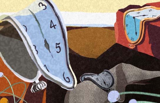
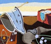
Accordingto Pablo Picasso, “Art is a lie that makes us realize truth.” Art in its very nature does not provide an accurate depiction of reality. In fact, oftentimes, it may even provide a twisted version of reality as can be appreciated by the surrealist paintings of Salvador Dali, or the magical realism present in Gabriel Garcia-Marquez’s writing. Hence, art can indeed be a lie about the very nature of reality. However, by actively spending time with the “lies” of great artists, we may begin to see the world at a deeper level. For example, the malleable, almost liquid-like clocks in Dali’s “The Persistence of Memory” may seem strange at a cursory glance. However, a keen eye may realize the very purpose of these illusory clocks are to reveal the truth that time may not be universally linear – it bends, stretches and collapses in dreams and memory. If we are therefore convinced by Picasso’s idea that “art is a lie that makes us realize truth”, we may be unsurprised to learn that art (or at minimum, artistic thinking) has fueled some of the most important discoveries in chemistry; the structure of the atom, the structure of the benzene ring, and the development of the polymerase chain reaction (PCR).
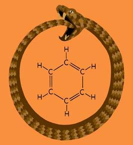
Perhaps the most famous implementation of Picasso’s definition of art lies in August Kekulé’s visualization of the benzene ring structure in 1865. Since its extraction from compressed coal gas by Michael Faraday in 1825, chemists throughout the 19th century wanted to understand the intricacies of benzene formation. This was largely due to the mysterious nature of benzene’s reactivity. Benzene’s chemical structure was known to have a formula of (C6H6) which made it a polyene, meaning, having many double or triple bonds. Unlike other polyenes known at the time which were highly reactive, benzene was not, which generated a lot of interest in understanding its enigmatic structure. In 1865, August Kekulé made the greatest initial leap in visualizing its structure. Having had architectural training prior to transi-
tioning to chemistry, Kekulé had what he described as an “irresistible need for visualization.” Through a dream, he imagined the initial structure of benzene, visualizing its cyclical structure as a snake eating its own tail (a symbol in ancient cultures known as the ouroboros). This dream led him to characterize benzene as a regular hexagon with a hydrogen at each corner. As Picasso would say of art, this structure of benzene would turn out to be a “lie.” However, it was not too far from the truth, and in fact, would lead Kekulé to the true structure of benzene several years later. Indeed, in 1872, Kekulé published the “true” structure of benzene that is known today – a mixture of cyclohexatrienes in rapid equilibrium.
Today, the knowledge that atoms are the building blocks of all life and matter is ubiquitous. We may even know that atoms mainly consist of negatively charged electrons orbiting in distinct shapes around a positively charged nucleus. Although this knowledge may seem obvious in the modern world, when Niels Bohr first developed his hypothesis for the structure of a hydrogen atom in 1913, the nature of how electrons orbited around a nucleus was still unclear. At the time, the prevailing notion was based on the Rutherford model, which postulated that electrons orbit around a nucleus like planets around the sun. However, the Rutherford model suffered from a critical flaw brought about by knowledge of classical physics; if electrons orbit around a nucleus, they should emit energy continuously and eventually spiral into the nucleus of the atom. However, it was observed that hydrogen emitted energy at specific wavelengths, and therefore the Rutherford model needed amendments. In a dream, Bohr imagined electrons orbiting around the atomic nucleus at fixed, discontinuous energy levels. Much like an observer at a museum interrogating a surrealist painting, Bohr pondered upon the incomplete works of his predecessors and improved upon them using his own imagination, thereby “realizing truth” as Picasso would imply. Bohr’s improvements of Rutherford’s model indicated that electrons may emit only specific wavelengths of energy, which was precisely what is observed when examining emission spectra of various elements. The dream-induced artistic influence earned Niels Bohr the Nobel prize in Physics in 1922.
Another example of imagination revealing scientific truth is Kary Mullis’ discovery of the polymerase chain reaction (PCR) in 1983 — a technique that would revolutionize molecular biology. While driving along the California coast at night, Mullis was reflecting on how to amplify specific segments of DNA. He suddenly envisioned a cyclical process involving heat to separate DNA strands, primers to mark the target region, and DNA polymerase to build new strands. This method could amplify a small fragment of DNA millions of times in a matter of hours. Although his colleagues at the time initially dismissed the idea as too simple to work, Mullis’ PCR transformed molecular biology completely. Today, PCR is indispensable to many facets of biological science, including basic science, genetic research, forensic science, and medical diagnostics. In Picasso’s terms, Mullis’ mental conception of PCR may, by itself, be considered art. At the time, his imagination may have seemed like a lie; an artistic distortion of practical reality. However, examining the technique at a deeper level reveals a profound biological truth regarding the necessary and sufficient factors for DNA replication. The discovery of PCR earned Kary Mullis the Nobel prize in Chemistry in 1993.
These breakthroughs, from Kekulé to Mullis, each demonstrate how artistic thinking enables science to leap beyond what is visible or logically obvious. What may appear, at first, to be fiction or fantasy often becomes the key to unlocking scientific truth. As Picasso suggested, it is through these “lies” that we come closer to understanding the deeper realities of our world.
“Art is a lie that makes us realize truth.” - Pablo Picasso

Written by Boyan Tsankov
Designed by Zoeen Carter
Before pictures Science Illustration
Cryo-electron microscopy, photon counting-computed tomography, quantum imaging – these are a few modern developments that have allowed scientists to capture microscopic details at ultra-resolution. Researchers today have a vast arsenal of tools to visualize phenomena, from describing how cells communicate at a molecular level to large scale technologies like deformation imaging that map the rigidity of the Earth’s crust and mantle. But how were scientific observations documented before these advanced techniques? Prior to modern technology, how did scientists record the complexities of the human body or observe distant celestial bodies?
Before the digital age, scientists were fundamentally artists. Early scientific figures were not simply visualizations of data – they were the data itself, captured meticulously through hand-drawn illustrations. Before photography became widespread, images used in scientific journals like Nature were wood engravings that were inked and set for printing – a task requiring an incredibly high degree of skill and precision. Illustrators and engravers often worked closely with or were themselves scientists, carefully emphasizing specific details. Looking back at this period, scientific drawings served as more than records – they reflected what the observer chose to illustrate and emphasized the features they considered most significant.
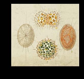
One example of the subjectivity of early scientific art is Ernest Haeckel’s idealized drawings of radiolarians, a group of marine organisms with intricate mineral skeletons. His depictions were highly stylized, emphasizing symmetry and aesthetics, sometimes over factual details. Despite these liberties, Haeckel’s images remained the gold standard for the field of marine biology until the advent of scanning electron microscopy nearly a century later.
The concept of the scientist-artist was exemplified by Leonardo da Vinci, who extensively studied human anatomy in addition to his well-recognized role as a painter. By his death in 1519, da Vinci had compiled over 5,000 pages of incredibly detailed notes and illustrations in anatomy, physics, geometry, and philosophy. His depictions of human organs were groundbreaking at the time; using glass casts, da Vinci accurately modelled the internal structure of the heart’s pulmonary artery. His anatomical sketches were exceptionally accurate – some, like his cross-sections of the human skull
Figure 1. Illustrations of radiolarians by Ernst Haeckel (1862).
have established themselves in medical education. Da Vinci’s sketches remained the forefront of anatomical understanding until Andreas Vesalius published De Humani Corporis Fabrica in 1543, a revolutionary text marking the introduction of “veridical representation”, a new paradigm focused on accurately representing anatomical structures as they truly exist. With the Fabrica came a change in using illustrations as visual records to essential didactic tools.
Galileo Galilei, although not primarily trained as an artist, effectively used artistic techniques in his astronomical studies. In Sidereus Nuncius (The Starry Messenger, 1610), Galileo included etchings of the moon based on his telescopic observations. He was among the first to depict the moon’s surface as rugged and cratered, directly challenging the Aristotelian belief that heavenly bodies were smooth and unblemished. His illustrations used a technique known as chiaroscuro, which contrasts light and shadows, to emphasize the moon’s topography — ridges, craters, and mountains — much like a Renaissance painter would. Beyond aesthetics, his drawings served explanatory and persuasive purpose. Galileo went on to draw star clusters like the Pleiades and Orion to demonstrate the unprecedented power of the telescope. His depictions of sunspots using sequential drawings functioned as modern time-lapse images, visually demonstrating that the sun’s rotation was imperfect, again refuting long-held beliefs.
The introduction of photography in the late 1800s marked a turning point in scientific imaging as wood engravings and hand-drawn sketches gave way to glass lenses and silver halides. Photography was embraced for its perceived objectivity – photographs were trusted to capture observations with an unbiased clarity that the eye could not. Photography’s
scientific relevance grew dramatically throughout the 20th century. When Kathleen Lonsdale pioneered the technique of X-ray crystallography to measure a sample’s atomic and molecular structure, her X-ray diffraction photographs were crucial to the discovery of DNA’s structure by James Watson and Francis Crick. The key piece of evidence was ‘Photograph 51’, which showed the diffraction pattern of DNA.
As we move further away from the stylized drawings and hand-made models of da Vinci’s and Haeckel’s eras towards AI-enhanced imaging and 3D rendering, one might question if the artistic process has been lost in today’s technology. Historically, art was an essential means of representation, revealing not only scientists’ observations but also their unique perspectives, philosophies and interpretations of the world. The deliberate and meticulous nature of traditional scientific illustration has since given way to modern methods prioritizing efficiency and objectivity. However, rather than disappearing, the role of art in science has taken a different shape. Artistic insight and creativity certainly still have a place in the scientist’s toolkit –today, researchers and medical illustrators must carefully choose colors, textures, and perspectives to highlight biological processes like cell interactions or neural pathways, making complex data accessible and engaging. The ability to effectively visualize data remains an inherently artistic skill, reminding us that science communication is and will always be an interplay between technical precision and creative expression.
Written by Rishabh Johri
Designed by Preya Patel
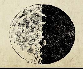
Figure 3. Sketch of the moon by Galileo Galilei (1610).
Figure 2. Anatomical drawing by Leonardo da Vinci showing a detailed cross-section of the human skull (1489).
The Hardi Cinader Prize: Celebrating Graduate Students Crafting Scientific Breakthroughs with Artistry & Precision
The Hardi Cinader Prize is a well-recognized award for those in the Department of Immunology at the University of Toronto. Each year, this award is given to a graduate student pursuing doctoral studies in Immunology. The award selects the student who “best reflects the goals and life of Dr. Cinader”, with a specific emphasis on scientific prowess and fascinations in both the arts and sciences. Dr. Cinader, more commonly known as Hardi Cinader, was himself a widely accomplished scientist with a deep interest in Canadian art. While the prestige and monetary value of this award are well recognized by graduate students and faculty members alike, we often do not lend enough thought to the origins of scholarships like the Hardi Cinader Prize, many of which act as legacy awards. Who was Hardi Cinader, and why does his name grace such a prestigious award? As time passes, it is important that we continue to remember and honour those who helped build the Department of Immunology into what it is today. So, without further ado, let’s talk about Dr. Cinader!

Dr. Bernhard (Hardi) Cinader was born March 30, 1919, in Vienna, Austria. He received his Bachelor of Science from the University of London in 1944-1945 and subsequently completed his PhD in Biochemistry at the University of London Lister Institute of Preventative Medicine in 1948 – talk about scholarly! Records indicate that he served on the scientific staff of the Lister Institute in 1950.
After achieving the title of Doctor of Science (D.Sc.) in Immunology from the University of London in 1958, Cinader immigrated to Canada, where his scientific career took off. Cinader became Head of the Immunochemistry subdivision of the Ontario Cancer Institute in Toronto before being promoted to Professor of Medical Cell Biology, Medical Genetics and Clinical Biochemistry at the University of Toronto in 1969. Around the same time, Cinader acted as a founding member and the first President of the Canadian Society for Immunology (CSI) from 1966-1969 while simultaneously playing an instrumental role in establishing the International Union of Immunological Societies (IUIS). His achievements continued into 1969, serving as the first IUIS President. Also around this time, Cinader became instrumental in the early days of what would become the Department of Immunology. He became the Director of the Institute of Immunology from 1971-1980, helping to foster its development until 1984 when Immunology attained academic status within the Faculty of Medicine as a fully-fledged Department.
Cinader was an incredibly accomplished scientist. His research was instrumental in guiding our understanding of many key immunological topics. He investigated foundational concepts including the production of cytokines , human antibody classes, antibody catalysis (the ability of antibodies to destroy pathogens), the first descriptions of mouse antibody classes (allotypes), and more. His work has been published across numerous reputable journals and he has been a pioneering figure in immunology research in Canada.
Beyond his scientific passions, Cinader was a wellknown patron of Indigenous Canadian art and artists. In 2001, shortly after his passing, he bequeathed his collection of First Nations’ art to the Royal Ontario Museum (ROM). His collection housed over 600 paintings, drawings, prints, sculptures, ceramics, textiles, quill and beadwork, which can now be seen on rotation at the ROM. He has been recognized as an authority on First Nations art. Together, the many aspects of Cinader’s life including his research, his artistic passions, and his teaching and mentorship are remembered. When described, many may have called Cinader “extraordinary” or “exceptional” for excelling beyond the standards of scientific research and engaging heavily with the community.
Traditionally the Cinader Prize is awarded at the Immunology Department Holiday Party (often sandwiched between a lighthearted faculty roast filled with scientific wit and the department busting a move on the dance floor). Any graduate student expected to earn their PhD in the succeeding year or who have received their PhD in the preceding year are eligible for the award. This award is not to be confused with the Hardi Cinader Graduate Scholarship in Immunology, the Cinader award lectureship granted through CSI, or the Bernhard Cinader Award given by CSI – which Cinader acted as the inaugural award recipient for at the first CSI meeting held in 1987.

It is important to remember the vast impact Dr. Cinader had on his community. From his extensive contributions and leadership endeavours, he was an exceptional figure in our collective history. His lasting legacy should remind us that while being driven in our academic endeavours is important, we will be remembered as more than the sum of our parts, and we should pursue our passions unapologetically.
Written by Raven Stoddart
Designed by Yinhan Dechi Castro


Humans have sought to mathematically describe and characterize the structure, patterns, and evolution of natural organisms and processes for over two millennia. The ancient Greek philosopher Pythagoras (570-495 BCE), who famously developed the Pythagorean theorem, helped to pioneer and disseminate the idea that natural phenomena, or “the cosmos”, could be explained using numbers and numerical relationships. Whether it is the ridged pattern of a pinecone, the ripple effect of a stone dropped in a pond, or the branching patterns of a tree or a neuron…, natural structures, growth patterns, and events are woven into mathematical relationships that help us to clarify and predict elements within a complex world.
Imagine this: You are breeding a pair of rabbits, which are raised in ideal conditions that enable them to give birth to one male and one female offspring. This pair of offspring, in turn, becomes fertile in one month and also gives birth to one offspring of each sex. Consider that with every new pair of rabbits reaching fertility, the older pairs are still breeding. If this cycle continues, how many pairs of rabbits will you end up with in a year? (See answer at the end of the article.)
This was a thought experiment proposed by the mathematician Leonardo Bonacci in the 13th century. The total number of breeding rabbits at the end of each month corresponds to what is now known as the Fibonacci sequence, a pattern by which each element is the sum of the two immediately preceding numbers. Starting with 1 and 1, as x1 and x2, respectively, it continues as 2, 3, 5, 8, 13…∞
The mathematical equation is written as:
Numbers within the Fibonacci sequence are commonly observed in nature. One example is the number of petals in flowers: lilies and irises (3 petals), butter cups and wild roses (5 petals), and delphiniums (8 petals). Other plant structures,

Solving for x-istential
like sunflower seeds and pinecones, consist of spiral arrangements; the number of seeds in each spiral reflects a number in the Fibonacci sequence. While not a deliberate design, natural organisms may have “adopted” the Fibonacci sequence to maximize occupancy within a given space. For plants, this would mean arrangement of petals and leaves to maximize exposure to sunlight and other nutrients. Nonetheless, flower species exist that do not follow this pattern, such as poppies (4 petals). Clearly, other genetic, environmental, and evolutionary pressures lead to variations.
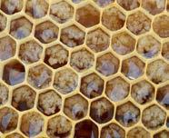
In addition to the Fibonacci sequence, other geometric shapes and patterns exist in the natural world. Fractal patterns, for one, are a potentially infinite repeating series in which an entire object is made up of smaller units resembling its likeness. Such mathematical patterns are ubiquitous in nature: the arteries of our circulatory system consist of large branching blood vessels, that extend into similar smaller branching units, called capillaries; similarly, ferns and broccoli consist of smaller units that mimic their overall structure. Tessellations, or tiling patterns, are also frequently observed in nature, with notable examples including the repeated hexagon structure of honeycombs, pineapple skin, turtle shell patterns, and fish scale arrangements. With regards to the honeycomb, its hexagonal arrangement permits maximal honey storage in its wells, while minimizing the wax required to coat the perimeter. Mathematicians have recently classified another 3-dimensional tiling pattern of “soft cells”, which lack corners; these tiling patterns are found in multiple biological structures, including nautilus shell chambers and muscle fibers.

Patterns within Nature
While the specific advantages of this shape are unclear, this ignites a conversation to understand how organismal adaptation and evolution favour the flexibility and resilience of certain structural patterns over others.
While the mathematical concepts above describe the structure of living things, math in nature extends beyond geometry and shapes. From predicting the transmission of minuscule viruses to planetary motion, math can be used to model natural phenomena. For instance, epidemiologists employ math to trace and predict patterns in infectious disease transmission. Vast quantities of clinical and surveillance data can now be collected from populations to allow for more precise estimation of biological, behavioural, and environmental traits that contribute to infectious disease spread. These data are interpreted using mathematical concepts like pathogen offspring distribution, which is the number of people infected by the original infected person, and generation time distribution, which is the length of time it takes for successive infections to occur from one individual to the next. These models help us to predict the severity of disease outbreaks and define key parameters, such as the number of infections that must occur before an outbreak can no longer be contained without public health intervention.
Beyond human health, scientists employ math to describe a variety of environmental occurrences. Tidal patterns are caused by the gravitational pull of the Sun and Moon on
the Earth, in combination with other factors like the Earth’s rotation, the wind, and atmospheric pressure. An alignment of the Sun, relative to the Moon, can create exceptionally high or low tides (also known as “spring tides”). The gravitational pull exerted by either the Sun or the Moon, relative to the Earth, can be represented using an equation that depends on the gravitational constant, G, the mass of the two objects, and the distance between them. By incorporating the gravitational force and other parameters, geographers can use sophisticated mathematical modelling to more accurately predict tidal patterns. Understanding tidal patterns are important in activities such as navigation, fishing industries, and coastal engineering. Given the prevalence of math in the natural world, from infectious disease biology to oceanography, it would seem that Mother Nature was a formidable mathematician!
Many of the mathematical concepts discussed here provide a logical framework to understand how organisms could have evolved for survival. Inspired by nature’s wisdom, these mathematical concepts are used by artists and scientists alike in designing both art and man-made structures. Unravelling the parallels between art, science, and nature, through the incorporation of mathematical patterns, presents hidden beauty, order, and complexity in the universe. Continuing to view nature through a mathematical lens will help to enrich and foster in us a previously untapped appreciation and sense of wonder for the natural world.
Answer: 233 pairs of rabbits after 12 months
Written by Zi Yan Chen
Designed by Manjula Kamath

The Science of Music
Music is omnipresent in our lives. From wishing ‘Happy Birthday’ to our loved ones, to humming to our favorite tunes while doing daily chores, music finds a way to keep us company. Music allows us to convey a wide range of emotions beyond the limitations of language. And most importantly, it possesses the remarkable ability to influence our emotions and mood. But, how? Before delving into how music can affect our emotions, it is necessary to understand how we are even able to perceive it.


How do we perceive sound?
Sound originates from vibrating objects that produce sound waves, composed of moving air molecules. Music is made up of complex patterns of simpler sound waves.
There are several properties of sound that play a part in the perception of music. For instance, a piano and a guitar may produce the same note with the same frequency that sound distinct from each other. This is due to their quality of sound being different, also known as their ‘timbre’. In addition, the perceived loudness of sound is determined by the ‘amplitude’ of its sound waves. For instance, a sound wave with greater amplitude is perceived as louder.
The auditory system, including the outer, middle and inner ear, the auditory nerves, along with the brain are collectively needed to process sound. Sound waves are directed inwards by the outer ear, where they cause a thin membrane called the tympanic membrane to vibrate. These vibrations then cause three small bones called ossicles to vibrate. This transfers sound waves to the fluid-filled cochlea in the inner ear. Separate regions of the cochlea vibrate to different frequencies of the sound wave and contain sensory hair cells. These hair cells are connected to auditory nerves that transfer the signal to the brain.


How can music affect our emotions?
Music can affect our emotions irrespective of whether we are passively listening or actively singing along. The limbic system is a group of brain structures that controls emotions and behavior. It can be strongly influenced by music. Specifically, when listening to music, the amygdala, which is a small brain structure that is needed to process emotions, and the hippocampus, a part that governs memory formation, activate. By listening to a song associated with a good memo ry, an individual may reminisce about a moment of happiness from their past that might improve their mood in the present.
The reward system, which is a part of the limbic system, is also in volved when listening to music. The medial prefrontal cortex that is in volved in emotional regulation, gets rewarded, and memory lights up when people listen to familiar songs. It is also one of the regions that degen erates last in Alzheimer’s disease, which may explain why music is strongly connected to personal memories. In addition, when listening to music, the nucleus accumbens that is responsible for pleasure and reward-seeking behavior is also activated and produces the happy hormone, dopamine. This production of dopamine further contributes to the pleasure felt when listening to music, and the desire to continue listening.
How can music affect our cognitive function?
Music can improve cognitive function, for instance by reducing stress. Stress can negatively affect cognitive function and can cause various issues related to memory and concentration. It has been found that listening to music can decrease blood pressure, heart rate, and the levels of the stress hormone, cortisol. By decreasing stress levels and enhancing mood, music can also improve symptoms of depression and anxiety.
Music therapy, involving regular sessions with a certified therapist, in combination with standard treatment, can better alleviate depression symptoms and enhance quality of life.
Music may also help improve cognitive impairment, such as dementia. It was found that when patients with dementia listen to familiar songs from their youth, they can access memories and enhance recall. This may lead to a lower rate of cognitive decline as found in a study on patients with dementia who received music therapy in addition to the current standard of care.
In conclusion, music significantly impacts our well-being by affecting our emotions and improving cognitive function. Next time we put on our favorite song, we can all take a moment to appreciate just how much it can influence our lives.
Due to the positive effect of music on emotions, it can also help alleviate pain by acting as an effective distractor. The hypothalamus is a part of the brain that regulates key processes such as mood and hunger by releasing hormones itself, as well as by controlling the production of hormones from the pituitary glands. Listening to music can cause the production of endorphins by the hypothalamus and pituitary glands, resulting in pain relief as well as an improvement in mood. In addition, it may also dampen the perception of pain due to the increased dopamine production by the nucleus accumbens as mentioned previously.
Although listening to music has many benefits, it is important to choose the right music depending on your mood. If you’re having a bad day, picking tunes that are uplifting can improve your mood. Listen ing to melancholic or irritable music when you are already feeling upset can increase negative emotions like depression and anxiety. It is also important to consider the lyrics in songs, especially those related to violence and negativity, as they can also negatively affect our mood.
Written & Designed by
Music can not only affect our emotions but can also have an impact on our cognitive function.


Faizah (Numa) Sayeed
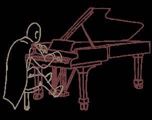
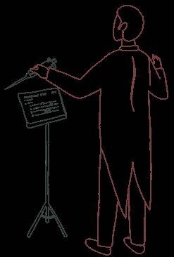
An Immunologist’s Suite
How Dr. Gabriel Victora Composed a Life in B Cells
Science and art are often presented as a juxtaposition, the former delimited by rationality and objectivity, while the latter exposing abstract thought and subjectivity. But what if these realms harmonized? Play your favorite song. Let it drift in the background and challenge this debate through the remarkable life of Dr. Gabriel Victora, a distinguished pianist and exemplary immunologist at The Rockefeller University. Science and art are often presented as a juxtaposition, the former delimited by rationality and objectivity, while the latter exposing abstract thought and subjectivity. But what if these realms harmonized? Play your favorite song. Let it drift in the background and challenge this debate through the remarkable life of Dr. Gabriel Victora, a distinguished pianist and exemplary immunologist at The Rockefeller University. His scientific achievements in B cell biology stand as a testament to the invaluable synergy of the arts and sciences. This article will delve into the lessons both fields offered for his success and life itself. His scientific achievements in B cell biology stand as a testament to the invaluable synergy of the arts and sciences. This article will delve into the lessons both fields offered for his success and life itself.
I.

II. Sherzo: Playful tunes and scientific turns
Allegretto:
The music behind Immunology
Growing up in Brazil amidst intellectually curious relatives, Dr. Victora’s career trajectory seemed set for music. Inspired by the spirited piano interpretations of his great-grandmother, he began playing the piano at a young age. At age 17, he moved to the United States where he pursued both his Bachelor and Master of Music at the Mannes School of Music in New York City, immersing himself in the strenuous life of a professional concert pianist. Yet over time, a quiet dissonance emerged. The intense regimen of practice, although rewarding, began to feel constraining, giving him the sense that something else was missing from the melody. Victora began to crave a different kind of creativity, one rooted in discovery and inquiry. Thus, he turned the page of the music sheet. The music paused, but another composition was just beginning.
Back in Brazil, Gabriel Victora found a new song to play. While translating research articles for his father, Dr. Cesar Victora, a renowned epidemiologist known for his work in child nutrition, he began to develop a growing appreciation for science. His duties soon expanded into translating medical journals. Although his interest in analytical reasoning had been apparent in the context of music theory, it was an internship in the laboratory of Dr. Jorge Kalil, an expert in allergy and clinical immunology, that shifted the tempo of his life. There, the curiosity crescendoed into conviction, prompting him to pursue a Master’s degree in Immunology in the Institute of Biomedical Sciences at the University of São Paulo.
During this formative period, Dr. Victora became fascinated by the intricate beauty of nature’s complexity. To him, science offered the opportunity to address many compelling questions that would benefit human health. But perhaps most transformative in his decision to pursue science was his early recognition of what he describes as “the importance of the free exchange of ideas”, a value he saw was essential to scientific progress and his own path. Just like how a musical phrase gains richness when it is passed between instruments, an idea or problem shared in a collaborative space can evolve in powerful ways with new meanings and textures. Science flourishes when voices are layered rather than isolated.
III. Allegro Molto: A fast movement towards lymphocyte dynamics
Venturing into science was not easy; it meant stepping into the unknown. But the discipline fostered by years of musical training primed him for his future success. He continued his scientific journey in the laboratories of Dr. Michael Dustin at New York University and Dr. Michel Nussenzweig at Rockefeller University, and later as a Whitehead Fellow at the Whitehead Institute for Biomedical Research at Massachusetts Institute of Technology. There, Dr. Victora turned his attention to one of the immune system’s most virtuosic players: B cells, and their own unique instruments, antibodies. He started investigating how this type of white blood cell develops in our bodies to produce antibodies, small molecules that specifically recognize and target foreign substances, allowing for protection following infection or vaccination. And so, it
began his life’s major composition: the symphony of studies in B cell dynamics.
Currently, Dr. Victora is the Laurie and Peter Grauer Professor and head of the Laboratory of Lymphocyte Dynamics at The Rockefeller University. His research has earned international recognition, marked by a prolific record of publications and numerous awards. Victora’s laboratory is focused on understanding the evolutionary processes that shape how B cells refine the binding ability of antibodies through mutation and selection. To tackle such complex questions, they utilize a broad arsenal of strategies, from traditional methodologies in immunology to cutting-edge technologies. Dr. Victora has found a space in which his creativity has clearly flourished.
In addition to obtaining valuable insights that could be considered art by themselves, the value of his scientific approach offers a lesson deeply rooted in his background as a musician. In a field defined by unexpected findings and shifting hypotheses, one of Dr. Victora’s most resonant pieces of advice is: “Make your own way to address the problem you ought to address.” As a musician, repetition paves the way to mastery, and such discipline prepares you for future challenges and failures. In parallel, scientific progress often depends on perseverance through failure. Whether it is a novel protocol, a new experiment, an unforeseen error, Dr. Victora reminds us: “Practice until it clicks… You keep going until the errors dissolve.” In both music and science, resilience is not optional, it is foundational.
In his inspirational career journey, Dr. Gabriel Victora continues to prove that science can be art, and art, a form of science. Dust off your instrument (or your microscope). Consider the world not just as a series of problems to solve, but as a composition waiting to be written, presented, and shared. After all, immunity has a rhythm, and sometimes science sings.
Notes of resilience
Ana Sofia Mendoza Viruega
Written & Designed by
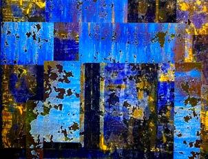
Michelle Letarte Michelle Letarte Art & Science
Building Community With D

r. Michelle Letarte is a Professor Emerita of the Department of Immunology at the University of Toronto. Her laboratory at The Hospital for Sick Children focused on understanding Hereditary Hemorrhagic Telangiectasia Type 1, a rare genetic disorder caused by mutations in the endoglin gene, which was discovered by her team. Alongside scientific research, she has devoted more than thirty years to artistic creation, with a focus on abstract painting. In this interview with IMMpress Magazine, Dr. Letarte reflects on her artistic journey and the creative passions that have shaped her life beyond the lab.
How did your artistic journey begin? I didn’t take art classes or make any art throughout high school, my Bachelor’s, PhD, or post-doc. But, I have always appreciated art. Especially during my post-doctoral fellowship in Oxford, I enjoyed going to art galleries all over Europe and appreciating different art forms. Then in 1975, when I started my lab here in Toronto, I decided to spend at least one evening
a week doing something different. So, I enrolled in OCADU (Ontario College of Art & Design University, then OCA) and took part-time classes for twenty-two years.
Which classes did you take in OCAD? I took classes in ceramics, textiles and sculpture. I also attended workshops provided by different artists or by other colleges, such as the Haliburton Art School. I remember one artist in particular gave a rust imprint demonstration. It is a simple process of wrapping a rusty metal object in an old sheet, dipping it in 50% vinegar, and leaving it outside for a week to mature. This leaves a rust pattern imprint which can be used as a background for a new piece of artwork. I enjoy exploring different media and techniques – which is reminiscent of doing experiments in the lab.
How did you later transition into painting? I didn’t start painting until 1990. My partner was a graphic designer and very good at drawing. I asked him, “Why don’t you paint?” And he replied, “I will only paint if you paint.” And I said, “Okay.” This is how I started painting – and since then, painting has been an important creative outlet for me.
With science, you must be precise and there’s not a lot of room for mistakes. One base pair difference
can be a mutation, right? In contrast, art is freeing. It is an expression of oneself. You can play with different materials, textures, colours – the possibilities are endless. It is overall a very satisfying and absorbing process, during which I forget about science for a few hours.
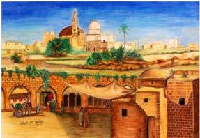
Your exhibition themes are often based on different countries – what inspired you to paint about your travels? I started travelling to exotic countries by getting involved with the International Union of Immunological Societies (IUIS). I chaired the Education Committee for twelve years, then served as the co-chair for another five. Our mandate was to organize immunology courses in low to middle income countries – mainly in Africa and Latin America. My colleagues and I would teach these very interactive courses and afterwards, take a vacation. That is how I ended up in Egypt, Morocco, Tunisia, Benin, Kenya, South Africa, Zambia, Colombia, Mexico and many more. During these travels, I use my cell phone to take photos. I am particularly fascinated by prehistoric caves and old walls. They tell ancient stories and show layers of history, partly in ruins and with moulds and shrubs growing on them – it is a great source of inspiration.
the gallery operates on membership fees and is run by the members themselves with a very active board. I served as chair of the board during the pandemic, and I am currently chair of membership. We have over fifty members and in the past year, we have managed to buy our own space with federal and municipal grants along with contributions from over 250 donors.
The gallery is not only a place of exhibition but also a community. The social aspect is just as important as the artistic side. It is very fulfilling to interact with other artists and work together to organize exhibitions, artist talks, and other events.
What are your current painting projects and what advice do you have for other scientists? I am preparing an exhibition on Antigua, Guatemala. The ancient capital is filled with old convents, cloisters, and churches dating back to the Spanish Conquest. During a recent vacation, I took 400 photos of old walls in Antigua. I am
“...art is freeing. It is an expression of oneself...a very satisfying and absorbing process, during which I forget about science for a few hours.”
currently completing artworks based on the photo transfers of selected images.
“I enjoy exploring different media and techniques - which is reminiscent of doing experiments in the lab.”
What is your process of starting a painting during these travels? When I take photos of an old wall, for example, I can see the painting with its composition, colours, and the emotions that it conveys. In the last five years, I have been doing a lot of photo transfers. After I print out my selected photos, I glue them inverted onto a board and peel off the paper pulp, which leaves behind the ink layer. This way I get the direct impression of the old wall; I then add layers of acrylic paint, pigments, and textures to create an abstract composition.
As a member of the Propeller Art Gallery, can you tell us more about it? Propeller Art Gallery was started thirty years ago by OCADU students who had no place to exhibit. They started a co-op -- meaning that
As for advice, I would say “don’t be afraid to go outside of your comfort zone and try new ventures”. You need to have interests other than science to keep your mind rested and sharp. Sometimes, the best ideas about science come not when you are sitting at your desk but when you are out for a walk or involved in other activities.
Written & Designed by
Jennifer Ahn
Shaping identity with
Science & Art: M ahmoud E l- M aklizi M ahmoud E l- M aklizi
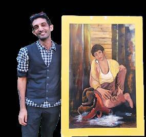
Mahmoud El-Maklizi has been a long-standing member of the Department of Immunology at the University of Toronto. After completing his PhD in Dr. Cynthia Guidos’ lab, he has continued his research as a postdoctoral fellow in Dr. Clint Robbins’ lab. In addition to immunological research, Mahmoud is dedicated to the visual arts. IMMPress Magazine sat down with him to discuss his artistic journey and how arts and science coexist in his life.
How did your artistic journey begin? My art journey has not been continuous. I started painting at the age of twelve and, continued until I was around sixteen to seventeen years old. Once I started my undergraduate degree in science, though, I did not paint at all. It wasn’t until after my PhD that I picked up art again.
When I was younger, my health condition made it challenging for me to experience life like others did. So art was
a necessary outlet to contend with the world around me. I remember my first painting was of these ducklings that were printed on a blanket I had as a child. When my parents saw the painting, they thought it was pretty good for an elevenyear-old and enrolled me into art lessons right away.
Why did you stop painting once you went to university? I actually got accepted to art school for a degree in architecture. But, I also liked math, physics, and biology. In fact, my health condition motivated me to zone in on biology almost obsessively. I was hyper-fixated on trying to understand and potentially fix my condition.
So during my undergrad, I majored in biology with a minor in math, physics and chemistry. I didn’t totally give up on art, though. In zoology courses, we’d have assignments that involved drawing specimens and labelling their features. I enjoyed doing these biological illustrations and making them as detailed and accurate as possible.
How did you get back into art after your PhD? Towards the end of my PhD, I was motivated to attain both perfection and accuracy in science – but eventually my brain needed a break. Also, a good friend of mine requested a painting for his birthday and that was a way to get back into making art.
What is your mindset towards science like now? I’m more hyper-focused on it now than I was ever before. I take science extremely seriously. I also tend to be hyper-focused on anything I do, which is why I choose to do very few, selective things. Once I start painting, for example, I become totally absorbed. I’d paint from 7 a.m. to 10 p.m., completely immersed for days, chasing every detail until the piece felt complete.
What are your painting subjects & how have they evolved? I grew up in Egypt and back home, there is a much larger sense of community than here in Canada. In Egypt, I’d always be surrounded by extended family members and people in my neighbourhood. This naturally inspired me to paint groups of people in the beginning. As I got older, I transitioned into painting individuals and sceneries that were meaningful to me. Also, I used paintings to express myself – and sometimes, seeing myself so clearly in the work felt too raw and revealing. But when I look back at those older paintings now, I can trace exactly how I was feeling in those moments.
What are the overlaps between art & science for you? With both art and science, you might create something—a painting or a scientific paper— with a specific intention, but the outcome can be interpreted differently depending on who’s engaging with it. Personally, I’ve always felt that biology is more similar to art than physics or math. Physics and math can become increasingly abstract the deeper you investigate, and the results are often intangible. But with biology and art, the process is more hands-on, and the outcomes can be visualized and grasped more clearly.
You recently showcased your work at our department’s 40th year anniversary. What was the experience like for you? It felt really good to be involved in the department in a different way and to have the chance to share my artwork. The last exhibition I had before this was when I was fifteen
years old – which is quite crazy considering how young and inexperienced I was then.
Our department community has also been very supportive. Jen [Gommerman] was one of the first people in Canada who encouraged me to start painting again, along with Dr. Michelle Letarte. At the time, I was in the beginnings of my PhD, and Jen [Gommerman] was the Graduate Coordinator. I used to bring back Christmas gifts from Egypt, and I’d often include a picture of my own paintings or drawings to make the gift more personal. That’s how people began to discover that I painted. I still remember Jen and Michelle encouraging me to seriously get back into painting, and their support – along with that of many others – motivates me to stay connected with my artistic side.
Who are your artistic mentors & what advice do you have for other artistic graduate students? I’ve always admired Leonardo da Vinci—his work reflects the level of detail and technical precision I aspire to in my own art. As for advice, I’d recommend building art into your routine, which is something I’m still working on myself. You don’t need to draw every day, but even setting aside 30 minutes to an hour can make a difference. Taking photos of things you’d like to draw later can also be a great way to stay inspired. And honestly, the
...when I look back at [my] older paintings now, I can trace exactly how I was feeling in those moments.”
hardest part is figuring out what you want to say through your art. I think it’s important for anyone interested in the arts to spend time reflecting on that.
Written & Designed by
Jennifer Ahn

Beyond Left and Right: Logic &
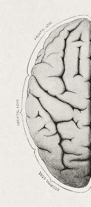
The human brain, with its elegant bilateral symmetry, has long been depicted as a battleground of opposites. On one side: the left hemisphere, logical, analytical, verbal. On the other: the right, creative, intuitive, emotional. This split-brain narrative, popularized in the mid-to-late 20th century, has seeped into every thing from personality quizzes to TED Talks, branding people as either “leftbrained” or “right-brained”, “rationalists” or “visionaries”.
In truth, however, the brain doesn’t draw such rigid lines between logic and creativity. Far from operating in isolation, the hemispheres are in constant dialogue, connected by a dense bundle of fibers called the corpus callosum. Most complex tasks, from composing a symphony to crafting a scientific hypothesis, light up networks across both sides of the brain. Creativity and logic aren’t rivals pulling us in opposite directions. They are collaborators.
While certain aspects of creativity are linked to increased activity in the right lateral prefrontal cortex, a region involved in generating ideas and combining bits of information in new and original ways, that is just one part of a much larger picture. Creative thought also depends heavily on the default mode network, a collection of brain regions across both hemispheres that become active during mind-wandering, daydreaming, and internal reflection.
But even the boldest ideas need structure. That’s where the executive control network steps in, particularly the left dorsolateral prefrontal cortex, which handles goal-setting, working memory, and critical evaluation. This region doesn’t just shut down during creative thought. In the most generative minds, it works in tandem with the default mode network. Recent functional neuroimaging studies show that artistically creative individuals exhibit simultaneous activation of both networks, toggling between spontaneous ideation and deliberate refinement of those ideas. Innovation, it turns out, is a conversation between different regional brain networks.
In recent years, the neural bases of creativity, including artistic creativity, have become a topic of interest. A recent study investigated how creativity, specifically planning an artwork,
The Neuroscience of
Creativity
affects brain connectivity by comparing professional artists to control participants. Using functional MRI, the researchers found that during the creative task there was stronger functional connectivity between the default mode network, which is associated with generating novel ideas, and the executive control network, which is involved in evaluating and selecting those ideas. This enhanced connectivity was especially pronounced in professional artists.
These findings provide a neural explanation for the observation that even the boldest ideas need structure. The executive control network, particularly the left dorsolateral prefrontal cortex, supports goal-setting, working memory, and critical evaluation and does not simply shut down during creative thought. Instead, it works closely with the default mode network allowing for a dynamic balance between spontaneous ideation and careful analysis. Thus, artistic creativity emerges from a conversation between these two brain networks rather than from one acting alone.
Moreover, this dynamic interplay fuels creativity beyond art. It applies to everything from a scientist’s experimental design to a poet’s revision process or even a graduate student rewriting a manuscript. Whether troubleshooting a method, interpreting messy data, writing a thesis chapter, or brainstorming a new experiment, our brains constantly balance structure and fluidity, focus and creative freedom.
The myth of the left-brained logician and the right-brained artist doesn’t just oversimplify neuroscience. It undersells us. Innovative thinking comes from blurring this made-up boundary and refusing to fit in a hemisphere-shaped box. Creativity needs constraint. Logic needs imagination. The best ideas come when the whole brain shows up to work.
Written by Mahdieh Golzari-Sorkheh
Designed
by Milea DiPonzio

The combined impact of art and science on society & the power of mixing fact with feeling
Today, it is not uncommon to view the arts and the sciences as mutually exclusive disciplines. We tend to imagine scientists and artists residing on opposite ends of a spectrum, each offering fundamentally different contributions to society. In an era of rapid technological advancements, STEM (science, technology, engineering, mathematics) is frequently recognized as the primary driver of progress while the arts are sometimes overlooked. While it’s true that these two disciplines shape our world in unique ways, their impact can be equally powerful — and arguably even greater when they work in unison. This article will explore the intersections between art and science, and how both work together to deepen public understanding, provoke new ideas, and inspire meaningful changes within our society.
Art and science are catalysts for action
Throughout history, science has provided key solutions to complex global problems. Take the COVID-19 pandemic, for example. Efforts within the scientific community to develop novel vaccines, model viral transmission, and shape effective public health policies were vital to saving millions of lives. But despite these efforts, widespread misinformation and distrust hindered public compliance. To bridge this gap, artists stepped in. Projects like the “COVID-19 Poster Project” and murals painted across cities around the world depicted scientific guidelines through art, reminding people to wear masks, wash hands, and support healthcare workers. Rather than viewing public health regulations as burdensome orders, these artworks invited the public to understand them as shared responsibilities for the greater good.
In a similar way, art has served to enhance public understanding of climate change. While the scientific data backing climate change is well-established, it can often appear abstract to the general population. Artists have contributed to
making climate change data more accessible by translating them into emotional experiences. For instance, Olafur Eliasson’s Ice Watch in 2014 placed pieces of glaciers from Greenland on the streets of London where they melted in real-time, allowing the public to tangibly observe the impact of rising temperatures on arctic ecosystems. In addition, Photographer Gregg Segal’s 7 days of garbage imaged American families surrounded by a week’s worth of their trash, confronting us with the issues of overconsumption in today’s world. Initiatives such as these provoke a sense of responsibility, causing us to rethink daily habits and demand more accountability from powerful groups to shape environmental outcomes.
These examples demonstrate that while science may offer solutions, it is often art that shapes how we perceive, interpret, and ultimately respond to the challenges we face on a societal level.
Art inspires scientific discovery
In addition to being a call for action, art has also played an important role in shaping scientific thought. From Leonardo da Vinci’s detailed anatomical drawings to Charles Darwin’s sketches supporting his theory of evolution, visual artistic representation has been crucial for inspiring new ideas.
An interesting contemporary example of this took place during the creation of Christopher Nolan’s Interstellar (2014), where the film’s producers collaborated with physicist Kip Thorne to create a scientifically accurate depiction of a black hole. In the process of creating the film, the team uncovered new scientific insights regarding how light behaves when in close proximity to black holes, which eventually led to the publication of a research paper in Classical and Quantum Gravity.
Beyond informing research, science fiction also possesses the ability to inspire curiosity and interest in STEM, especially within younger generations. Many scientists, engineers, and astronauts have credited their early interest in science to fictional books, shows, and movies. Gary Blackwood, a program manager at NASA, credits the 1960s show Star Trek for fueling his interest in science, and quotes “Star Trek really gave me a sense of wonder—going to other stars on a spaceship, the stars flying by, that image on the screen. Star Trek always spoke to that greater vision, that greater achievement, what mankind could become.”
Art influences our interpretation of science
Art in the form of science fiction has also served as a platform to explore the future of science and its role in society. The TV show Black Mirror explores avenues of emerging technology, like the dangers of social media and unregulated virtual reality while The Humans depicts the currently relevant issues of integrating artificial intelligence and robots into society. These cinematic pieces push audiences to imagine a world where these technologies are implemented, testing the waters for novel ideas. They creatively bring ethical considerations to the surface, pushing us to ask not just how these technologies are made, but if they should be implemented in the first place. As quoted by NASA scientist Mae Jemison, “What really good science fiction does is to allow you
to reflect on yourself, your values and your beliefs…It uses a fictionalized science as a mechanism to push us to think about what we’re doing.”
Conclusion
At their core, artists and scientists often share a common objective: to challenge conventional ways of thinking and uncover new perspectives that allow us to enhance our experience as human beings. When we mix fact with feeling, we not only enrich knowledge but inspire change for the future. Keeping both the arts and the sciences alive isn’t just valuable; it’s necessary for maintaining an informed, thoughtful, and driven society.
Written by Milea DiPonzio
Designed by Baweleta Isho
Painting and Science: S What im P re SS ioni S m an d Photogra P hy Teach Us About innoVation
Before Photography: Painting as a Window to the World
Before the invention of photography, painting was humanity’s most powerful visual storytelling tool. Whether capturing the grandeur of royal courts, recording historic events, or preserving the likenesses of loved ones, painting served as both documentation and interpretation. Artists held a vital role as visual historians, and great technical skill was required to faithfully represent reality on canvas.
But painters were not entirely alone in this pursuit of realism. For centuries, many had turned to an ingenious optical device known as the camera obscura.
Camera Obscura: The Artist’s Secret Assistant
The camera obscura, Latin for “dark chamber,” was an early imaging device dating back to antiquity, but it became especially important in the hands of Renaissance and Enlightenment-era artists. It worked on a simple principle of optics: light entering a darkened space through a small hole or lens would project an inverted image of the outside scene onto a surface inside. In the 17th and 18th centuries, portable versions were developed, allowing artists to trace the outlines of landscapes, architecture, and people with impressive accuracy.
Far from being seen as cheating, the use
of camera obscura was celebrated by many for aiding in the quest for realism. Artists like Vermeer and Canaletto are believed to have used it to achieve their remarkable detail and perspective. It was not just a technical tool, but a bridge between art and science, an early demonstration of how optics, mathematics, and creativity could intersect.
Despite the help of such devices, however, the process of painting was still painstaking. Creating a single portrait or scene might take days, weeks, or even months. And no matter how accurate the drawing, the artist still had to build light, texture, and emotion through their own hand.
The Disruption of Photography
In the early 19th century, pioneers like Nicéphore Niépce, Louis Daguerre, and William Henry Fox Talbot developed ways to chemically capture light on a surface, fixing an image directly from reality. What once took a painter hours to observe, and render could now be caught by a lens in seconds.
By the mid-1800s, the daguerreotype portrait studio had become a booming business, especially in urban centers like Paris. By 1849, over 100,000 Parisians were sitting for portraits each year. Thanks to innovations by photographers like André Adolphe Disdéri, these portraits could be reproduced multiple times, shared, mailed, and cherished widely. Portrai-


ture, once the privilege of the elite, became available to the middle class.
For painters, this technological leap was existential. If a machine could now do the work of recording faces, light, and landscape with unmatched precision, what purpose was left for painting?
Critics, patrons, and even certain artists feared that the rise of photography would render painting obsolete. Many believed that the emergence of this new technology marked the death of painting. But history would soon prove otherwise.
The Birth of Impressionism: A Creative Response
But instead of dying out, painting evolved. Artists like Claude Monet, Edgar Degas, and their contemporaries turned away from competing with photography’s realism. Instead, they leaned into what photography couldn't yet do: capture the fleeting light, the feeling of a moment, and the subjective experience of seeing.
Impressionism, which emerged in the 1870s, focused on light, color, motion, and emotion rather than detailed likeness. These artists were inspired by the visual effects of photography such as blurred movement caused by long exposures, but chose to paint how a moment felt, not how it looked through a lens. In fact, the first Impressionist exhibition in 1874 was held in the studio of Nadar, one of Paris’s most famous photographers. Art and science were already in deep conversation.
Photography freed artists from the burden of perfection. It encouraged experimentation, spontaneity, and ultimately, a revolution in artistic thinking.
Lessons for Today: AI and the Creative Future
Today, we are facing a similar moment with the rise of artificial intelligence. Just as photography once challenged painters, AI now raises
questions about the future of programmers, designers, and writers. If a machine can generate code, write prose, or create artwork in seconds, what happens to human creativity?
But history offers us a hopeful answer: technology doesn't kill art. Instead, it transforms it. Rather than replacing human ingenuity, innovations like AI could free us from repetitive tasks and open new avenues for creative exploration. Just as photography gave rise to Impressionism, AI might usher in new styles, new questions, and new forms of expression.
Maybe It's Not the End
When faced with radical new tools, we have a choice: resist or evolve. The Impressionists chose to evolve. In doing so, they changed the course of art forever. We now stand at a similar crossroads.
So instead of fearing that AI will kill creativity, maybe it's time to ask:
What new forms of beauty will we create once we let go of the need to replicate reality?
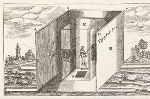
“Athanasius Kircher’s camera obscura”, illustration from Kirscher’s Ars magna lucis et umbrae (1671) reproduced in Josef Maria Eder’s Ausführliches Handbuch der Photographie (Detailed handbook of photography, 1905)
Written & Designed by
Tianning
Yu


AI Art 1.o An AI-generated picture worth a thousand words
Artificial intelligence, or AI, is now at the forefront of technology and is becoming increasingly available to the general public. Along with its rise in our current culture, there also comes new arguments for and against using AI. Tools like DALL-E, Stable Diffusion, Midjourney, and Google Veo can create realistic images and videos in minutes, conjuring awe yet simultaneously provoking strong controversy about its utility and ethics.
Even textbooks have pictures
It can be argued that the democratization of any tool or technology has a positive effect on moving society forward. AI tools provide not only convenience, but also inclusion and accessibility. Generative AI removes the barriers faced by individuals who do not have artistic training, eliminates prohibitive requirements for expensive art software or materials, and can turn a personalized concept into reality. Moreover, the ability to adjust on a bulk scale means that for any given product, brainstorming can be significantly faster, and many prototypes or mock-ups can be made easily. The images made by AI also do not necessarily have to be a final product – it can be used as a springboard for new ideas from which an artist can modify and refine further.
Scientific illustrators are a group of people particularly impacted by this recent shift in AI use. Universities offer specialized Biomedical
Illustration programs in which students receive training in both artistic skills and scientific knowledge, and they represent a unique intersection between art and science. Unsurprisingly, there is consternation about whether AI has a role in biomedical illustration. Despite this, some illustrators argue that there are defensible uses of AI. For example, AI-generated images can be a massively useful tool for those in biomedical fields. In medical training, imagery that is (currently) free of copyright and does not violate patient privacy laws can be

generated easily. Concepts like protein folding or cellular metabolism can be abstract and difficult to convey but are more easily expressed through animations or interactive visual elements. Research articles that are tedious to read and laden with scientific

jargon become easier to grasp and more exciting to the audience when the educator is able to create visuals that suit their topic. Important public health information, like how vaccines work to prime immune cells, or educating an individual about a disease affecting them, can be made more accessible by explaining with images rather than words.
Two sides to every coin – the pitfalls of AI imagery
expertise that artists accrue in their training. Scientific illustration programs at universities are uniquely specialized in training artists with a strong grasp of both the scientific facts they are interpreting and artistic skill. Scientific illustrators have reported a decline in work, forcing them to reduce fees to remain competitive. Moreover, there is concern in the wider art community that using AI ultimately erodes artistry.
When scientists publish manuscripts, authorship is critical to acknowledge the time and effort that individual members contribute to the work. But AI is different: who receives the credit? The products that AI generates are not original; the wealth of training data that AI uses is generated through decades of work from humans, often without consent from the original creators. While not specifically regarding scientific illustration, there have already been lawsuits filed against AI art companies for copyright infringement, highlighting the need for legislation and regulation surrounding copyright and intellectual property with regards to AI-generated content.
Promise or peril?

As with any novel tool, there must be skepticism and caution about the ethical concerns. The ability to generate nearly any image also means that misinformation can be created and spread just as easily as real, factual science. Deepfakes spreading inaccurate or misleading information are becoming increasingly abundant and more difficult to detect or differentiate by those watching. Recently, an AI-generated figure was published in a peer-reviewed journal which caused an uproar in the scientific community because of its depiction of an egregiously anatomically inaccurate mouse, ultimately leading to the retraction of that paper. In a period of time when public trust in science is rapidly diminishing, the use of AI imagery, either unintentionally wrong or with malicious intent, could even further undermine confidence in and devaluation of the specialized

There is no denying that AI is here to stay. While some believe AI use is being adopted too hastily, others view the use of AI as a supplementary or assistive tool that enhances or speeds up previously tedious and time-consuming work. Some believe that AI tools will eradicate jobs for artists, and others are confident that AI will always lack the “human touch.”
Some argue that the use of AI imagery is acceptable if it is explicitly disclosed. Still, there remains the question of what can be considered fair use of training material, or what could be considered as copyright infringement. Until there is more consensus and regulation in how AI can be ethically trained and used, the contention will persist.
Written by Annie Pu
Designed by Tianning Yu

When Science & Creativity Join Paths
Bioart is an art form that intertwines the beauty of art with biology, using live cells and unique chemical processes as a medium. This movement advances the traditional boundaries of art and opens new dialogues about our relationship with the world around us.
At its core, bioart is the creation of art through the manipulation of biological processes and living materials. “ ”
This includes working with living organisms, such as bacteria, fungi, tissues, and images or data from biomedical research. Gaining traction in the last several decades, bioart dates back to the early 1900s to a revolutionary scientist. Before he discovered penicillin, Alexander Fleming was more than a scientist – he was an artist. Unlike other painters, however, he painted with microbes. Using paper soaked in culture medium, he inoculated different species of microbes that had different colours to create images of whatever occurred to him such as soldiers, ballerinas, and houses. Obsessed with discovering new strains of bacteria with different colours, he continued to collect unique microbes, ultimately leading to his Nobel Prize winning discovery of penicillin as well as the lysozyme.
Fleming’s germ art continues to this day, giving rise to bioart competitions such as the American Society for Microbiology Agar Art Contest. Evolving beyond agar on petri dishes, bioartists use a plethora of scientific techniques to create art using aquatic organisms, bioluminescent bacteria, slime mold, and even genetically modified organisms. The art form now extends further to innovative techniques such as cultivating bacteria on film negatives by Erno-Erik Raitanen and in vitro culture of mammalian cells in bioreactors by Oron Catts and Ionat Zurr.
In our modern era, many view the concepts of art and science as divided, separate entities with contrasting approaches to explaining life. Science has a reputation of
being technical, structured, and complex while art is viewed as an emotional and sensory experience. Despite these perceived differences, both fields emerged from a need to understand and explain how the world works, our relationship with the world, and the meaning of life. As we continue to push the boundaries between art and science, bioart instigates critical conversations about the implications of biotechnology, longterm consequences of manipulating our environment, and scientific ethics.
In his artwork aptly titled “GFP Bunny,” Eduardo Kac genetically engineered an albino rabbit to glow bright green when exposed to blue light. First presented in 2000, this transgenic artwork is more than just the rabbit or the visual aesthetics; this work challenges the traditional notion of art to incorporate dialogue on the societal perception of genetics, the ethical implications of genetic modification, and the concept of normalcy in our culture.
However, one may question the ethics of bioart itself for engaging in scientific experimentations and mammalian
Rendition of Fleming’s microbial painting
Figure 1.


alterations. In the purely scientific context, these alterations can be interpreted as “playing God.” However, some artists contend that bioart is a medium that merges scientific concepts with aesthetics to convey messages, elicit reactions, and incite discussion.
In 2000, artist Marta de Menezes collaborated with a scientist, Paul Brakefield, to modify the wing patterns of butterfly through small physical wounds on the wing discs of the caterpillar. The intent was to explore the differences between natural and artificial features. In 2002, Brandon Ballengée and scientist Stanley Sessions generated frogs with malformed limbs, with the intent to grow awareness of environmental issues. Both these examples highlight the neglect in care, responsibility, and long-term consequences of these manipulated creatures afterwards and whether these manipulations were necessary to begin with. For example, Kathy High’s work “Embracing Animal” in 2005 involved the care of retired breeding rats originally genetically engineered to study autoimmunity, demonstrating the possibility of self-expression without irreparable damage to lifeforms. Inarguably, the public display of bioart engages a wider audience in cultural discourse on the impact and ethics of advancing science.
Some bioartists have even taken to modifying their own body for their creations. Stelarc is an Australian performance artist who explores the integration of our physical form with technology. Using his own body as the medium, Stelarc has centered his work around medical prosthetics, capturing
attention for his “Ear on Arm” - the surgical implantation of an ear constructed with porous polyethylene on his own forearm. The artwork has incited discussion of the ethics of carrying out clinically unnecessary procedures. Already controversial, Stelarc unsuccessfully attempted the implantation of a microphone in the ear, resulting in infection and removal. Undeterred, Stelarc continues to be driven by the concept that we’ve evolved beyond our bodily architecture, challenging the physical limits of the human body and the definition of being human.
Technology continues to advance, shaping our interaction with the world, changing our perception of reality, and influencing society at all levels. Bioart not only questions the intersection of nature and technology but amplifies public debates and scientific dissemination. These fascinatingand perhaps jarring - pieces of artwork cross boundaries to remind us to consider important questions and challenge traditional notions that may limit our viewpoint as our world continues to evolve.
Written by Siu Ling Tai
Figure 2.
Alba, the fluorescent bunny
Figure 3.
Stelarc’s Ear on Arm
Designed by Sophie Sun
the inseparable nature of art and science: Does the “Renaissance man” still exist today?
Science
and art have co-existed as disciplines throughout human history. Both are manifestations of the human imagination and require a modicum of creativity and skill in observation. From intricate star-shaped geometries employed by architects during the Islamic Golden Age and the application of materials science in manuscript illumination and lettering, to the emergence of natural history as a fledgling science during the artistic Renaissance period in Europe, the two disciplines frequently intermingled.
As an early scientist, it was one’s job to accurately depict natural phenomena through artistic illustration, which was invaluable to studying and classifying the natural world. Flemish artist Peter Paul Rubens, through his insightful human anatomical drawings, and Charles Darwin, through his accurate ornithological depictions in “On the Origin of Species,” both demonstrated how science and art could be indistinguishable at times, and that to be a scientist required skills in many other disciplines. Perhaps the most renowned example of the “Renaissance man”, which was a person who was knowledgeable in many things, was Leonardo Da Vinci, who was a painter, sculptor, engineer, botanist, and scientist.

By the 19th century, however, science and art have diverged into separate cultures, as methodologies started to differ; science was becoming more specialized, governed by organization and rules,

while art was becoming more personalized. Schools and universities separated the disciplines, making it difficult to study both. While artists have developed their own schools of thought and methods, diverging from the classical art that also stemmed from observation, many contemporaries continue to draw inspiration from science to spread beauty of the natural world as well as to evoke wonder of the “unknown.”
But what about scientists? Do they still draw inspiration from their artist compatriots?
Contrary to the popular opinion that science and art are polar opposites, many scientific discoveries today continue to draw on artistic principles. For example, the Foldscope is an origami-based paper microscope; with its low cost and ease of assembly through paper folding, it opens windows for collecting scientific images in remote regions. Slavic ornaments, which utilize elaborate geometric patterns, have inspired the design of more efficient sound absorbers for the manipulation of low-frequency sound. A painting called “Court Ladies Preparing Newly Woven Silk” from the Tang Dynasty in China, depicting a lady with a blue-green tattoo on her forehead, was the inspiration for a printable and self-adhesive nanogel-based tattoo that monitors UV radiation from sunshine and Vitamin D synthesis of an individual.
Despite the cultural separation of science and art into individual disciplines, they remain interconnected. At its core, science can be thought of as a form of
art and vice versa. Both scientists and artists study and interpret the world they live in, albeit using different modes of expression, with similar goals of expressing truth. As Albert Einstein once said, “The most beautiful thing we can experience is the mysterious. It is the source of all art and science.” Postulating that art and science arise from the same essence, he also states “The greatest scientists are always artists as well.” Therefore, while not officially dubbed as artists, the “Renaissance man” is still embodied by scientists today, highlighting the creative dimensions of scientific work.
Written by Deeva Uthayakumar
Designed by Baweleta Isho
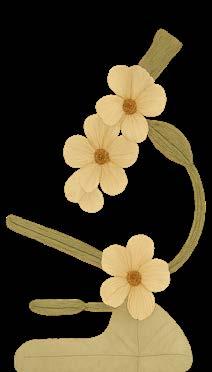
Entrée:

The Science of Culinary Arts
After a long week of work, don’t you want to treat yourself to a good meal? Maybe you’re not too confident in your culinary arts…but it’s a good thing you’re such a great scientist! Let’s create a menu optimized with some science-based tips.
Side
dish:
crunchy veggies or soft, velvety potatoes?
crispy chicken wings or a perfectly seared steak?
Crispy chicken wings
Tip: Add baking powder and salt to your batter or spice rub for extra crispiness.
The science: When you’re sweating in front of the kitchen stove, an important reaction happening before your eyes is the Maillard reaction. This is a chain of reactions between amino group on proteins with the carbonyl group of reducing sugar. Proteins and sugar engage in a complex chemical dance, spurred on by the symphonic harmony of heat, humidity, and acid, creating a chef d’oeuvre of intense flavour, aroma, and colour. Adding baking powder facilitates the Maillard reaction by breaking peptide bonds and raising the pH of the surface of your chicken wings, helping achieve that juicy, crispy bite.
But perhaps you’re looking to replenish your iron and want to make a hearty steak dinner instead…
Perfectly seared steak
Tip: G enerously salt your steak and let it sit at room temperature before patting it dry and searing it in a sizzling hot pan.
The science: Generously salting your steak draws out moisture in the meat, creating a brine on the surface which is then reabsorbed for extra flavour. Like with the chicken wings, achieving a good sear on the steak also relies on the Maillard reaction. For optimal results, leave the steak at room temperature and pat it thoroughly dry before searing in a hot pan. This ensures that the surface of the steak reaches 280-350°F (138-178°C), the optimal range for the Maillard reaction to occur. If moisture remains on the surface, the temperature cannot rise above the boiling point of water, resulting in a lackluster sear on your very expensive cut of meat.
Next on the menu: what about a side dish?
Tip: Add an acid to optimize crunchiness OR add a base to optimize quick cooking and softer bites.
The science: Vegetable cell walls are held together by a polysaccharide glue called pectin, which is sensitive to changes in acidity and tends to break down in alkaline environments. Adding baking soda to boiling water can shorten cooking time for softer potatoes. In contrast, acidic environments slow pectin breakdown and allow veggies to retain shape and crunch while cooking. A splash of lemon juice or vinegar not only adds flavour but can give your veggies a toothsome bite.
Now let’s finish up the meal with a refreshing drink.
After dinner drink: a fun cocktail or a strong spirit? How will you keep cool in the summer heat?
Tip: Use crushed ice if you want to chug and cool down, and use large ice cubes if you’re looking to sip and savour.
The science: Crushing ice reduces its surface area and increases melting- cooling down your drink faster while also diluting it. This is great for cocktails that are best served cold, with an alcoholic taste that you want to mellow out. Think mojitos, Moscow mules, and mai tais. Not a fan of cocktails? For drinks you wish to savour without worrying about it becoming diluted, large ice cubes with less surface area will have a longer melting time. This is ideal for an old fashioned, negronis, or a simple dram of your finest whiskey.

With these tips in mind, happy cooking! And more importantly, happy eating!
Vera Lynn Written & Designed by
BODY HORROR AND HORRIFIC BODIES:
the horror genre can tell us about responsible science.
BODY HORROR AND HORRIFIC BODIES: what

t first glance, it would seem that the horror genre and science are natural opposites. After all, horror is driven by a fear of the unknown, of things that lurk in the shadows and go bump in the night, while science seeks to demystify and explain the unknown. This is the central conflict surrounding science and horror, and yet, horror has repeatedly drawn inspiration from science, realized through on-screen depictions of experiments gone wrong, and grotesque violations of nature. Yet, throughout all these onscreen depictions of scientific horror, a key question remains: what is horror trying to tell us about science?
Mad scientists and the boundaries of science
No trope characterizes the intersection between science and horror better than the mad scientist, and no two words better epitomize this trope than “It’s alive!”.
Frankenstein is one of, if not, the most influential horror work of all time, telling the story of a mad scientist who manages to bring to life a monstrous creature one stormy night, an act that inevitably leads to his downfall. Now famous for its critique of scientific ethics, mob mentality, and the nature of monsters, a key aspect that seems to have been lost in the shuffle is Mary Shelley’s original inspiration for the story. Specifically, she was furthered by the emerging field of galvanism and electrophysiology, and experiments that soght to stimulate of dead muscle by electrical currents.
Similarly, in Vincenzo Natali’s 2009 movie Splice, two scientists splice human and animal DNA to generate a human-animal hybrid that, once again, predictably leads to their downfall. While not entirely a one-to-one retelling of Mary Shelley’s Frankenstein, it retains much of the same (pun intended) DNA, updated with the advent of genetic engineering and cloning. In this movie, rather than body parts, it is lines of genetic code being metaphorically sown together. More interesting is the fact that Vincenzo Natali was inspired by a real-life experiment: the 1996 Vacanti mouse experiment, whereby a human ear was grown on the back of an immunodeficient mouse in order to study organ and tissue regeneration for transplantation.
Using science as a backdrop and real-life experiments as inspiration, both of these stories illustrate cultural anxieties surrounding scientific ethics. While they do not outright
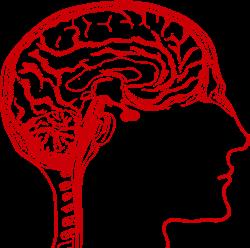
condemn scientific investigation, these stories ask how far we are willing to go in service of progress, and what of the horrific bodies that are created in its wake. They show, through the death of Victor Frankenstein’s friend at the hands of his monster, and through the havoc wrought by the hybrid monster Dren in Splice, that science without ethics has the potential to lead to disastrous outcomes. But while these pieces of media place scientists as the central protagonist, what of the monsters that have been created?
Cellular Anxieties turned bodily horrors
Rather than focusing on the horror of creating something monstrous, body horror focuses on the horror of becoming something monstrous. It is a subgenre of horror interested in distortions of the human body, and the physical and mental consequences of this transformation. While in some cases the cause is supernatural and otherworldly, two of the most influential pieces of body horror cinema choose to root this transformation in simple biology.
In David Cronenberg’s The Fly (1986), Seth Brundle, a young scientist played by Jeff Goldblum, unwittingly merges with a housefly following an accident involving a teleportation device. What follows is the grotesque metamorphosis of his body into a human-fly hybrid, a phenomenon that the film interestingly attributes to the mixing of human and fly DNA, and therefore, RNA, protein, and body.
Moving one step up the biological ladder, in John Carpenter’s The Thing (1982), the source of this transformation is no longer genetic, but cellular. The film follows an Antarctic research team that comes face-to-face with an alien creature with the ability to imitate its victims. This ability, as illustrated in a computer screen in the movie, stems from the alien’s cells being able to devour and mimic its hosts cells, allowing it to eventually consume the host body.
Once again, these movie monsters are grounded in biology; malformations of the body occur at the very fundamental building blocks of life, prodding at the perceived immutability
of biological law. They challenge our trust in science and reflect our fears of losing control of our biological selves, of the body being exploited or rebelling against its intended purpose, and the consequences thereafter. It is unsurprising then, that body horror commonly invokes the imagery of tumours. Cancer, as well as the mutations that cause it, are a real-life parallel to the very fear that body horror capitalizes on. The fear of losing control of an internal, uncontrollable growth that our body and immune system should be able to keep in check reflects many of the similar themes present in body horror.
A cautionary tale of horror and science
It is interesting then, that in some ways, body horror can be seen as reflecting the same themes as the mad scientist trope, only through a more personal sense. Where stories of mad scientists ask the question “What have I done?”, body horror asks the question “What has been done to me?”. This dichotomy only further humanizes the creatures of mad scientist stories, and provides us a discomfortingly personal glimpse into the consequences of irresponsible and careless meddling with biology.
Meanwhile, science, as the grisly subject of this push and pull between victimization and victimhood, grounds these ideas in real-life concerns. In an era of unprecedented scientific development and engineering, questions about the long-term effects of experimental treatments, inclusivity of treatment groups, and the historical exploitation of marginalized people (e.g., the use of Henrietta Lacks’ cells) are extremely pertinent. In some ways, these stories should make us continuously question the validity and ethics of our research as scientists, though this is not to say that all scientific research is bad. Rather, it stresses that more scrutiny and a better understanding of the ethics of scientific experimentation are needed in order to do proper and responsible science.
This ethical reflection is captured at the end of Frankenstein, where Victor Frankenstein, the scientist responsible for the monster, overwhelmed by guilt, wishes he had never created his monster to begin with. It is in our best interest to heed his warnings.
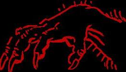
Written by Christopher Ryan Tan
Designed by Victoria Sephton
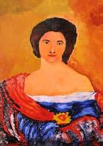
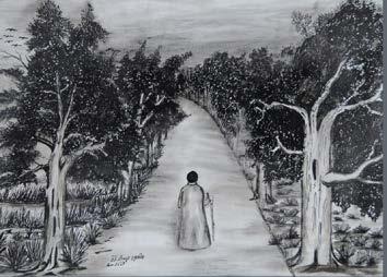
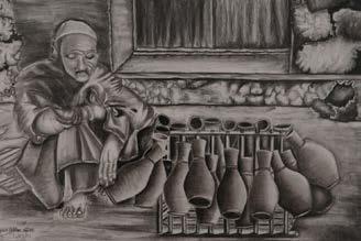

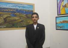
MAHMOUD El-Maklizi
MICHELLE Letarte
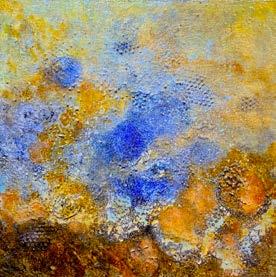
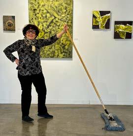
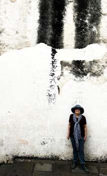
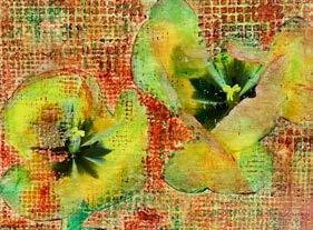
Department of Immunology
University of Toronto
1 King’s College Circle
Toronto, ON M5S 1A8
Canada

U of T Symphony Orchestra rehearsing at Convocation Hall (March 1952)
Photo Credits: University of Toronto Archives.