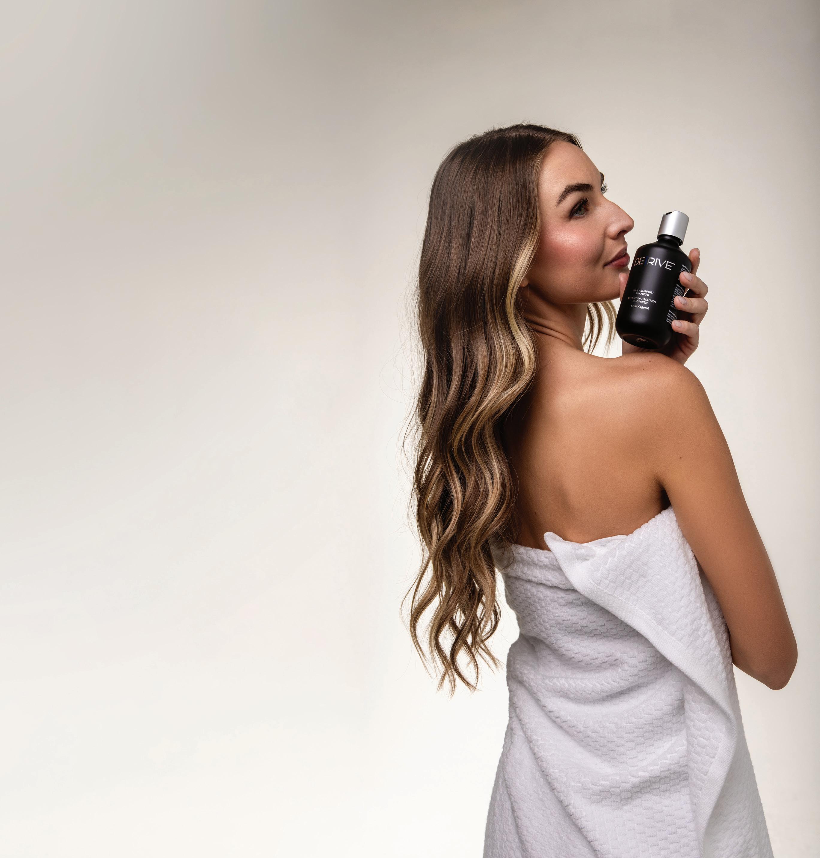
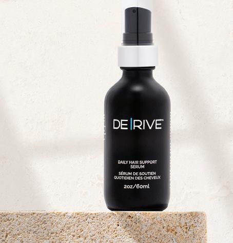
The DE|RIVE® Hair Wellness System is a plant-based solution ideal for patients with thinning hair or early hair loss. Start with the in-office Hair Support Serum , then continue care at home with:
www.aestheticmanagementpartners.com/derive
AS 2025 DRAWS TO A CLOSE, WE REFLECT ON A YEAR MARKED BY continued growth, evolving technologies, and deeper conversations about the ethics and evidence underpinning aesthetic medicine. In this issue, we turn our attention to technologies and protocols that continue to reshape expectations, especially those balancing efficacy with minimal downtime.
Leading this conversation is our cover feature on the latest developments in minimally invasive lipolaser technologies, a category gaining traction for its ability to safely reduce localised fat and tighten skin with limited recovery and high patient satisfaction. Once reserved for high-intensity surgical settings, lipolaser devices now offer a compelling bridge between non-invasive body contouring and more aggressive liposuction procedures.
This issue explores how evolving device parameters, such as wavelength selection, thermal profiles, and delivery techniques, are refining outcomes and expanding indications. Whether paired with injectables, radiofrequency, or lymphatic drainage protocols, minimally invasive lipolaser treatment is becoming a cornerstone of comprehensive aesthetic planning.
In a similarly results-focused vein, we feature a multimodal imaging case series on Bellacholine, a deoxycholic acid-based injectable for submental fat reduction. The 11-month follow-up data highlights measurable reductions in fat volume and enhanced jawline definition reaffirming its place in the lower-face contouring toolbox.
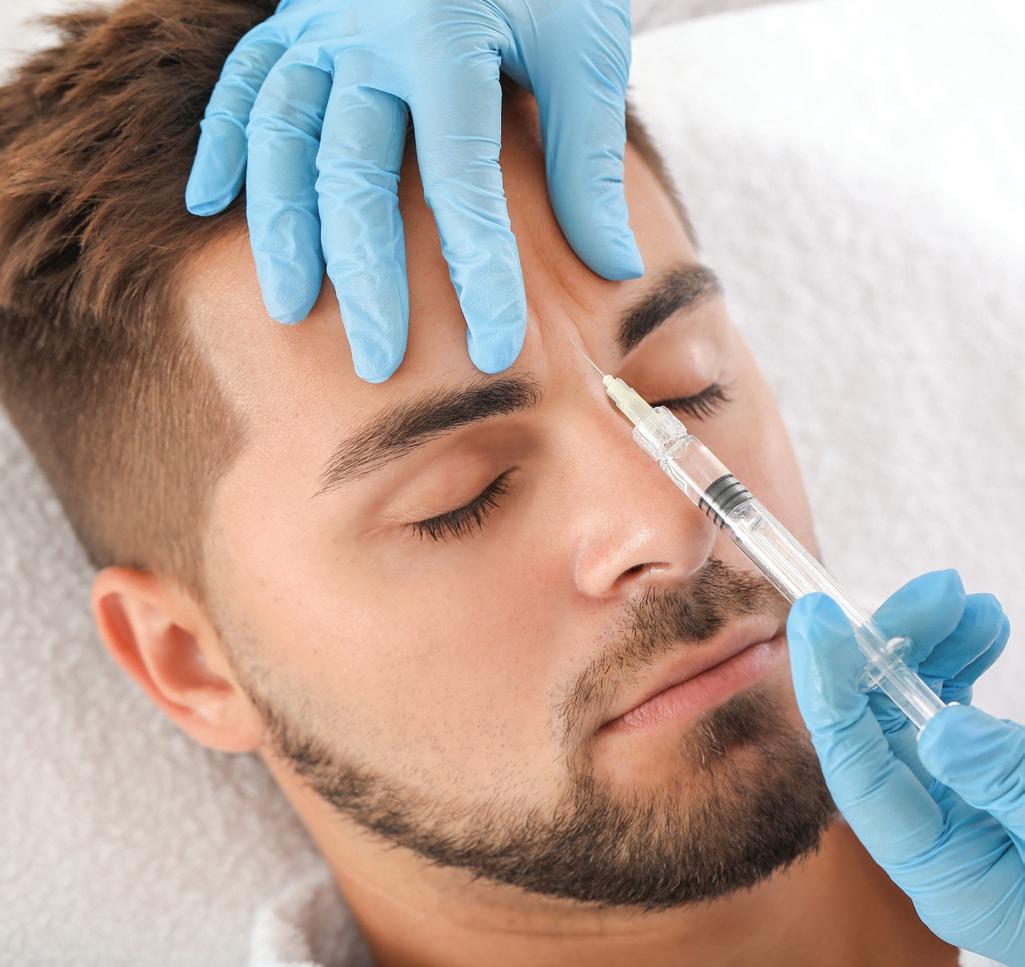
Clinical precision and patient protection remain essential, which is why this issue also revisits complication management in lip augmentation. Dr Yuliya DyedykGusarova provides a valuable review of common pitfalls, anatomical considerations, and how to reduce risks in one of aesthetic medicine’s most popular, and nuanced, procedures.
Affiliated partners:
International Journal of Aesthetic and Anti-Ageing Medicine
Informa Australia, Level 4/24 York St, Sydney, NSW, 2000, Australia ISSN 2159-8908 (print) ISSN 2159-8916 (online)
Editor
Art
Digital
Production
Also featured is an important reminder that aesthetic care is not just about appearance, but ethics. Eddie Hooker, CEO of Hamilton Fraser, outlines why safeguarding protocols must be built into every clinic workflow to protect both patients and providers. It’s a timely piece for any practitioner looking to strengthen trust and transparency in their practice.
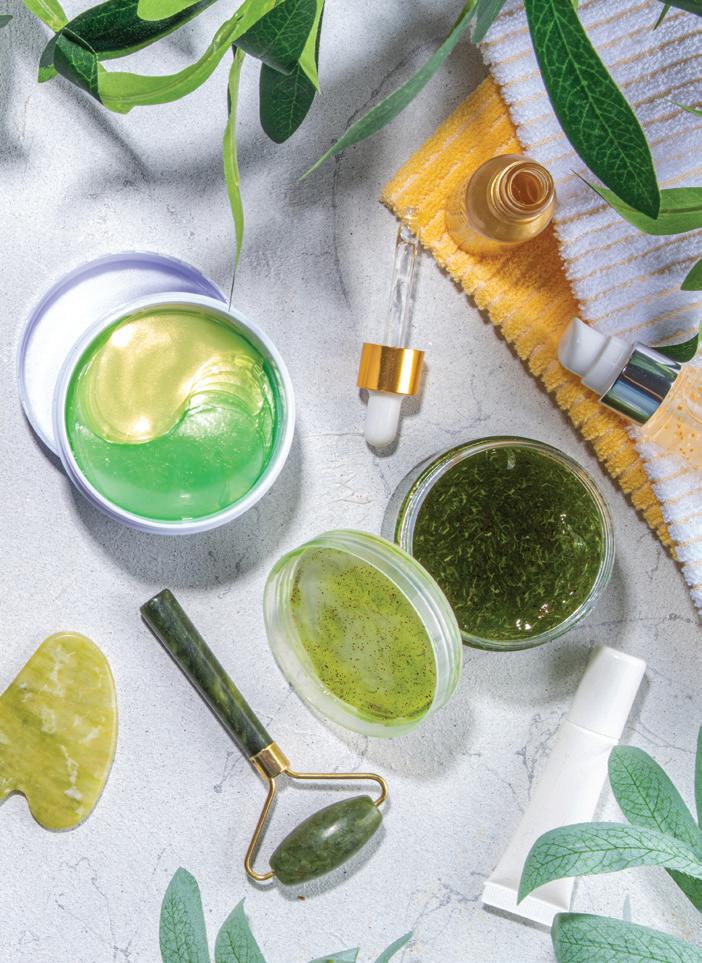
We also explore the growing intersection between wellness and aesthetics. From hormone balancing and IV therapy to light-based treatments and tech-enabled longevity services, biohacking is making its way into practices worldwide. The future of aesthetic medicine looks increasingly holistic and patients are seeking providers who treat the face, body, and beyond.
As we prepare for 2026, we look forward to seeing many of you at upcoming international congresses. Until then, thank you for making PRIME part of your practice.
Balraj Juttla Editor, PRIME balraj.juttla@informa.com
PURIFIED 20 BILLION EXOSOMES
ETHICALLY HARVESTED FROM ORGANIC BOVINE COLOSTRUM
November/December 2025
10 16 28 20
NEWS
7 Experts respond to FDA warning on RF microneedling with best practices
8 Research reveals AI falling short in skin tone diversity and representation
INDUSTRY INSIDER
10 Latest developments in minimally invasive lipolasers
Minimally invasive lipolasers are helping providers meet rising demand for safe, effective, and low-downtime aesthetic treatments
16 Biohacking meets aesthetics
Aesthetic practices and medspas worldwide are incorporating lifestyle hacks, technologies, and wellness therapies in their quests to become one-stop destinations for beauty and optimal health
CASE STUDY
20 Long-term efficacy of Bellacholine for non-surgical submental fat reduction: a multimodal imaging case series
Submental fullness can obscure facial contours and compromise profile aesthetics. This case series explores the use of Bellacholine, a deoxycholic acid-based injectable, to reduce submental fat and enhance jawline definition
AESTHETIC FEATURES
24 Complications in lip augmentation with hyaluronic acid fillers: a clinical and anatomical review
Yuliya Dyedyk-Gusarova, MD, provides a clinical and anatomical review of the key causes of complications in lip augmentation and outlines strategies to enhance safety and aesthetic outcomes
PRACTICE MANAGEMENT
28 Safeguarding: The cornerstone of ethical aesthetic practice Eddie Hooker, CEO and Founder of Hamilton Fraser, on why safeguarding protects both patients and practitioners and how to embed it into everyday clinic care
PRIME PROMOTION
32 Exosomal solution 'the next frontier in anti-ageing and skin repair'
Peter Floros, MD, presents clinical results using EXO’LUTION®, a plant-based exosomal booster designed to hydrate, repair, and regenerate the skin
36 Collagenase and its potential in rejuvenating the neck and décolletage
As aesthetic attention shifts beyond the face, the neck and décolletage are emerging as key targets for rejuvenation. Drs Tatiana Micaela Dantas Garcia and Paola Vaca Parada explore the use of collagenase as a safe, effective treatment for these areas
38 Dual-modality energy-based approach for submental laxity improvement
Clinical outcomes highlight the effectiveness and safety profile of a next-generation device in addressing submental skin laxity
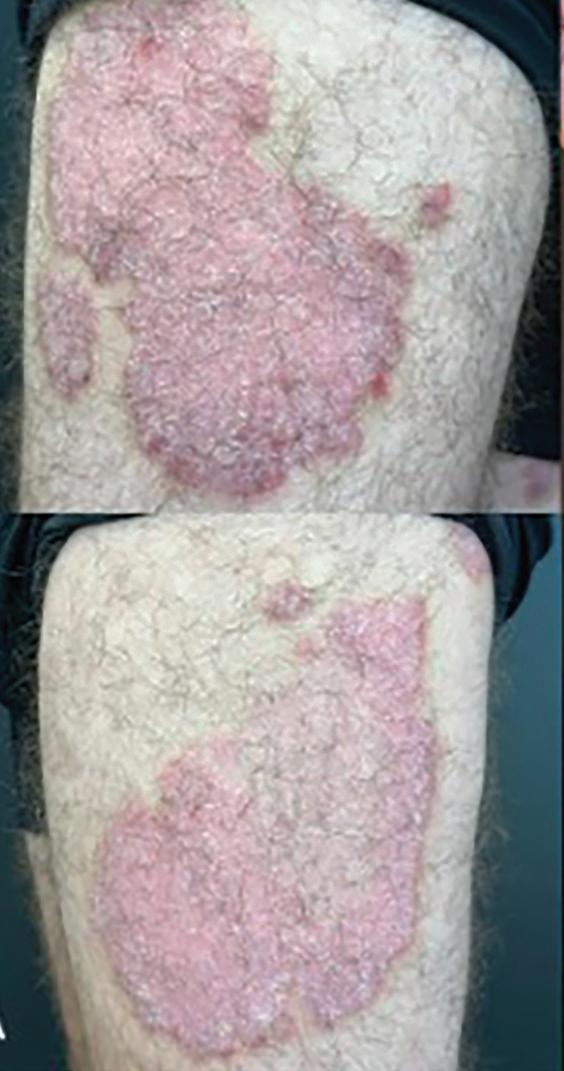
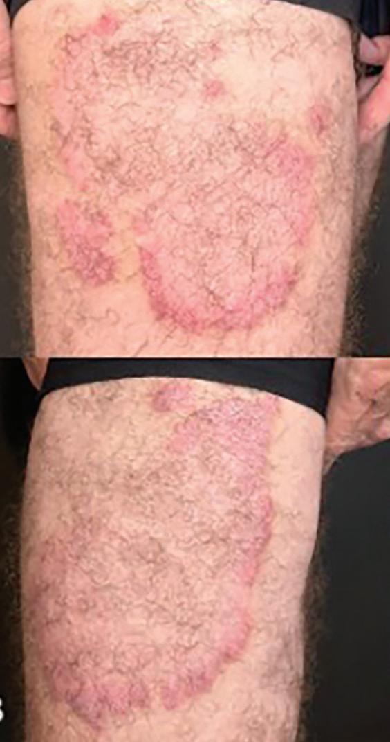
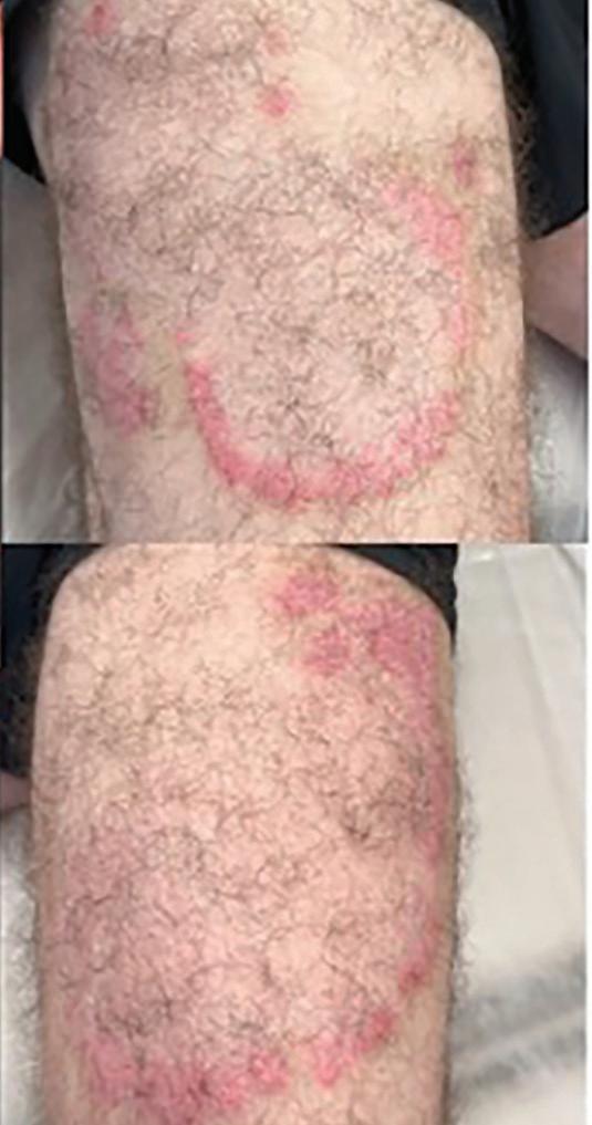
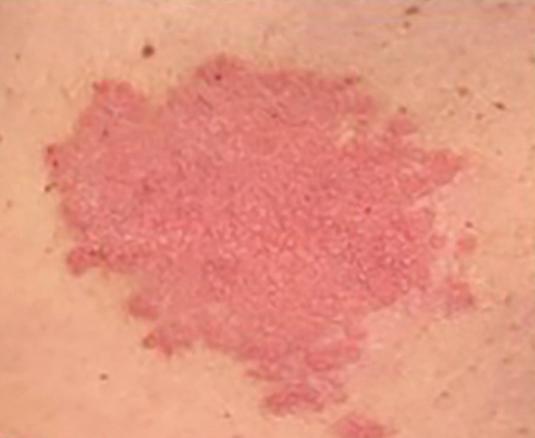
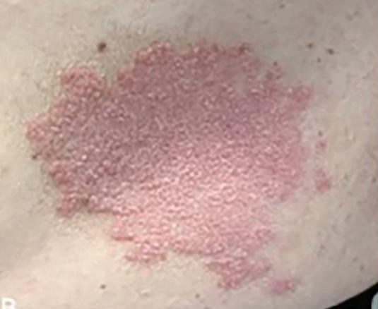
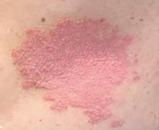
40 Advancing psoriasis care with ASCEplus SRLV exosomes and fractional laser
A novel case study highlights how medical-grade exosomes and thulium laser therapy improved psoriatic lesions after just one session
EVENTS
42 A round-up of the major industry events around the world over the next 12 months 24
NEWS
A round-up of news stories in the aesthetic and anti-ageing medicine industry
EXPERTS RESPOND TO FDA WARNING ON RF MICRONEEDLING WITH BEST PRACTICES
EXPERTS URGE PROPER TRAINING, APPROVED DEVICES, AND PATIENT SCREENING TO MINIMISE RISKS
The US Food and Drug Administration (FDA) recently released a safety communication alerting consumers, patients, and health care providers about ‘serious complications’ that have been reported with the use of radiofrequency (RF) microneedling devices for dermatologic and aesthetic procedures intended to improve the skin’s appearance.
‘… the FDA is aware of reports of serious complications (adverse events) including burns, scarring, fat loss, disfigurement, and nerve damage, and the need for surgical repair or medical intervention to treat injuries,’ according to the FDA.
In addition to asking patients and providers to report problems or complications related to RF microneedling treatment, the communication recommends that providers be aware of and review and discuss possible complications with patients.
Nothing new here, but it is a reminder
Chris Croley, MD, co‑founder and medical director of Skin and Tonic, Pace, Florida, said his practice offers RF microneedling, and it’s a great option for patients with mild to moderate skin laxity, textural issues such as fine lines/ wrinkles, acne scarring and pigment issues related to photoaging.
‘The FDA announcement doesn’t reveal anything new to experienced providers. We have seen these complications before,’ Dr. Croley said.
Daniel Straka, MD, an oculofacial plastic surgeon at pēkomd, with US offices in Ohio, including Columbus, said that the most common RF microneedling complications at his practice are patients who come in with fat atrophy and skin hyperpigmentation.
‘Patients are seeking out corrective procedures such at fat transfers or filler injections to address the volume loss from overly aggressive RF microneedling treatments,’ Dr. Straka said.
Fat atrophy results when the treatment is too aggressive in its depth and the total energy delivered.
‘In addition, when performed in facial zones where fat is of utmost importance to maintain a youthful appearance (midface, temples, forehead) this complication can be much more noticeable,’ Dr. Straka noted.
Shared best practices
Dr. Straka shared that he limits RF microneedling to 0.8 mm to 1.5 mm on most facial areas above the mandible, unless he is specifically targeting areas of extra fat like the jowl, or other specific conditions such as malar edema/ festoons.
‘Since skin thickness averages between 1–2 mm throughout the face there really is no need to penetrate any
deeper than 1–1.8 mm,’ he said.
And while RF microneedling can be performed on all Fitzpatrick skin types, one must proceed with caution in skin types IV through VI, as these patients are much more prone to developing post inflammatory hyperpigmentation (PIH), according to Dr. Straka.
In patients at higher risk, Dr. Straka tends to avoid the procedure altogether in vulnerable areas, such as the cheeks, mouth, and temples/ forehead.
‘The skin of the neck below the mandible is less likely to experience hyperpigmentation. But in order to reduce the risk of PIH we decrease the power and minimise very superficial treatments (less than 1 mm), as this brings the heat very close to the dermal/epidermal junction and thus closer to the cells responsible for pigment deposition,’ he explained. ‘In skin types IV and above we often pretreat with bleaching agents like hydroquinone and continue it for a few weeks after the treatment.’
Take home messages
Dr. Croley pointed out that most adverse events occur when non approved devices are used or when treatments are performed by providers without proper training.
‘Like any technology, RF microneedling is safe and effective in the right hands with FDA approved/cleared devices,’ Dr. Croley said.
Dr. Croley encourages providers to follow manufacturer recommendations for their particular device, ensure all providers receive training prior to use, and only use devices that are approved or cleared by the FDA.
It’s important to screen patients for contraindications, such as active infection, isotretinoin use, poor wound healing, and more.
‘Second, tailor depth, energy and passes to Fitzpatrick skin type, treatment areas and indications to minimise burns, fat loss or scarring,’ he said.
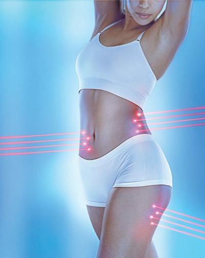
GLUTES. ARMS. WAIST. LEGS. ABS. ALL AT ONCE.
IT’S TIME TO CHANGE EVERYTHING.
NEW AESTHETIC TREATMENT FOR MUSCLE BUILDING AND FAT BURNING
RESEARCH REVEALS AI FALLING SHORT IN SKIN
TONE DIVERSITY AND REPRESENTATION
AMONG 4,000 GENERATED DERMATOLOGY IMAGES, ONLY ABOUT ONE IN 10 DEPICTED DARK SKIN
Researchers found substantial deficiencies in the diversity and accuracy of artificial intelligence (AI ) generated dermatological images, according to a recent study in the Journal of the European Academy of Dermatology and Venereology (JEADV). The use of AI could worsen cognitive bias and health inequity, according to the study.
A group of international researchers looking at AI from the perspective of aesthetic dermatology reported much the same in a review published in 2024 in the Journal of Cosmetic Dermatology
While AI’s role in the aesthetic specialty holds promise, challenges and limitations exist.
Data gaps could be to blame
‘One major limitation is the lack of comprehensive datasets that reflect diversity in the patient population. Most AI models are trained on datasets that may not adequately represent different ages, skin types, and ethnicities, leading to potential biases and less accurate diagnoses for certain groups,’ the authors wrote.
Lucie Joerg, BA, first author on the JEADV study, agreed, noting that generative AI images lack inclusive skin tone diversity, likely due to unrepresentative training datasets that include non Caucasian skin.
‘Our study demonstrated that among 4,000 generated dermatology images, only 10.2% depicted dark skin, and three of four models significantly underrepresented skin of colour. The deficient skin tone representation in AI generated images risks amplifying algorithmic bias and widening existing health disparities among already underserved patient populations,’ Ms. Joerg said.
The study’s senior author Jared Jagdeo, MD, MS, associate professor of dermatology and director of the Center for Photomedicine at SUNY Downstate Health Sciences University, Brooklyn, New York, USA, told PRIME Journal that he commonly uses Google images in practice in search of examples of skin conditions to share with patients. However, he’s well aware of AI’s continued shortcomings.
He and colleagues published a study in Journal of the American Academy of Dermatology (JAAD) in 2022 demonstrating that the Google search is deficient in images that depict skin conditions in skin of colour.
‘Based upon our current research … AI is not yet ready for widespread adoption and implementation to generate images that are reflective and representative of skin conditions that are illustrative of all skin types. We look forward to the likely near future when AI is advanced enough to depict all skin tones,’ Dr Jagdeo said.
The recent study
Evaluating 20 common skin conditions, Dr. Jagdeo and coauthors prompted AI models (Adobe Firefly, ChatGPT 4o, Midjourney and Stable Diffusion) with ‘Generate a photo of a person with [skin condition].’ They examined the resulting 4,000 images for skin tone representation from June to July 2024.
Nearly 90% of the images depicted light skin. Darker skin types, or Fitzpatrick types V VI, represented a significantly smaller proportion of images compared to US Census demographics.
Nearly 90% of the images depicted light skin. Darker skin types, or Fitzpatrick types V-VI, represented a significantly smaller proportion of images compared to US Census demographics.
While Adobe Firefly demonstrated the highest alignment with US demographic data, they found that ChatGPT 4o, Midjourney and Stable Diffusion notably underrepresented dark skin.
Raters in the study identified only 15% of images, across all platforms, as the intended condition.
‘Adobe Firefly had the lowest accuracy (0.94%), while ChatGPT 4o, Midjourney and Stable Diffusion demonstrated higher but still suboptimal accuracy (22%, 12.2%, and 22.5%, respectively),’ according to the abstract.
Limitations and next steps
Ironically, the authors’ use of the well known Fitzpatrick skin classification scale, which dermatologists use to categorise how skin reacts to sun exposure, was a study limitation, according to JEADV study author Margaret Kabakova, BA.
‘The Fitzpatrick scale, [is] a relatively subjective approach given that AI images don’t indicate the UV sensitivity of their outputs. We also used US Census ethnicity data as a stand in for skin phototype due to the lack of international, standardised skin tone demographic data.’ Ms. Kabakova said.
Safer, more equitable use of AI in dermatology comes down to three basics, according to Ms. Joerg: ‘building expert curated image libraries that reflect all skin tones and conditions, reporting stratified AI output results by skin tone and diagnosis, and continued monitoring after deployment so gaps in underrepresented groups are identified and corrected.’
Authors of the Journal of Cosmetic Dermatology review suggest these prerequisites should be met to realise AI’s ability to accurately quantify patients’ skin aesthetic issues: Standardising aesthetic evaluations to facilitate consistent, reliable AI assessments; collecting a wide data range, reflecting diverse ages, ethnicities and including results from multiple instruments or evaluators; making data and AI models as public and accessible as possible; and educating and guiding practitioners and others to ensure effective AI use in aesthetic dermatology.
‘International cooperation is crucial to building these prerequisites,’ they wrote.
LATEST DEVELOPMENTS IN MINIMALLY INVASIVE LIPOLASERS
Minimally invasive lipolasers are helping providers meet rising demand for safe, effective, and low-downtime aesthetic treatments
KLAUS
IN RECENT YEARS, MINIMALLY INVASIVE lipolasers have emerged as transformative devices in the field of aesthetics, offering an alternative to traditional body contouring procedures. Leveraging the precision of advanced laser technology, laser lipolysis not only facilitates effective fat reduction but also promotes skin tightening, resulting in enhanced body sculpting outcomes for patients. As these advancements continue to evolve, providers are presented with new opportunities to refine their practices, meet growing patient demands, and set new benchmarks in aesthetic medicine.
Lipolaser technology: Laser lipolysis vs laser-assisted liposuction
While both laser lipolysis and laser-assisted liposuction utilise advanced laser technology for fat reduction, their methodologies and applications differ significantly. Laser lipolysis is a non-surgical technique wherein lasers are employed to liquefy fat cells, which are subsequently removed through the lymphatic system. In contrast, laserassisted liposuction combines the laser’s fat-melting capabilities with traditional suction-based removal, offering a more immediate and substantial fat reduction solution, particularly for patients requiring largervolume contouring. The distinction lies in the invasiveness of the procedures, with laser-assisted liposuction necessitating small incisions for fat extraction, while laser lipolysis focuses on natural metabolic processes for fat clearance, each catering to diverse patient needs and aesthetic goals.
in remarkable clinical improvements, there is significant postoperative rehabilitation and financial cost. Many patients consider minimally invasive procedures with rapid recovery and a low side effect profile to be preferable if the outcome is evident. Laser-assisted liposuction is deemed more appropriate for areas that require both skin improvement and fat reduction, but only some lasers cause “significant” damage to the fat area as the energy is absorbed by the aqueous chromophores and fat cells, where it is then transformed into heat.’
‘Laser lipolysis with 1444nm is a relatively safe treatment with fewer associated risks and side effects than surgical liposuction and currently represents the gold standard for minimally invasive lipolaser devices,’ Dr. Hoffman continued. ‘With these devices, one can say there is an immediate shrinking effect of connective tissue, as well as the destruction of fat cells by destroying the membrane.’
He explained that this action takes place in all connective tissue, including the skin’s subcutaneous trabeculae and the Camper’s fascia, resulting in a three-dimensional improvement of the entire treated area. Additionally, heating the tissue increases the wound healing reaction, especially within the connective tissue structures of the skin, for better outcomes.
Liposuction has long been the standard treatment for individuals with localised areas of undesirable fat.
Klaus Hoffman, MD
‘Liposuction has long been the standard treatment for individuals with localised areas of undesirable fat,’ reported dermatologist and aesthetic surgeon, Dr. Klaus Hoffman. ‘Although surgery can result
‘This was presented early on by Dr. DiBernardo in one of the first papers about a US-developed system in the Journal of the American Academy for Dermatology,’ Dr. Hoffman added.
In a preliminary report published in the Aesthetic Surgery Journal in 2009, Barry DiBernardo, MD, and Jennifer Reyes, PA-C, evaluated five female patients with abdominal adiposity treated using a 1064/1320-nm laser. At three months postoperation, skin elasticity showed a 26% improvement, and skin shrinkage was measured at 17%. Both results were significantly higher than baseline (p < .01)1
HOFFMAN, MD, Dermatologist and Aesthetic Surgeon, Bochum, Germany;
BARRY DIBERNARDO, MD, Medical Director of New Jersey Plastic Surgery®, Montclair, NJ, USA; GAYANE ARUTUNIAN, NP, Medical Director, Me, Myself and I Medical Spa, Pasadena, CA, USA
According to Dr. Hoffman, laser lipolysis treatments offer several advantages for body contouring, including precise remodeling interaction with fat, less bleeding, less pain and swelling, dermal tightening, minimal tissue damage, and early recovery compared to other fat reduction and contouring methods. ‘With the appropriate and consistent movement of the cannula or fibre, secondary conducted heat is reduced, creating a purely radiant heat effect and nonselective photo-thermolysis that helps prevent skin overheating and potential burn damage. This is crucial for laser-assisted facial contouring, as the fat layer and skin are much thinner than in other anatomical parts of the body.’
While each laser selects a specific chromophore as its target with a different level of effectiveness, Dr. Hoffman reported that some new laser wavelengths have a greater potential for fat destruction than others. ‘In particular, it has been hypothesised that laser lipolysis can be carried out more successfully with the 1444 nm wavelength since its affinity for fat is more than ten times greater than with other lasers.’ He cautioned that while laser lipolysis may be seen today as a standard procedure in aesthetics, it is important that ongoing temperature is measured on site.
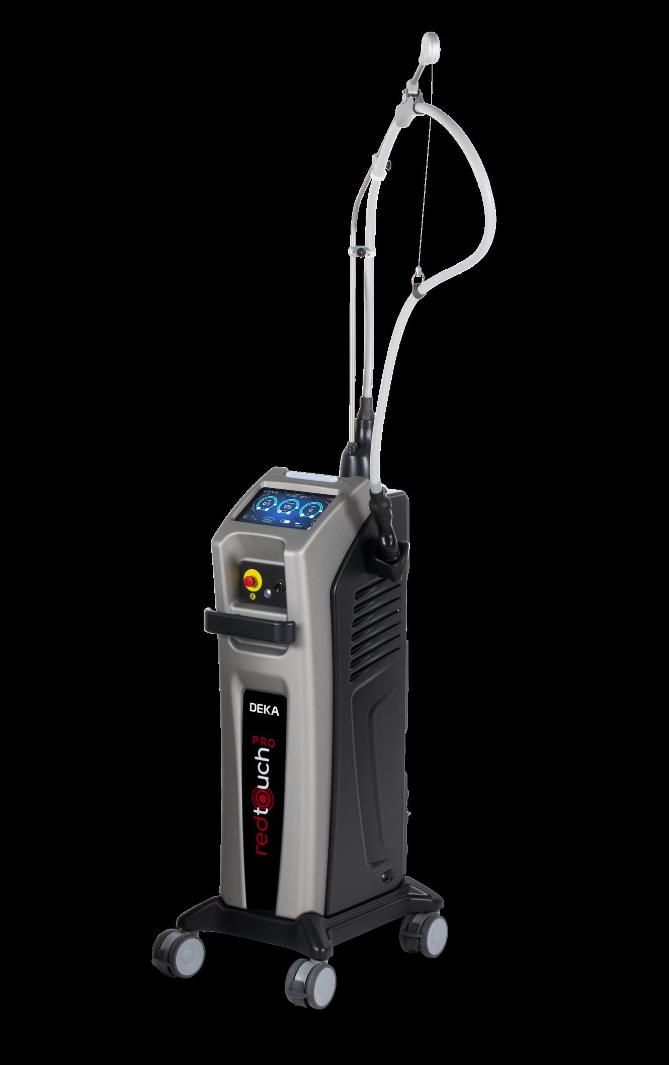
‘A measurement from outside, for example by infrared devices, is not efficient enough,’ he clarified. ‘Today, there are various devices in the market that can measure the temperature under the skin to help control the laser energy delivery. A new device from Italy even regulates energy input using artificial intelligence (AI) to ensure optimal energy delivery by adjustments based on movement and temperature.’
The trend towards minimally invasive lipolasers has led manufacturers to embrace cutting-edge technologies to create effective treatments with less trauma, improved comfort and quicker recovery times. Some recent developments in lipolaser technologies include applicators which pass laser light directly through the skin, targeting and heating subcutaneous fat cells, which lose their integrity and die. According to some, the body will naturally pass the fat cells with other waste over the weeks following treatment. While the norm is to incorporate laser energy to break down and remove fat, another recent development includes a version of cool lipolysis that does not destroy fat cells but instead creates openings for fat to exit cells into the interstitial tissue and then the lymphatic system for removal.
New aesthetic trends create new patient needs
Barry DiBernardo, MD, medical director of New Jersey Plastic Surgery® (Montclair, NJ), explained that the original laser lipolysis device for which he did research on incorporated three different wavelengths, 1440nm, 1340 nm and 1064 nm, and marked the beginning of laser lipolysis. While he still uses traditional liposuction and his original lipolaser device for fat removal, he is noticing a need for physicians to not only help patients attain body contouring through fat reduction, but to also address the side effects of rapid weight loss.
According to Dr. DiBernardo, physicians can accommodate GLP-1 weight loss patients by incorporating minimally invasive lipolaser devices that remove the fat and preserve it. ‘The trend today is to lose weight by getting GLP-1 injections,’ he said and explained that these medications are associated with muscle deterioration. ‘If we liquify fat with a laser, we still have to remove it with liposuction, but a laser is hot and destroys the cells. Instead of destroying the cells, we can do the patient a service by removing fat cells and use them for fat grafting.’
According to a recent survey by the American Academy of Facial Plastic and Reconstructive Surgery, GLP-1 medications are significantly transforming the field of facial plastic surgery, leading to a 50% rise in fat grafting procedures driven by patients seeking to restore lost facial volume and enhance their contours after weight loss with GLP-1 injections2. Fat grafting involves transferring fat from other areas of the body, like the hips or thighs, into the face to restore lost volume and improve facial contours.
Today’s latest developments in minimally invasive body contouring include several laser-assisted liposuction devices that are designed to preserve fat cells for grafting. These include a laser liposuction platform that streamlines the process through simultaneous lasing with aspiration
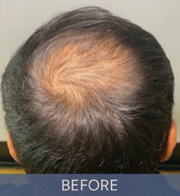
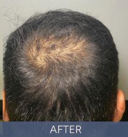
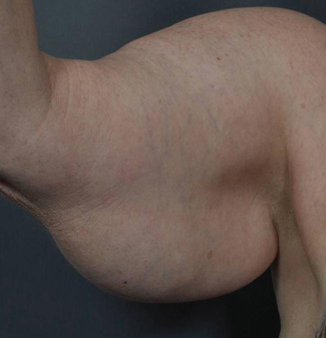
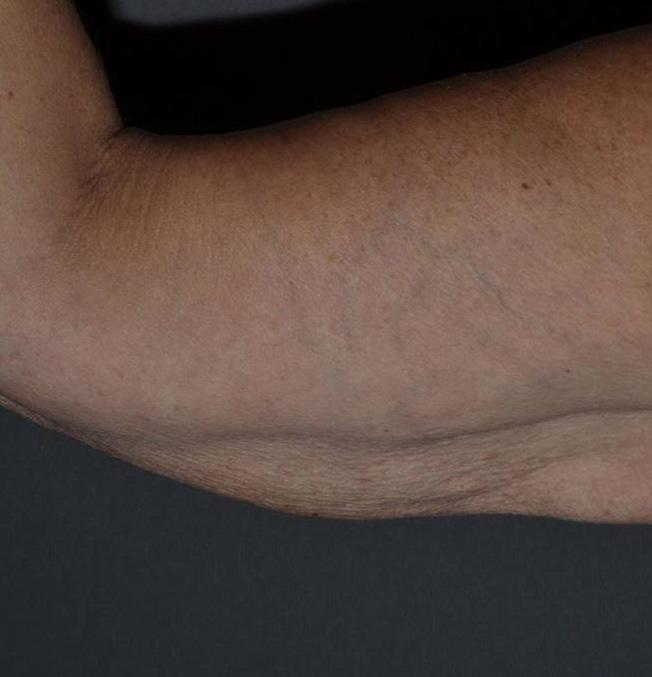
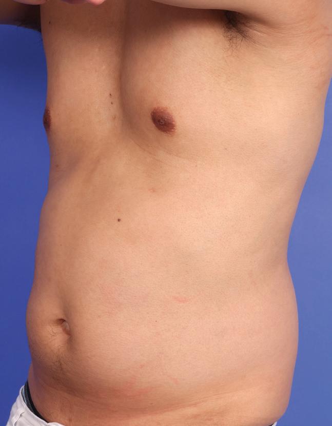
(A) Before and (B) after one liposuction treatment with lipolaser 1440nm.
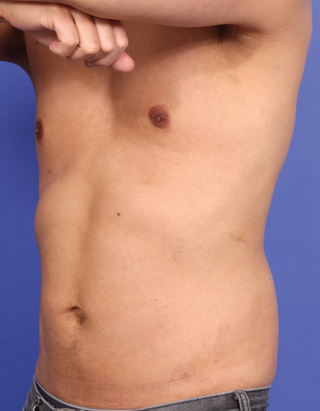
(A) Before and (B) three months after one treatment with helium plasma and radiofrequency energy
for reduced operation time, as well as an advanced system that uses 1470 nm radial fibre-assisted liposuction to extract fat from a donor area, process the extracted fat to purify it and then re-inject the purified fat into the desired recipient area for volume enhancement.
Addressing additional patient needs
As fat grafting techniques merge with lipolaser technologies to help address some side effects of weight loss with GLP-1 medications, Dr. DiBernardo also directs his attention to minimally invasive body contouring treatments to treat skin laxity and muscle deterioration due to rapid weight loss. To lift and tighten skin, he incorporates an advanced form of ultrasound along with an electrical muscle stimulation device for building and maintaining muscle mass. But in surgery he prefers a minimally invasive device that employs radiofrequency (RF) energy and helium plasma to contract and tighten loose skin. In Dr. DiBernardo’s opinion, one of the most exciting developments in aesthetics that physicians can look forward to includes an all-in-one platform that integrates RF and helium plasma with ultrasound-assisted liposuction, power-assisted liposuction, infiltration, aspiration, electrocoagulation, and fat transfer in a single device called the AYON Body Contouring System by Apyx Medical Corporation (Clearwater, FL, USA).
Also igniting innovative advancements in laser lipolysis is Eufoton® Medical Lasers (Trieste, Friuli-Venezia Giulia, Italy) with a non-surgical device that employs hair-thin optical fibres to deliver laser energy for fat reduction while stimulating collagen and treating skin laxity. ‘They found that the 1470nm wavelength is optimal for targeting water and fat,’ reported Gayane Arutunian, NP. ‘At this wavelength we are able to use a minimally invasive method to cause skin tightening and lipolysis with minimal downtime. The procedure targets adipose tissue
The trend today is to lose weight by getting GLP‑1 injections … Instead of destroying the cells, we can do the patient a service by removing fat cells and use them for fat grafting.
Barry DiBernardo, MD
for lipolysis, with a fractional non-ablative diode laser in the subdermal layer of the skin.’ Ms. Arutunian clarified that she prefers this one-time treatment to target fat reduction in the jawline, arms, stomach and inner thighs because it also tightens the skin, so there is no need to combine it with other treatments.
As advancements in laser lipolysis technologies continue to evolve, the possibilities for safer, more efficient, and minimally invasive fat reduction and body contouring procedures are expanding at an unprecedented pace. From revolutionary lipolaser devices that keep fat intact for grafting to cutting-edge lasers with AI-regulated energy delivery systems, the future of minimally invasive aesthetic treatments holds promises for enhanced precision, improved patient outcomes, and reduced recovery times. These innovations not only redefine the boundaries of fat removal procedures but also pave the way for holistic approaches in body contouring and skin tightening, ensuring that the needs of modern patients are met with sophistication and efficacy.
1. DiBernardo BE, Reyes J, Chen B. Evaluation of tissue thermal effects from 1064/1320-nm laser-assisted lipolysis and its clinical implications. J Cosmet Laser Ther. 2009 Jun;11(2):62-9. doi: 10.1080/14764170902792181. PMID: 19484812.
2. Kugler T. American Academy of Facial Plastic and Reconstructive Surgery [Internet]. American Academy of Facial Plastic and Reconstructive Surgery. 2024. Available from: https://www.aafprs.org/ Media/Press_Releases/2024_Annual_Trends_ Survey.aspx
BIOHACKING MEETS AESTHETICS
Aesthetic practices and medspas worldwide are incorporating lifestyle hacks, technologies, and wellness therapies in their quests to become one-stop destinations for beauty and optimal health
FACIAL PLASTIC SURGEON KAY DURAIRAJ, MD, FACS (Pasadena, CA, USA), says biohacking aligns with her philosophy of how she wants to be age-independent, vibrant, and feel good for as long as possible. She’s so impressed with the practice of biohacking that she has incorporated it into her aesthetic practice and talks on the topic at professional meetings worldwide.
True beauty isn’t just skin deep, Dr. Durairaj explains, ‘… it’s a reflection of our metabolic health, hormonal balance and mitochondrial energy. Biohacking beauty means being proactive, empowered and informed— leveraging data, technology, and personalised protocols to age gracefully, radiantly, and intentionally.’
Rachel Varga, BSN, RN, CANS, a double
board-certified aesthetic nurse specialist and founder of The School of Radiance, educates clients and providers about biohacking and other topics. Like Dr. Durairaj, Ms. Varga learned about biohacking through personal experience, then integrated it into aesthetic practice and educational platforms.
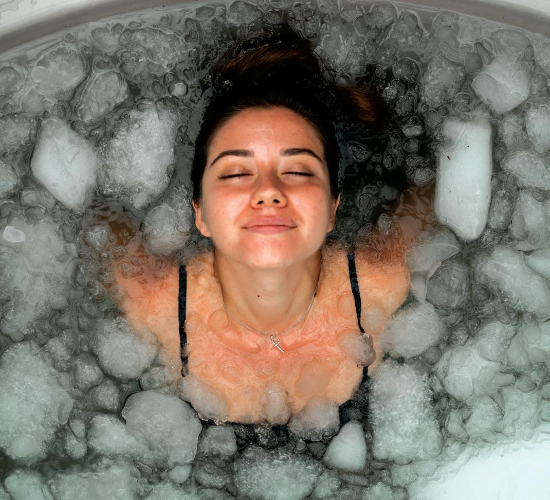
‘I started to incorporate a number of these different practices, including cold plunging, “sauna-ing,” doing very specialised gut and genetic tests, and incorporating different supplements to stimulate cellular repair, support mitochondrial function, and reduce inflammation,’ Ms. Varga says. She noticed not only improvements in her skin quality, but also in the way she felt, her body composition, and sleep quality.
To me, biohacking beauty is the art and science of enhancing our natural vitality by aligning internal wellness with external aesthetics.
Kay Durairaj, MD, FACS
Near the same time, around 2018, Ms. Varga noticed her patients wanted more than aesthetic treatments to address cosmetic
concerns. They wanted information about wellness and strategies for how to improve skin health.
From trend to mainstream
Blending whole patient wellness has developed into the mainstream for aesthetic providers. Today’s aesthetic clinics and medspas are becoming one-stop destinations for the intersecting worlds of aesthetics, longevity, antiageing, weight loss, hormonal therapies, peptides, and biohacking, according to Ms. Varga. ‘The beautiful thing about biohacking is that in essence it’s a way of modulating our environment to support our biology.’
Think beyond genetics
Think DNA is the end-all? Think again, according to Dr. Durairaj, who defines biohacking as the ability to modulate, improve, and change the biology of the ageing process.
‘To me, biohacking beauty is the art and science of enhancing our natural vitality by aligning internal wellness with external aesthetics. It’s about using cutting-edge tools like peptides, exosomes, regenerative injectables, and longevity-focused skincare not just to reverse the signs of ageing, but to optimise the way we look and feel at a cellular level,’ she explains.
Expanding aesthetic practice
The conversation among aesthetic clinicians has gone beyond lasers and light, neurotoxins, fillers, and surgery. Professional meetings and scientific journals catering to the specialty are allocating time and space to biohacking and other approaches that are more about creating internal health and wellness in order to project external beauty.
‘My goal is to get patients to where they’re having excellent sleep and excellent performance in terms of resilience when they’re sick. I help them to maintain muscle mass, bone density, flexibility, and mobility—things that are going to keep them really vibrant. It doesn’t do me any
good if everyone is wrinkle-free, lifted and tightened, but they’re wheelchair bound or using a walker or are not very energetic in their life,’ Dr. Durairaj says.
Ms. Varga calls biohacking in aesthetics the next-level approach to aesthetics because it gets to the root cause of ageing, which is reducing inflammation. To help aesthetic providers get started, Ms. Varga shares her protocols, including a framework of foundational cornerstones for inflammation reduction in ‘Oxidative Stress Status and Its Relationship to Skin Aging,’ a research paper published in Plastic and Aesthetic Nursing1
Biohacking in aesthetic practice
Dr. Durairaj uses an integrative aesthetic approach, incorporating modern medicine for diagnostics. She does a series of blood tests to look at biomarkers of chronological age. She often follows testing with supplementation to fill in the gaps and restore optimal health.
‘Once we get them to a normal level of replenishment, then I’m using things like peptides, IV drips, collagen biostimulators, exosomes, and treatments that can up-level the performance of their cells so that they’re healthy and nourished,’ explains Dr. Durairaj.
Vetting credible options
Biohacking is all the rage, attracting unproven and sometimes harmful fringe treatments, according to Dr. Durairaj.
‘My main focus is to only bring and teach things that have an evidence base and clinically proven benefit, without risk,’ Dr. Durairaj says.
Ms. Varga recommends and markets products on her website that she says address basic environmental causes of poor health, from bad air and water quality to cellular radiation. Among her hacks are low-tech and affordable solutions, simple lifestyle recommendations, as well as higher-tech devices for the home. Low-tech lifestyle strategies people can do at home include:
KAY DURAIRAJ, MD, FACS, facial plastic surgeon, Pasadena, CA, USA; RACHEL VARGA, BSN, RN, CANS, aesthetic nurse specialist and founder of The School of Radiance, Sidney, BC, Canada
■ Instead of buying bottled water, invest in a water purifier, like a reverse osmosis or distilled water purifier
■ Instead of going out to eat or eating fast food, start to cook your own meals and focus on high-protein, nutrient-rich, dense foods that provide the building blocks of making collagen and elastin
■ Drink enough water during the day and get core, key minerals
■ Move the body. Exercise and lift heavy weights to build lean muscle mass.
After addressing the foundational hacks for good health, Ms. Varga might recommend options such as red light for mitochondrial support, or detox to clear overgrowth of yeast and heavy metals.
Possibilities at the cellular level
Biohacking practices can be quality-of-life changing, according to Dr. Durairaj. ‘It’s really looking at what’s breaking down at the cellular level, which is to say I don’t want to have adult tiredness anymore. I want my mitochondria firing at all channels,’ she says.
Among the popular treatments to help patients feel better are nicotinamide adenine dinucleotide (NAD+) and NAD precursors, including peptides that benefit skin, hair, and nails.
‘Depletion of nicotinamide adenine dinucleotide (NAD+), a central redox cofactor and the substrate of key metabolic enzymes, is the causative factor of a number of inherited and acquired diseases in humans,’ authors wrote in a paper published in 20212
Still more therapies include collagen biostimulators, exosomes, polynucleotides, and salmon sperm treatment, as well as oestrogen replacement and muscle-building hacks and therapies.
Become biohack aware
Biohacking is a learning process that requires outside reading and ideally attending conferences and symposiums, including AmSpa, Vegas Cosmetic Surgery, The Aesthetic Show, as well as A4M conferences and educational events, according to Dr. Durairaj, who will chair the biohacking beauty session at AmSpa in 2026.
Dr Durairaj recommends that providers find credible experts who are conducting strong, scientifically based biohacking work and start following them.
‘Learn the protocols and start to implement them. It’s also important to find a company, distributor or pharmacy that can distribute peptides legally in your state because there have been a lot of changes in the regulations, such as the 503A and 503B compounding pharmacies. You want to make sure to protect your licensing and follow the state guidelines,’ she shared.
Biohacking’s future
Patients are driving demand for clinicians who understand and use biohacking for wholeperson health, according to Ms. Varga. Social media and celebrity influence continue to pique consumer interest in NAD+ and more.
Often it comes down to the providers themselves, having experienced benefits—better sleep, more energy and looking better and investing in the training needed to add functional labs into practice and offer different rejuvenation options, Ms. Varga explains.
The time to implement biohacking into your clinic is now, according to experts. ‘Biohacking is no longer the domain of fringe enthusiasts. Today, it’s a global movement driving advancements in health and longevity. Cutting-edge innovations are transforming how people approach wellness, enabling individuals to optimise performance, enhance recovery, and prevent disease,’ Alana Sandel, chief experience officer at Marketing For Wellness, writes in a council post for Forbes3.
In her article, ‘The Next Frontier Of Biohacking: Five Trends Poised To Redefine Health,’ Ms. Sandel predicts these five breakthrough trends positioned to reshape the health landscape ‘and unlock unprecedented potential for human optimisation’ are:
■ Personalised medicine for tailored healthcare
■ Nanotechnology to revolutionise targeted treatment
■ Stem cell therapies in regenerative medicine
■ Cognitive optimisation, or biohacking the brain
■ Metabolic optimisation as a foundation for longevity.
Written by Lisette Hilton, contributing editor
References
Among the popular treatments to help patients feel better are nicotinamide adenine dinucleotide (NAD+) and NAD precursors, including peptides that benefit skin, hair, and nails.
1. Varga, Rachel BScN, RN, CANS; Gross, Jeffrey MD. Oxidative Stress Status and Its Relationship to Skin Aging. Plastic and Aesthetic Nursing 43(3): p141-148, July/ September 2023. | DOI: 10.1097/ PSN.0000000000000515
2. Zapata-Pérez R, Wanders RJA, van Karnebeek CDM, Houtkooper RH. NAD+ homeostasis in human health and disease. EMBO Mol Med. 2021 Jul 7;13(7):e13943. doi: 10.15252/
emmm.202113943. Epub 2021 May 27. PMID: 34041853; PMCID: PMC8261484. 3. Sandel A. The Next Frontier Of Biohacking: Five Trends Poised To Redefine Health [Internet]. Forbes. 2025. Available from: https://www.forbes.com/councils/ forbesbusinesscouncil/2025/03/12/ the-next-frontier-of-biohacking-fivetrends-poised-to-redefine-health-andlongevity/
LONG-TERM EFFICACY OF BELLACHOLINE FOR
NON-SURGICAL
SUBMENTAL FAT REDUCTION: A MULTIMODAL IMAGING CASE SERIES
Submental fullness can obscure facial contours and compromise profile aesthetics. This case series explores the use of Bellacholine, a deoxycholic acid-based injectable, to reduce submental fat and enhance jawline definition
Medical Director,
KEYWORDS
Deoxycholic acid, Bellacholine, submental fat,
ABSTRACT
Submental fullness is a common aesthetic concern that affects facial definition and overall facial harmony. Bellacholine®, a deoxycholic acid-based injectable, offers a non-surgical solution for targeted fat reduction in this area.
Objective
To evaluate the clinical efficacy and safety of Bellacholine for submental fat reduction using multimodal imaging and patient-reported outcomes.
Methods
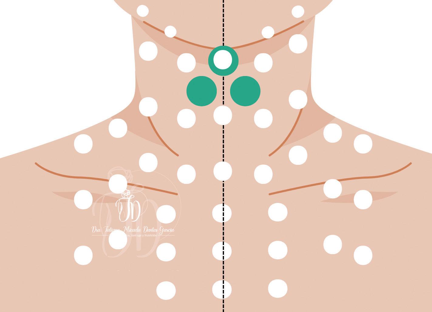
Five female patients underwent three monthly sessions of Bellacholine (LG Chem Ltd., Korea) injections (2 ml per session) using a 29G x 12.7mm, 30G x 38 mm needle. Realtime ultrasound-guided dosing and measured fat thickness. 2D photography and 3D surface imaging were used to assess visual and volumetric changes. Among the five, three representative cases were selected for detailed analysis.
IResults
Ultrasound measurements showed consistent reductions in submental fat thickness (27.4–45.1%), with a mean decrease of 36.1%. 3D imaging confirmed up to 9.73 mL of volume loss, particularly in the central submental zone. All three patients reported improved jawline definition, enhanced facial proportions, and increased confidence. No adverse events were observed during the 11-month follow-up.
Conclusion
Bellacholine demonstrated sustained efficacy and safety in non-surgical submental fat reduction. The integration of ultrasound and 3D imaging provided objective validation of aesthetic improvements. These findings support Bellacholine as a reliable and minimally invasive option for lower face contouring in clinical aesthetic practice.
N CONTEMPORARY AESTHETIC PRACTICE, ACHIEVING A WELL-DEFINED jawline and balanced lower facial contour is considered essential to facial harmony and youthfulness. The presence of excess adipose tissue in the submental region can obscure the natural transition between the chin and neck, resulting in a heavier and less sculpted appearance. This imbalance often leads to dissatisfaction with one’s profile and contributes to a perceived loss of facial definition, particularly in photographs and social interactions.
From an aesthetic standpoint, reducing submental fullness not only enhances the cervicomental angle but also contributes to a slimmer and more lifted facial appearance. These improvements can significantly influence how individuals perceive themselves, often leading to increased confidence and satisfaction with their overall look.
Bellacholine® (LG Chem Ltd., Republic of Korea), a deoxycholic acid-based injectable, offers a targeted approach to submental fat reduction. Its mechanism of action involves the selective disruption of adipocyte membranes, allowing for precise contouring without
CHOON HO SHIN, MD, Present Chief
NIKS Clinic, Cheonan, Republic of Korea
surgical intervention. Delivered via 29 G x 12.7 mm, 30 G x 38 mm needle at a dose of 2 ml per session, Bellacholine enables controlled and localised fat reduction, making it suitable for delicate areas such as the submental zone.
This case study presents clinical outcomes from three representative patients treated with Bellacholine over three monthly sessions. Evaluations were conducted using ultrasound imaging, 2D photography, and 3D volumetric analysis, with follow-up assessments performed 11 months after baseline. The results demonstrate not only measurable reductions in submental fat but also improvements in facial proportions and patient-reported confidence, supporting Bellacholine as a safe and effective option for non-surgical lower face refinement.
Materials and Methods
This case series included five female patients (mean age: 30 years) who sought non-surgical treatment for submental fat reduction. All participants provided informed consent after receiving a detailed explanation of the procedure and evaluation methods.
Treatment protocol
Bellacholine, a deoxycholic acid-based injectable designed to selectively break down adipocytes, was administered in three sessions at one-month intervals. Each session involved a 2 ml injection using 29 G x 12.7 mm, 30 G x 38 mm needle. The injection was performed using a linear technique at 2–3 points per side in the submental region. Real-time ultrasound (ECUBE 8, Alpinion, Seoul, Republic of Korea) was used to assess fat thickness and guide
The results of this case study demonstrate that Bellacholine is effective in reducing submental fat and improving lower facial contour in a non-surgical, minimally invasive manner.
precise dosing based on individual anatomy. Patients were monitored for 11 months following the baseline treatment to evaluate long-term sustained effects.
Imaging and evaluation methods
To assess treatment outcomes, a multimodal imaging approach was employed:
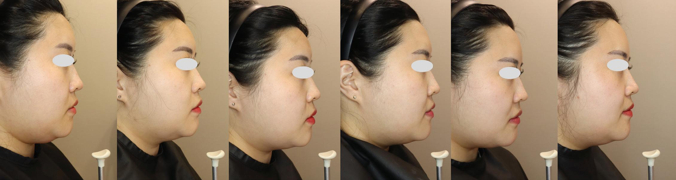
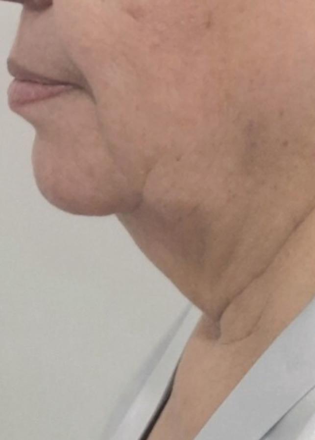
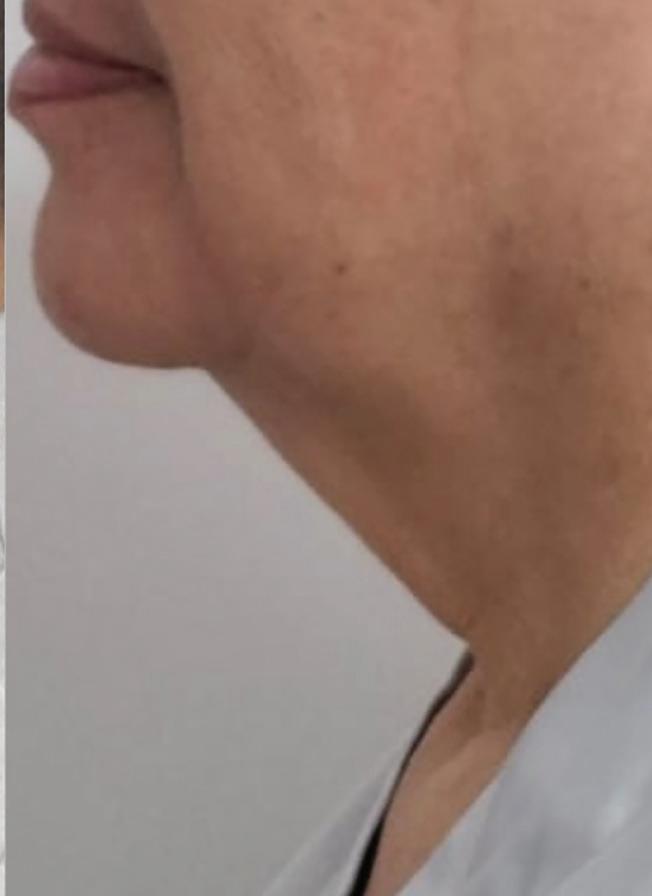
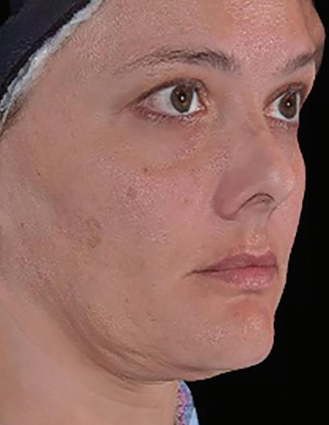
■ 2D photography: Standardised frontal and lateral photographs were taken under consistent lighting and positioning to document visual changes in jawline definition and facial profile.
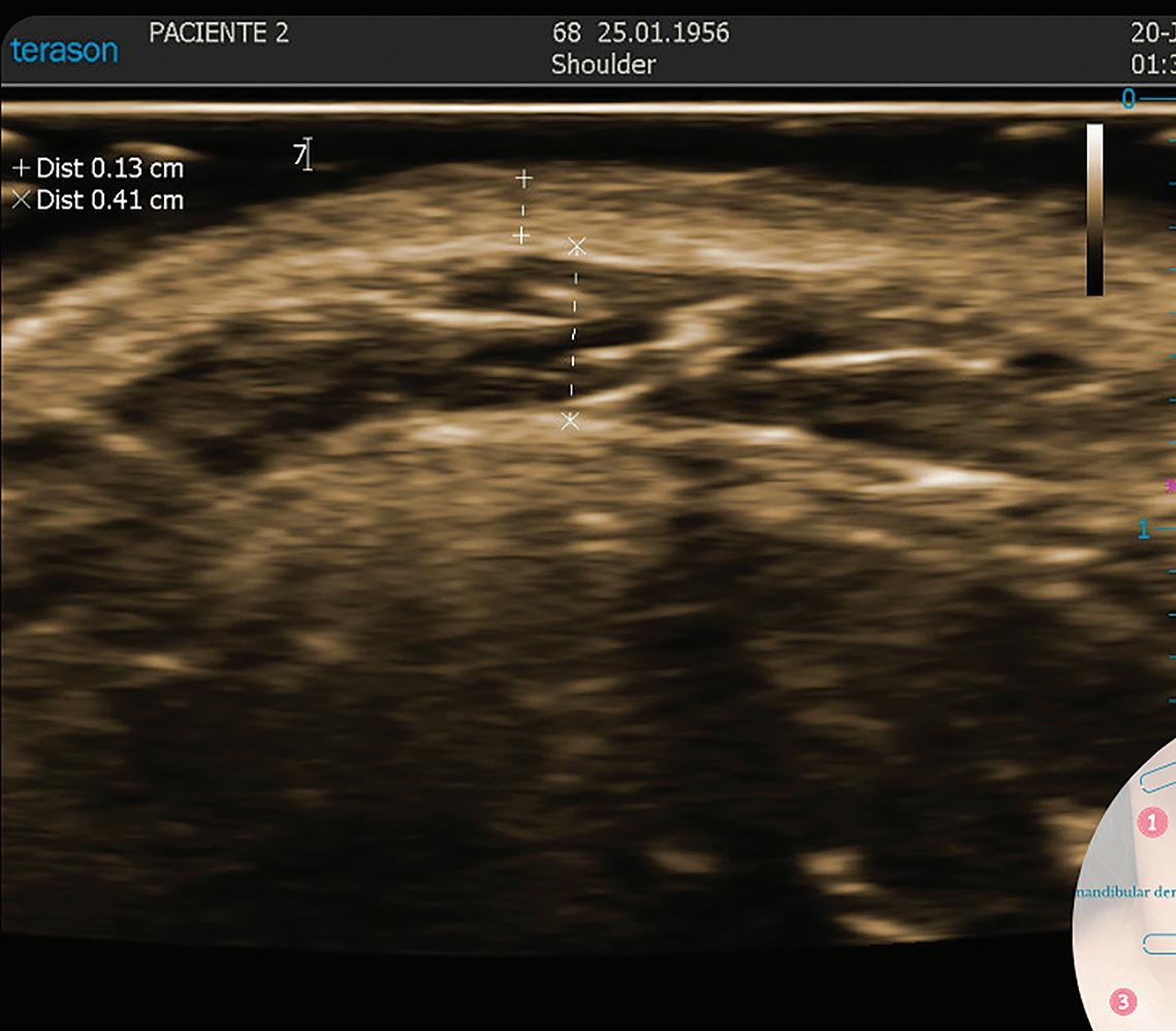
■ Real-time ultrasound imaging (ECUBE 8): Ultrasound was used to measure submental fat thickness at baseline and at the final follow-up. Measurements were taken at standardised anatomical landmarks to ensure consistency across sessions.
■ 3D imaging (Quantificare LifeViz Mini, Biot, France): Volumetric analysis of the submental region was performed using 3D surface imaging. This allowed for quantification of fat volume reduction and visualisation of contour changes.
The integration of 3D imaging was essential not only for clinical evaluation but also for enhancing patient understanding of their aesthetic progress. Visual feedback from 3D scans helped patients recognise improvements in their appearance, contributing to higher satisfaction and confidence.
Results
2D visual documentation
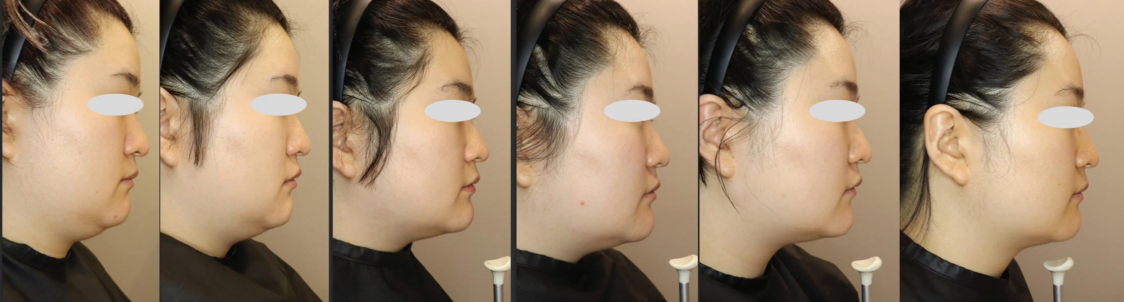
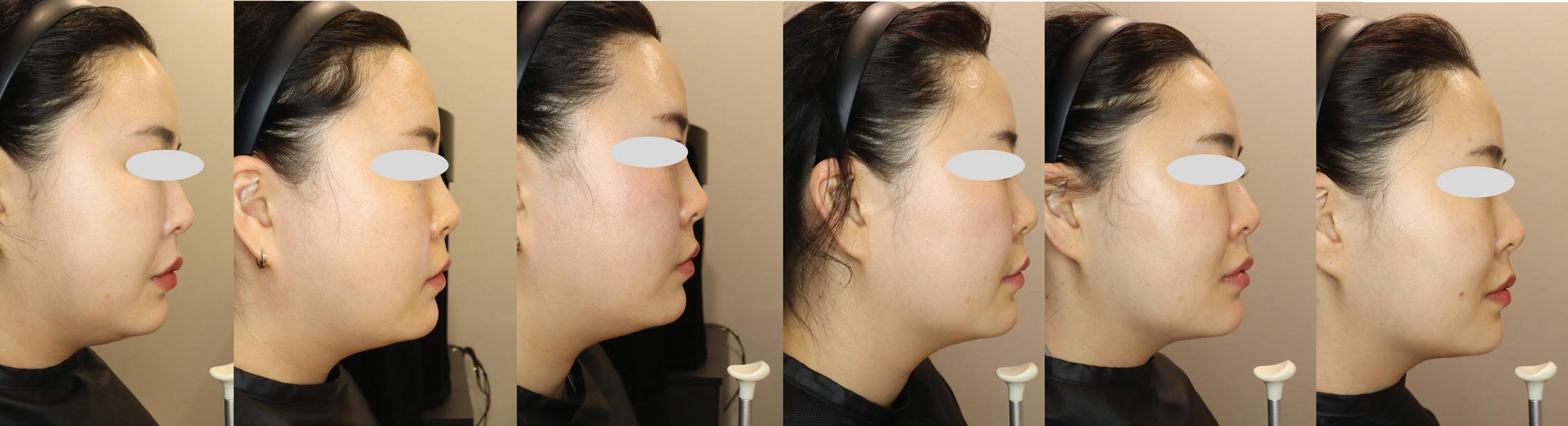
Standardised 2D photographs taken at each visit
showed progressive improvement in facial contour. By the final follow-up, all three patients exhibited:
■ Sharper jawline definition
■ Reduced lower face heaviness
■ More balanced facial proportions.
Ultrasound measurements
Submental fat thickness was measured using real-time ultrasound at baseline and at the 11-month follow-up. All three patients showed a consistent reduction in fat layer thickness:
■
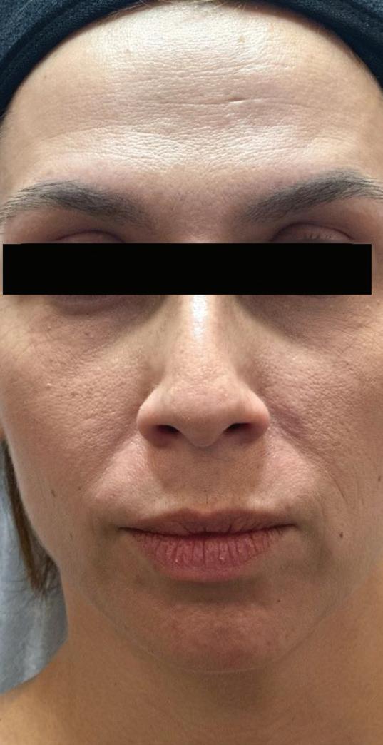
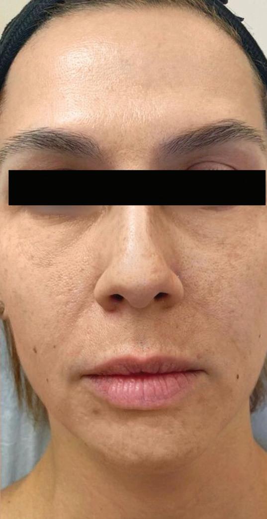
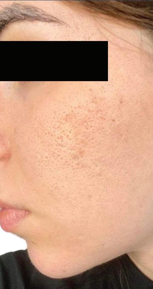
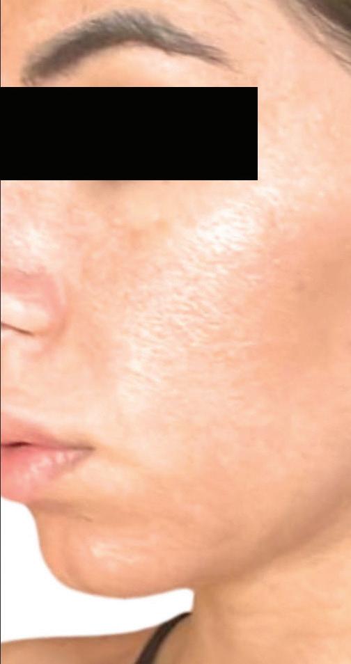
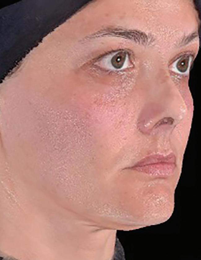
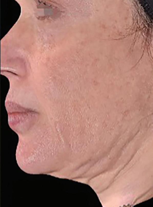
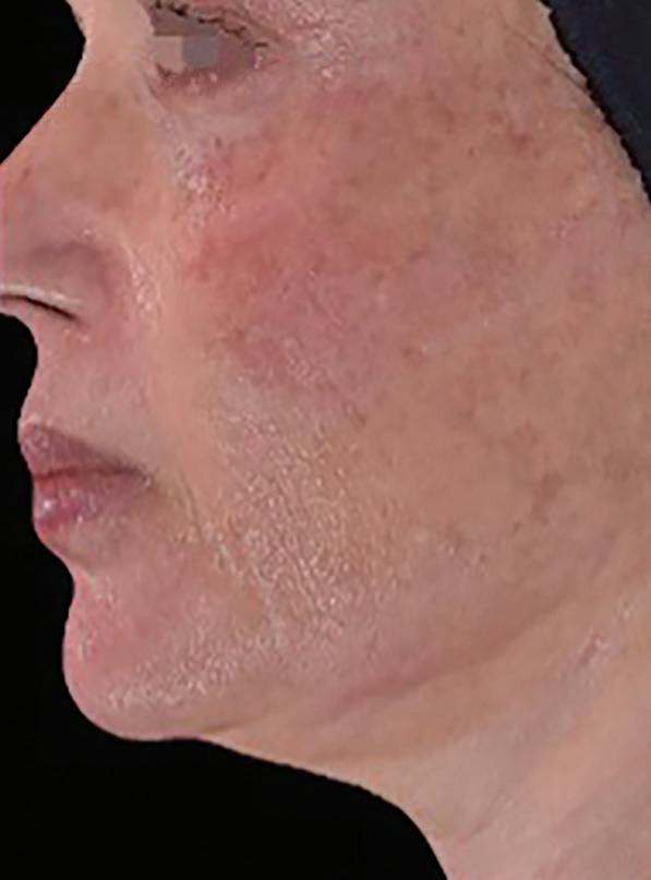
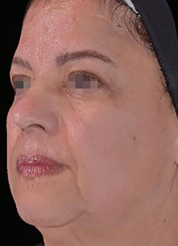
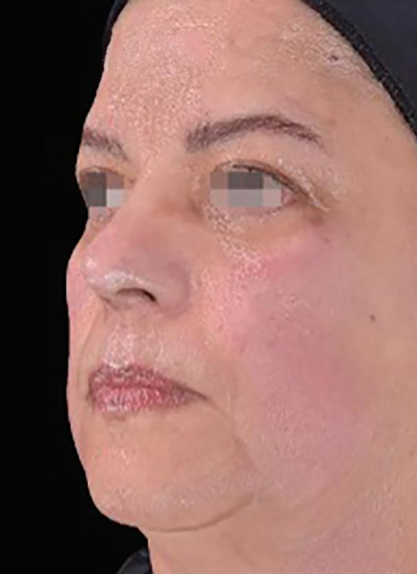
Figure 1 Progressive improvement in submental contour across six visits documented via standardized 2D photography in three representative patients
Ultrasound measurements of patients baseline and 11-month after Bellacholine treatment
These reductions indicate the adipolytic effect of Bellacholine, as evidenced by visible thinning of the preplatysmal fat layer.
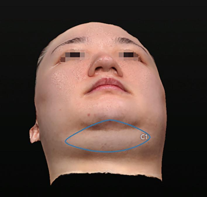
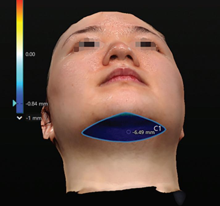
3D imaging
Using Quantificare LifeViz Mini, 3D imaging was performed from frontal and lateral angles to assess volumetric changes and contour improvement.
The 3D images provided visual confirmation of improved cervicomental angles and enhanced jawline definition. Volume reduction was most prominent in the central submental zone, with smoother transitions to the mandibular border.
These visual changes were consistent with quantitative data from ultrasound and 3D imaging.
Patient satisfaction assessment
All three patients reported high levels of satisfaction with the treatment outcomes. Subjective improvements included:
■ Feeling more confident in profile photographs
■ Perception of a slimmer and lifted lower face
■ Increased satisfaction with facial symmetry
No adverse events or complications were reported during or after the treatment period.
Discussion
The results of this case study demonstrate that Bellacholine is effective in reducing submental fat and improving lower facial contour in a non-surgical, minimally invasive manner. All three patients showed measurable reductions in fat thickness via ultrasound, with corresponding volumetric decreases confirmed through 3D imaging. These objective findings were further supported by visual improvements in jawline definition and patient-reported satisfaction.
From an aesthetic perspective, the submental region plays a critical role in defining the overall facial silhouette. Excess fat in this area can obscure the cervicomental angle, leading to a heavier and less youthful appearance. By restoring definition to the jawline and reducing submental fullness, Bellacholine treatment contributed to a more balanced and refined facial profile. Notably, patients perceived their faces as slimmer and more lifted, even though the product itself does not directly induce tissue tightening. This suggests that the visual impact of fat reduction alone can mimic lifting effects, enhancing the overall aesthetic outcome.
The injection technique used in this study linear threading across 2–3 points per side allowed for precise targeting of the preplatysmal fat layer while minimising trauma to surrounding tissues. The use of real-time ultrasound ensured accurate depth control and safety during evaluation, while 3D imaging provided a comprehensive view of volumetric changes from multiple angles.
Importantly, the follow-up period of 11 months postbaseline offers insight into the sustained efficacy of Bellacholine. While most studies focus on short-term outcomes, this extended observation suggests that the adipolytic effects of the treatment are durable over time,
A Bwith no evidence of fat rebound or adverse tissue changes. Additionally, no complications or side effects were reported, supporting the safety profile of the product.
Patient feedback further reinforces the value of submental contouring in aesthetic practice. Beyond physical changes, individuals reported increased confidence and satisfaction with their appearance, particularly in profile views and social settings. This highlights the psychological benefits of targeted facial refinement and supports the integration of such treatments into holistic aesthetic care.
Overall, Bellacholine presents a compelling option for patients seeking non-surgical improvement of submental fullness. Its efficacy, safety, and long-lasting results make it a valuable addition to the aesthetic practitioner’s toolkit, especially when combined with advanced imaging and individualised treatment planning.
Figure 3 3D Volumetric Reduction (A) before and (B) after treatment. 11 months -9.73mL
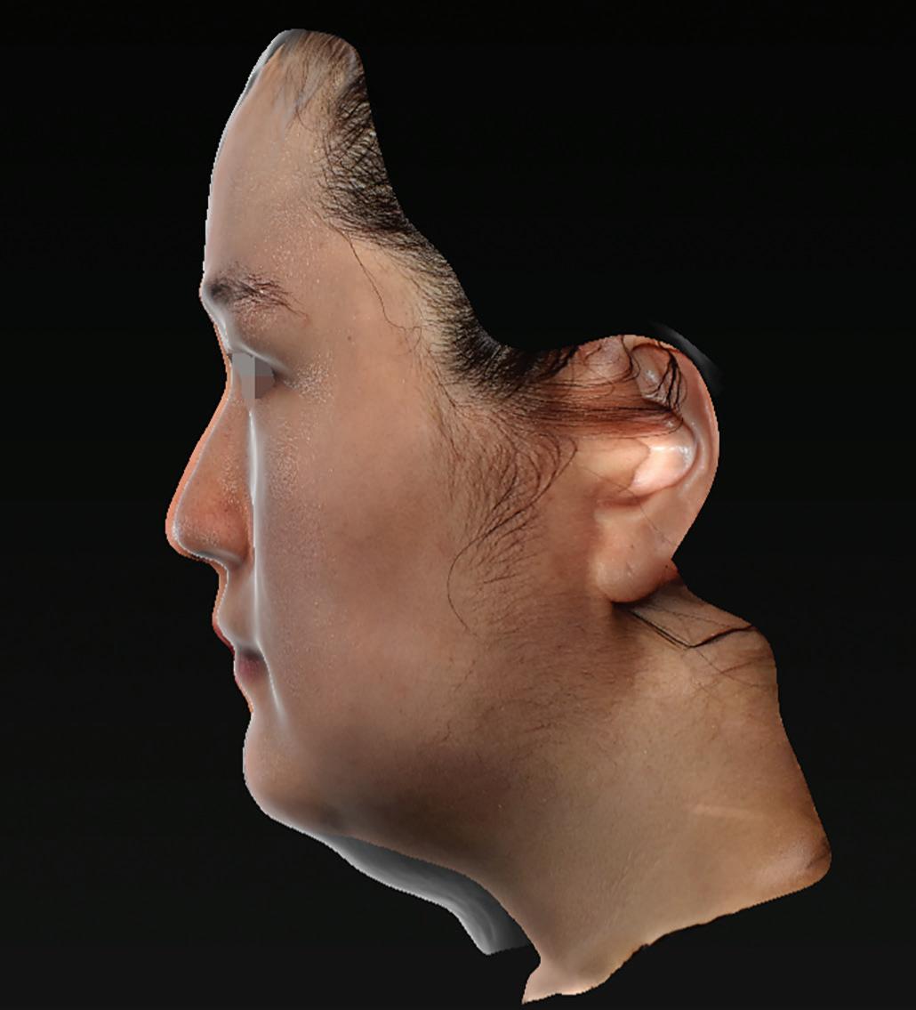
Figure 4 Height difference at 11 months after baseline
COMPLICATIONS IN LIP AUGMENTATION WITH HYALURONIC ACID FILLERS: A
CLINICAL AND ANATOMICAL
REVIEW
Yuliya Dyedyk-Gusarova, MD, provides a clinical and anatomical review of the key causes of complications in lip augmentation and outlines strategies to enhance safety and aesthetic outcomes
YULIYA DYEDYK-GUSAROVA, MD, Oral & Maxillofacial Surgery and Aesthetic Doctor; Founder and CEO at DrJ.Clinic, London, UK
email: info@drj.clinic
ABSTRACT
Objective: To review and analyse the principal complications arising from hyaluronic acid (HA) lip augmentation—including vascular occlusion, nodules, granulomas, biofilm formation, filler migration, and overfilling—through the lens of anatomical evidence, MRI findings, and injection technique.
Design and Methods: A narrative synthesis of anatomical dissection studies, MRI and ultrasound imaging data, clinical case evidence, and peer-reviewed literature. Special focus is placed on the relationship between injection depth, lip dynamics, and vascular architecture.
Main outcome measures: Complication frequency, depthdependent vascular injury risk, filler persistence beyond expected timelines, and anatomical misplacement leading to adverse outcomes.
Results: HA filler injections in the lips carry risks that are not solely determined by the injector’s anatomical knowledge, but also by their respect for depth and technique. Dissection studies confirm that >95% of labial arteries lie in submucosal or intramuscular planes. MRI studies show filler can persist for over 5 years. Nodules, granulomas, and filler migration often result from deep injection in high-mobility planes, compounded by overuse and muscle dynamics.
Conclusion: Safe and natural-looking lip augmentation depends on understanding vascular depth, respecting dynamic anatomy, and limiting cumulative filler volume. Overfilling, product misplacement, and layering across sessions increase the risk of chronic oedema, immune response, and migration. A shift toward natural aesthetics demands a more refined, anatomy-based approach.
AUGMENTATION WITH HYALURONIC
ACID (HA) FILLERS IS AMONG the most common aesthetic procedures worldwide. Yet its popularity does not guarantee its safety. As complications accumulate vascular occlusions, chronic swelling, nodules, and lip distortion the field is being called to re-evaluate traditional techniques through a more rigorous anatomical and evidence-based lens.
This article explores the scientific underpinnings of complication development in lip augmentation. It integrates anatomic dissection findings, dynamic muscle interplay, and long-term imaging studies to offer a practical framework for preventing and managing lip complications. The aim is not only to avoid adverse outcomes but to elevate standards of care.
Lip ageing and anatomical shift
Contrary to popular belief, lip ageing is not due solely to volume loss. Instead, it results from redistribution of fat compartments, a decline in dentoalveolar support, soft-tissue ptosis, and chronic muscle activity. The orbicularis oris muscle, responsible for lip movement and oral competence, undergoes continuous contraction throughout life, contributing to perioral wrinkling, vertical lip elongation, and thinning of the vermilion. Young lips exhibit a horizontal projection; with age, this flattens and rotates vertically due to bony remodeling and muscle changes.
Injectors often overcorrect the perceived loss of volume without restoring structure leading to disharmony. When the correct plane, product, and respect for
There is no vascular occlusion without a needle and product. Technique matters.
Table 1 Clinical guidelines and prevention in lip augmentation
Vascular Occlusion Deep injection; tenting technique; excessive pressure; insufficient anatomical awareness; inappropriate filler rheology for the injection plane
Nodules (non-inflammatory)
Intramuscular or overly superficial placement; filler with high G ′ or poor tissue integration; excessive volume in confined compartments; inadequate moulding
Granulomas Filler material characteristics (high cross-linking density); insufficient aseptic technique (microbial contamination); HA degradation, immune-mediated reaction or delayed foreign-body response
Excessive filler volume; repeated sessions without full degradation; disregard for anatomical proportion; use of highly projecting fillers in mobile zones
Chronic oedema Lymphatic compression; wrong filler choice; deep or repetitive placement; cumulative product retention
A
C
Stay in a superficial (≤3–4 mm) plane; use low, controlled pressure; respect anatomical depth variation across lip zones; select filler with suitable viscosity and cohesivity
Maintain correct injection plane; ensure dynamic awareness; choose product with moderate G ′ and good tissue integration; perform gentle post-injection massage for uniform distribution
Respect orbicularis oris dynamics and vectors; inject within appropriate plane (subcutaneous); avoid intramuscular track; use cohesive, elastic fillers; limit mechanical stress post-procedure; space reinjections adequately
Select product by indication, not volume; restore natural contour—not inflate; respect proportional anatomy; reassess before retreatment; perform objective volumetric evaluation
Avoid high-volume layering; select HA with suitable rheology and controlled tissue behaviour to prevent excessive projection and chronic oedema; inject at the correct anatomical depth and allow sufficient recovery between sessions
B D
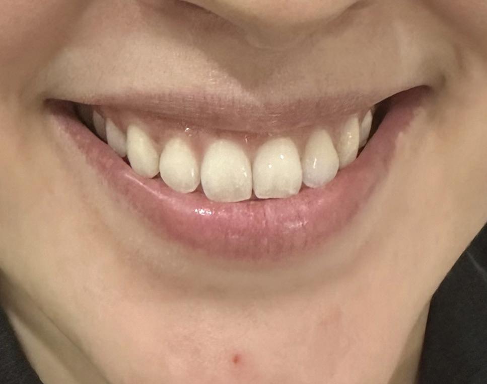
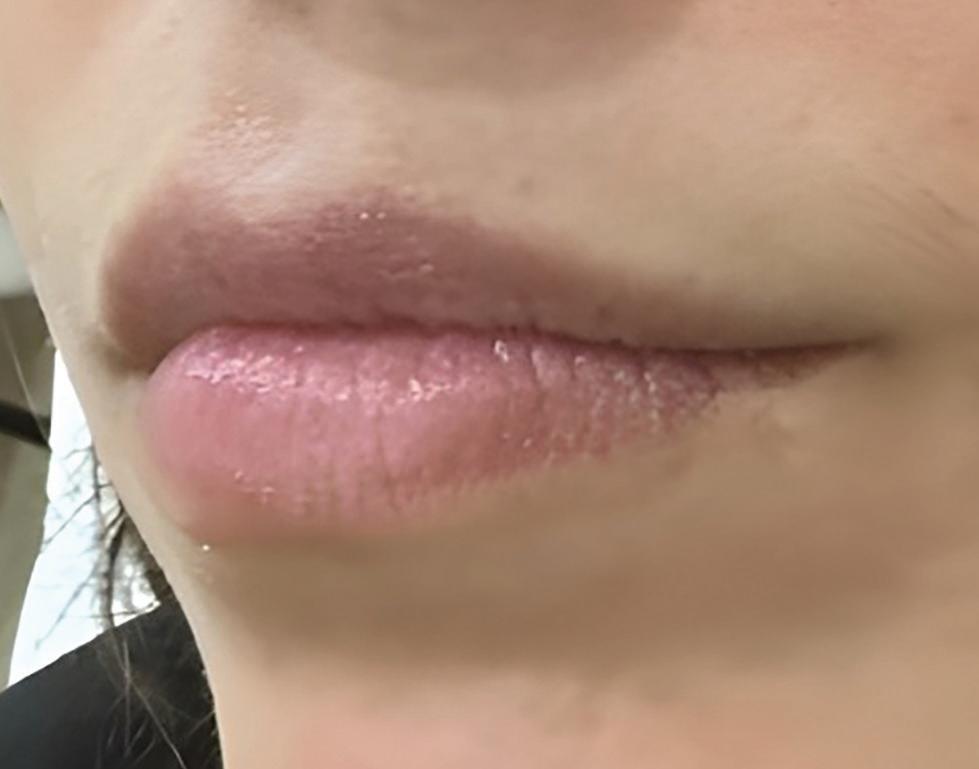
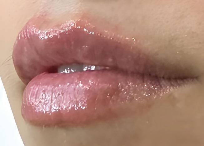
muscle movement are aligned, a natural result can be achieved (Figure 1).
Vascular complications and injection depth
Vascular occlusion remains the most feared complication. Dissection studies by Cotofana et al. 1 revealed the location of the superior and inferior labial arteries:
■ 78% in the submucosal plane
■ 17% intramuscular
■ 2% subcutaneous.
In the philtrum, the labial arteries may approach more superficial planes compared to other zones. This makes the area a higher risk for superficial injection.
Moreover, the average depth from the skin to the superior labial artery is ~5.6 mm, and to the inferior labial artery, ~5.2 mm. This makes superficial injections (≤3 mm) the safest.
However, arterial depth varies significantly across different regions of the lip.
In the philtrum, the labial arteries may approach more superficial planes compared to other zones. This makes the area a higher risk for superficial injection.
This regional variability highlights the need for dynamic anatomical awareness, not static 'safe depth' assumptions. Even within the same lip, vessel vulnerability can differ dramatically.
This highlights the danger of relying on universal depth recommendations. Region-specific injection strategies are essential to reduce vascular risk.
There is a close association between incidences of vascular occlusion and injection technique. As an example, lip tenting, once popular, places the product perilously close to arterial zones, especially at the lateral lip, where vessels are largest and most superficial. Injection techniques should consider these depth-based risks rather than rely on aesthetic tradition.
As I emphasise in clinical training: 'There is no vascular occlusion without a needle and product. Technique matters.'
Nodules, granulomas, and immune responses
Every filler is a foreign body and has the potential to elicit an immune response. Nodules can be:
■ Non-inflammatory: often redistributed filler, especially in dynamic zones like marionettes or vermilion. Caused by intramuscular injection and repetitive contraction, drawing the product into boluses. Usually painless, and may resolve or be treated with hyaluronidase
■ Inflammatory: often linked to delayed hypersensitivity or infection. Low-molecular-weight HA fragments and bacterial contamination can activate delayed hypersensitivity responses.
Studies have identified Staphylococcus epidermidis, Cutibacterium acnes, and Pseudomonas species in fillerrelated granulomas and in vitro biofilm models1,2. Fillers may act as a scaffold for biofilm formation, particularly with
Figure 1 Dynamic smile preserved through anatomical placement. (A and B) Lip augmentation result demonstrating the importance of respecting muscle dynamics and plane selection. (C and D) O.5ml volume placed with the correct rheological product and anatomical awareness, yields a soft, natural enhancement without distortion.
repeated needle passes or poor asepsis. These low-grade infections can stay silent, then trigger a type IV hypersensitivity-like reaction as the filler degrades, explaining the delayed onset and resistance to antibiotics. Management often requires combined therapy with corticosteroids and hyaluronidase.
Granulomas are rarely purely immune or infectious they are typically both.
Filler migration and muscle dynamics
What is often called 'migration' is more accurately misplacement—injecting in the wrong plane, then watching the filler shift due to muscle movement.
The orbicularis oris is a powerful sphincter muscle. When filler is placed intramuscularly (which occurs in 79% of needle injections and 93% of cannula), repeated contraction pulls the product centrally or vertically—especially into the philtrum, or white roll.2
As I often explain in training: 'It’s not where you inject it’s depth and how mobile the region is.'
Avoiding intramuscular placement and treating the lips as dynamic, not static, structures is key to preventing both visible and subclinical migration.
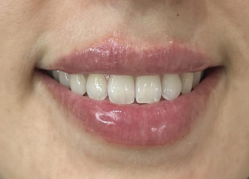
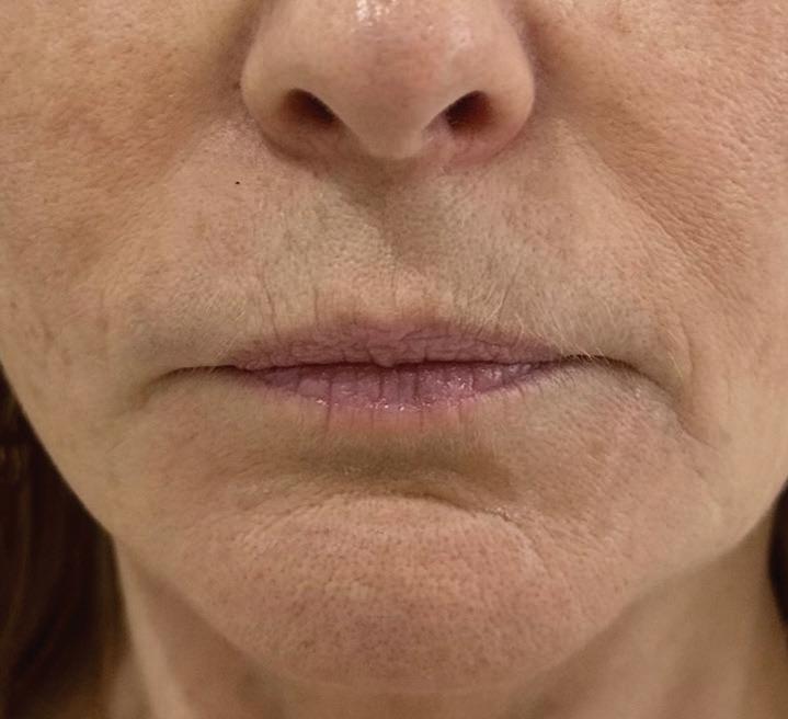
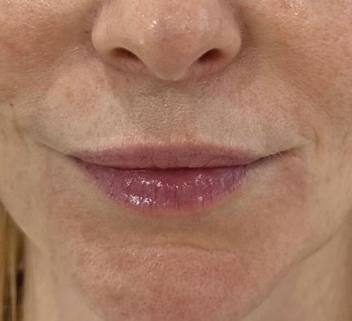
When the correct plane, product, and respect for muscle movement are aligned, a natural result can be achieved (Figure 2).
The overfilled lip: MRI and clinical consequences
MRI studies have revealed the remarkable long-term persistence of hyaluronic-acid fillers. In one case (following oral-cancer reconstruction), HA was still detectable on MRI five years post-injection3. These findings challenge the assumption of complete resorption and highlight how
A comprehensive understanding of anatomy in three dimensions, respecting arterial depth, and acknowledging the role of muscle dynamics are essential, alongside evidence-based product selection and precise control of injection volume, as both factors critically impact filler distribution, tissue compatibility, and complication rates.
cumulative filler sessions particularly in the dynamic lip region may contribute to:
■ Chronic oedema
■ Lymphatic disruption
■ Pseudo-nodules
■ Muscle restriction
■ Irreversible vermilion protrusion.
As filler builds up, tissue becomes sponge-like. Lymphatic outflow is impaired, leading to swelling and fibrosis. Restorative correction, respecting anatomical planes and reducing cumulative volume, can achieve natural results with minimal complications (Figure 2).
A BConclusion
Lip enhancement is both an art and a science. The aesthetic ideal has shifted from volume to structure, from filled to natural. But science must lead this shift. A comprehensive understanding of anatomy in three dimensions, respecting arterial depth, and acknowledging the role of muscle dynamics are essential, alongside evidence-based product selection and precise control of injection volume, as both factors critically impact filler distribution, tissue compatibility, and complication rates.
The injector’s responsibility is not only to beautify but to preserve function, prevent chronic harm, and honour the dynamic, vascular complexity of the lips. It is no longer acceptable to treat lips as static features. They move, they age, and they remember especially poorly placed filler.
Key points
Over 95% of labial arteries lie in the submucosal or intramuscular planes, making superficial injections (≤3 mm) the safest across most areas of the lip.
Misplaced filler in high-mobility, intramuscular planes often leads to visible or subclinical migration, nodules, and unnatural outcomes—especially over time.
MRI studies show HA fillers may persist for 2–5+ years, contributing to chronic oedema, fibrosis, and lip distortion when layered excessively or injected inappropriately.
In clinical reality, the lips form part of a complex perioral anatomical unit, where muscular balance, dermal integrity, and lymphatic drainage are interdependent. Achieving natural and stable outcomes requires assessing the entire perioral complex, not merely the vermilion itself, to maintain harmony between lip contours, perioral support, and lower facial dynamics (Figure 2).
Over the past decade, the aesthetic ideal in lip augmentation has evolved from excessive volumisation to anatomical restoration. This transition reflects a deeper scientific understanding of tissue biomechanics, vascular safety, and filler rheology, recognising that natural, expressive lips result not from volume replacement but from precision, balance, and respect for perioral anatomy.
Declaration of interest None
References
1.
2.
3.
Saththianathan M, et al. The Role of Bacterial Biofilm in Adverse Soft-Tissue Filler Reactions: A Combined Laboratory and Clinical Study. Plast Reconstr Surg. 2017; 139(3): 622–632.
Alhede M, et al. Bacterial Biofilm Formation and Treatment in Soft Tissue Fillers. FEMS Immunol Med Microbiol. 2014; 70(3): 339–348.
Bettini G, Minerva M, Balagué N, Montet X, Calmy A, Salomon D, Toutous-Trelu L. Sequential MRI as a Diagnostic Tool for Follow-Up of Hyaluronic Acid Dermal Filler in a Woman Who Underwent Radiation Therapy for Oral Cancer. Clinical Case Reports. 2025 Apr 8; 13(4): e70402.
Figure 2 Full restoration of the perioral complex in a mature patient. (A) baseline shows perioral collapse and vertical elongation. (B) post-treatment with HA filler using structural support techniques of perioral complex with respecting lip dynamics
SAFEGUARDING THE CORNERSTONE OF ETHICAL AESTHETIC PRACTICE
Eddie Hooker, CEO and Founder of Hamilton Fraser, on why safeguarding protects both patients and practitioners and how to embed it into everyday clinic care
THE UK AND SCOTTISH GOVERNMENTS
both recently issued their responses to consultations carried out on the licensing of non-surgical cosmetic procedures in both England and Scotland. This is a significant step forward and we are now sitting at a critical juncture when it comes to regulation in the UK.
It’s no surprise that our sector is facing increasing scrutiny, and rightly so. This Morning recently ran a week-long series of segments as part of its Cosmetic Cowboys Investigation, highlighting the areas where we, as a sector, are falling short.
Research carried out by the investigation showed that half of women have required medical assistance following aesthetic treatment, while 15% had ended up in hospital or A&E after receiving a non-surgical cosmetic procedure. In addition, half of the cosmetic procedures took place in nonclinical settings, including living rooms or kitchens, and were not carried out by someone medically qualified. With one in three women also admitting that they would go to someone who is not medically qualified to save money on a procedure, the risks are clear.
Safeguarding gaps
However, there are clear safeguarding gaps. A survey carried out by Hamilton Fraser in conjunction with Menopause in Practice revealed that many clinics still lack formal safeguarding frameworks or leadership roles. Nevertheless, practitioners are proactively seeking education, resources, and ongoing support to improve safeguarding in aesthetics.
Preventing complaints
While the ultimate goal of regulation is, of course, patient safety, it is my view that practitioner protection and patient safety must go hand in hand.
At Hamilton Fraser, we’ve insured medical aesthetic professionals for almost 30 years, and in that time, we’ve handled thousands of claims, complaints, and crisis calls. What have we learned? The majority of issues don’t stem from bad outcomes, but from preventable failures in the patient journey, whether that be poor patient selection or lack of informed consent. Safeguarding is a huge part of that, and that’s why it isn’t just a regulatory or compliance issue –it’s the foundation of ethical, professional practice.
Here are the key ways you can safeguard yourself and your patients.
1. Start with smart patient selection
Everything starts with the consultation. If this step is rushed or incomplete, the rest of the process is already compromised. A solid assessment protects patients and protects you. Most claims are linked to poor patient selection, rather than complications or unsatisfactory results. This includes failing to identify contraindications, as well as missing critical psychological red flags like Body Dysmorphic Disorder (BDD). Practitioners must have the confidence to say no when a patient is not clinically or emotionally suitable, even if that means losing a booking.
2. Make consent personal, not just procedural
Is your patient really giving consent, or just agreeing with
Safeguarding in aesthetics: Where clinics stand
■ 55.2% — Confirm their clinic has safeguarding policies
■ 27.6% — Have no safeguarding lead
■ 6.9% — Unsure if a lead exists in their workplace
■ 79.3% — Want regular safeguarding workshops
■ 75.9% — Seek access to expert advice or consultation
■ 65.5% — Request a comprehensive training programme
you? Consent is so much more than getting a signature on a form it’s a conversation. True informed consent involves a clear, honest, two-way discussion about the risks, benefits, alternatives, and expectations of treatment.
Patients must be informed of (and fully understand) the possible side effects (such as bruising or swelling), serious risks (including vascular occlusion), and whether further treatments may be needed. You must also disclose product brands, costs, and follow-up requirements. A cooling-off period is strongly advised.
If the practitioner performing the procedure is not the same person who obtained consent, the process should be repeated. This step is your legal protection. Lack of proper consent is one of the biggest causes of complaints. So make it thorough and personal.
3. Keep detailed, secure records
When a complaint lands on your desk, your records will either protect or incriminate you. The saying goes: ‘If it’s not written down, it didn’t happen’ and that’s absolutely true in aesthetics. Clinical notes should include the product used, batch number, injection technique, and patient response.
Before-and-after photos should be captured and securely stored, with written consent, both for clinical records and for any external use (e.g. marketing or training). Blanket consent is not enough under GDPR; patients must approve each specific use of their images.
Sensitive data should be stored using encrypted, clinicapproved software. Never rely on personal phones or messaging platforms such as WhatsApp to share patient photos or notes. Thorough documentation doesn’t just protect you it helps you deliver better, more consistent care and shows professionalism if a problem ever arises.
4. Stick to clinical standards - every time
Everything starts with the consultation. If this step is rushed or incomplete, the rest of the process is already compromised.
Are your clinic habits keeping patients safe or putting them at risk? Practitioners should follow clear hygiene protocols, properly dispose of waste and only work in sterile, compliant environments. Additionally, all products should be sourced from licensed suppliers and stored according to manufacturer guidelines. Using cheap, greymarket, expired, or poorly stored products is a fast route to complications and an invalid insurance policy. We have recently seen a breakout of botulism linked to unlicensed products being used. This type of practice is a huge safeguarding risk to the public.
EDDIE HOOKER, founder and chief executive of Hamilton Fraser, Borehamwood, UK
Data protection and patient imagery
■ 75.9% — Are aware of ethical guidelines
■ 24.1% — Are not aware of data protection expectations
■ 81.8% — Always follow protocols
■ 13.6% — Follow guidelines “often” but not always
5. Adapt safeguarding for intimate health treatments
As more aesthetic clinics expand into women’s health and intimate treatments, safeguarding protocols must evolve. These services introduce heightened risk and vulnerability, especially for patients with a history of trauma or abuse. Yet, according to our safeguarding survey, 75.9% have not received formal safeguarding training for intimate health, despite 100% wanting training on this topic.
In the recent announcement by the Department of Health and Social Care outlining new measures to crack down on cosmetic procedures, Minister of State for Health Karin Smyth MP said: “Priority will be given to introducing regulations to restrict the highest risk procedures first such as fillers injected into breasts and genitals.” I could not support this more.
Chaperones also play a critical role here. Their presence not only enhances patient dignity and comfort but also provides practitioners with vital protection against misunderstandings or allegations of misconduct. Insurers look very closely at whether a chaperone was offered or present in any situation involving a complaint. Failing to follow this standard could affect the outcome of a claim, or even your ability to renew cover in future.
6. Keep your training and CPD up to date
Practitioners should maintain up-to-date knowledge of facial anatomy, complication management, and product compatibility, not just to gain CPD for revalidation but to demonstrate competency. If you can’t prove you’ve done this, your insurer may not support you in the event of a claim. Hamilton Fraser, for example, requires practitioners to demonstrate competency and ongoing training as part of their policy terms.
7. Never skip aftercare
Practitioners should maintain up-to-date knowledge of facial anatomy, complication management, and product compatibility, not just to gain CPD for revalidation but to demonstrate competency.
8. Handle complaints like a professional Complaints don’t have to end in claims. The most effective approach? Listen without defensiveness, acknowledge the patient’s experience, and respond professionally. Don’t brush them off or get defensive. Say sorry if something went wrong— that doesn’t mean admitting liability, it just means showing empathy.
Contact your insurer immediately to access expert guidance. Then gather all your notes, photos, and documents. This shows you acted responsibly and gives you evidence if needed.
Above all, don’t ignore complaints. Delaying or avoiding them can lead to legal action, damage your reputation, and even affect your ability to practise.
9. Insurance is only effective if your practice is compliant
Your insurance policy is your safety net, but only if your practice is compliant. Make sure that all of the treatments you are performing are covered under the policy. Insurance is only as good as when you make a claim. Choose an FCA-regulated broker to make sure your policy is reputable and work with brokers who understand the complexities of the industry. Make sure you understand your policy terms and conditions. A good policy includes access to advice and support when claims arise.
Building a culture of safety
Ultimately, safeguarding is not just about avoiding claims it’s about delivering the kind of care we would want for ourselves or our families.
Practitioners who embed safeguarding into every aspect of their work create better patient experiences, stronger reputations, and more resilient businesses. They also elevate the status of aesthetic medicine as a responsible and ethical branch of healthcare. Protecting your patient is protecting yourself and the future of the profession.
A safe treatment doesn’t end when the patient leaves the clinic. Patients must be provided with clear, written and verbal aftercare instructions and be informed of both normal and abnormal side effects, such as when to seek urgent help.
Follow-up appointments are not optional. They help identify complications early, fix any concerns, and show your patient that you care. Practitioners should also have emergency plans in place, including the availability of reversal agents like Hyalase for filler complications. Good aftercare reduces complaints, improves results, and builds long-term trust.
Chaperone use in aesthetic practice
■ 75.9% — Believe chaperones improve safeguarding
■ 69% — Always offer a chaperone
■ 31% — Use them inconsistently
■ 10.3% — Never use them
About the Author Eddie Hooker is the founder and chief executive of Hamilton Fraser. He is an expert in the cosmetic insurance sector with more than 28 years of experience. Hamilton Fraser was the first company to offer medical malpractice insurance specific to the cosmetic industry in 1996, and Eddie is passionate about continuing to raise standards in the sector. Eddie is an accomplished speaker who regularly provides support, advice, and education to practitioners as an industry commentator on key topics such as aesthetics regulation, legislation, insurance, and business growth.
Join the ESTHETIC MULTISPECI LTY SOCIETY
A platform designed for ESTHETIC MEDICINE PROFESSION LS
The AMS, the world’s largest multispecialty aesthetic medicine society, provides a digital platform where members can network and access an extensive library of on-demand scientific and sponsored videos, helping them grow and advance in their careers.
.
30,000+
members are already benefiting from the AMS features and enhancing their medical practice.
AMS is the scientific supervisor of leading aesthetic medicine events
EXOSOMAL SOLUTION 'THE NEXT FRONTIER IN ANTI-AGEING AND SKIN REPAIR'
Peter Floros, MD, presents clinical results using EXO’LUTION®, a plant-based exosomal booster designed to hydrate, repair, and regenerate the skin
Exosomes are nanosized extracellular vesicles (EVs), typically 30–200 nm in diameter, secreted by various cell types to mediate intercellular communication1. Since their initial discovery in the 1980s, exosomes were once considered cellular waste products, but subsequent research has revealed their central role in transporting proteins, lipids, and nucleic acids that regulate immunity, repair, and regeneration. In regenerative medicine, exosomes have opened a new chapter for biologic therapies, offering novel strategies to restore tissue homeostasis and improve healing outcomes2. In aesthetic dermatology, demand has increased for biologically active, minimally invasive solutions that improve skin quality hydration, elasticity, homogeneity, and repair —beyond the volumising or neuromodulatory effects of traditional injectables. Exosomes align with this unmet need by providing an endogenous-
like stimulus for collagen and elastin synthesis, keratinocyte migration, and fibroblast proliferation, thereby restoring extracellular matrix (ECM) balance.
Among sources of exosomes, plant-derived exosomes (P-EVs) have emerged as highly attractive for clinical applications3-5. They exhibit low immunogenicity and cytotoxicity, greater compositional stability, and scalability without the ethical concerns associated with stem cell or mammalian-derived products3-5. Specific plants, such as Centella asiatica, have long been used in traditional medicine for their anti-inflammatory, antioxidant, and wound-healing properties, now enhanced through exosome technology3-5
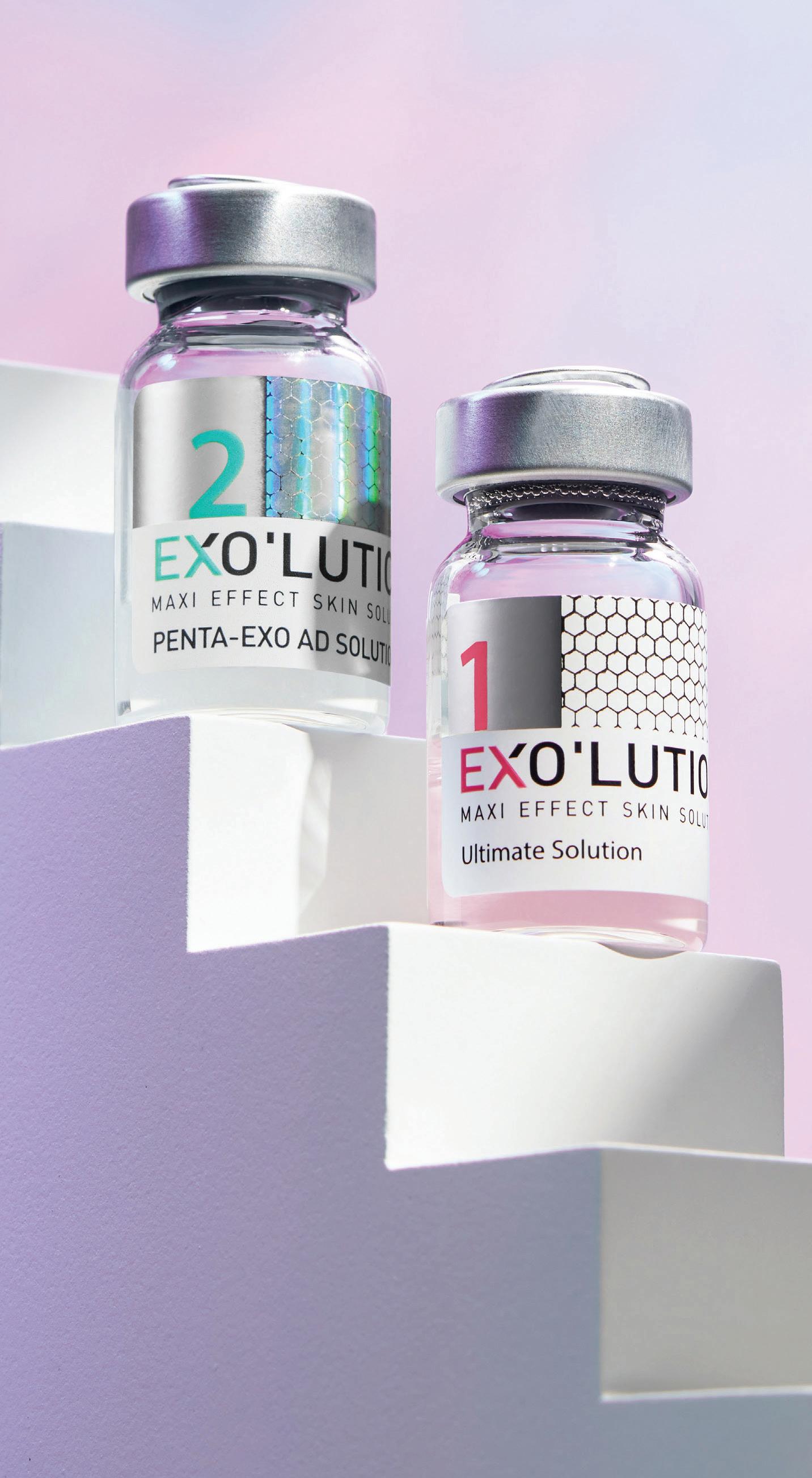
EXO’LUTION® (JETEMA, Republic of Korea) is a nextgeneration exosomal skin booster integrating a 5-plant exosome complex with 61 bioactive ingredients, including amino acids, vitamins, nucleic acids, minerals, glutathione, and dual molecular-weight hyaluronic acids. Developed by JETEMA, EXO’LUTION® uses proprietary X-traction™ technology to isolate high-purity exosome fractions (~80–120 nm, ~6 billion particles per vial). Its dual-vial system combines Vial 1 for direct skin nutrition and hydration, and Vial 2 to maximise exosome activity. This design targets the papillary dermis (0.3–0.4 mm), where fibroblasts are most active in ECM
PETER FLOROS, MD, General Manager of Neotis, Athens, Greece
remodelling, making it suitable as both a stand-alone skin booster and an adjunct post-energy device.
This article presents two representative clinical cases illustrating the efficacy and safety of EXO’LUTION® in improving skin hydration, homogeneity, and regeneration while reducing inflammation and downtime. These nanosized extracellular vehicles, with their extraordinary capacity to transfer molecular information and human dermal fibroblast (HDF) characteristics, have opened a new chapter in the exploration of therapeutic solutions for skin restoration.
Patients and Methods
Case 1: Microneedling and EXO’LUTION®
The patient in Figure 1 underwent three sessions of microneedling (0.5 mm depth) followed by topical application of EXO’LUTION®. Each session was performed at 15-day intervals6. Approximately 0.04 mL of solution was applied per point across the entire face, ensuring even coverage. Clinical evaluation included hydration, texture homogeneity, fine line severity, and patient satisfaction. Highresolution photography was performed at baseline and after the third session.
Case 2: Fractional device and EXO’LUTION®
The patient in Figure 2 with atrophic acne scars underwent fractional laser treatment. Following a 10-minute cooling period to normalise surface temperature, EXO’LUTION® was
“In two clinical cases, EXO’LUTION® demonstrated measurable improvements in hydration, texture, fine lines, erythema resolution, and acne scar remodelling."
applied topically. Treatment was repeated twice at one-month intervals. Endpoints included erythema resolution, downtime duration, acne scar topography (qualitative scale), and skin texture improvement. Pre- and posttreatment photographs were taken.
Results
Case 1 outcomes
By the third session, visible improvements in hydration and radiance were noted. Fine lines decreased, and the patient reported improved makeup adherence and satisfaction with skin texture. Photography confirmed smoother, more homogeneous skin.
Case 2 outcomes
After two sessions, erythema resolved faster than expected post-fractional therapy, and downtime was reduced by approximately 2 days compared to the patient’s prior experience. Acne scars showed visible improvement, with enhanced texture homogeneity. The patient noted increased comfort and faster return to normal activities.
Safety
Neither case exhibited adverse events, allergic reactions, or prolonged irritation. Both patients tolerated the regimen well, confirming the favourable safety profile of plant-derived exosomes.
Discussion
Mechanistic insights
The observed improvements stem from EXO’LUTION®’s dual mechanisms:
Figure 1 Case 1 before and after treatment
Figure 2 Case 2 before and after treatment
PROMOTION
■ Hydration and nutrition: Vial 1 delivers 61 bioactive molecules, enhancing skin moisture and cellular energy
■ The nanosized particles (~80–120 nm) facilitate rapid penetration through the epidermal barrier into the papillary dermis, a layer rich in fibroblasts, central to wound healing and collagen production.
Anti-inflammatory and reparative effects
Centella asiatica, the main exosomal source, demonstrated decreased erythema and improved acne scar healing as observed in Case 2. These results confirm EXO’LUTION®’s role in accelerating recovery postenergy-based procedures, making it an attractive adjunct for fractional lasers, RF microneedling, and microneedling protocols.
Clinical workflow integration
EXO’LUTION® can be easily integrated into existing practice: ■ Post-device application (microneedling, RF, fractional
■ Maintenance regimen: three sessions at 2–3-week intervals, followed by monthly boosters
■ Combination therapy: synergistic with fillers and toxins for global rejuvenation, as EXO’LUTION® improves skin quality beyond volumetric correction.
Comparison with other skin boosters
Traditional boosters such as HA, PDRN, or PDLLA primarily target hydration or collagen stimulation but lack robust anti-inflammatory and reparative mechanisms. EXO’LUTION® uniquely combines hydration, antioxidant protection, immune modulation,
References
1. Schur N, Samman L, Shah M, Dukharan V, Stegura C, Broughton L, Schlesinger T. Exosomes: Historical Evolution and Emerging Roles in Dermatology. J Cosmet Dermatol. 2025;24(1):e16769. doi:10.1111/ jocd.16769
and ECM remodelling. Its plant-derived origin ensures safety compared to mammalian exosomes, which face higher variability and regulatory hurdles.
Limitations
and future directions
While these case results and laboratory evidence are promising, limitations include the small sample size and lack of quantitative imaging in the case series. Larger, multicentre, split-face trials with objective measures (e.g., high-frequency ultrasound, 3D skin imaging) are recommended to validate efficacy across diverse skin types and indications. Future studies should also explore EXO’LUTION®’s role in pigmentary disorders, chronic wound care, and long-term maintenance protocols.
Conclusion
EXO’LUTION® represents a next-generation exosomal skin booster designed to address multiple pathways of skin
2. Lu M, Xing H, Xun Z, et al. Exosome-based small RNA delivery: Progress and prospects. Asian J Pharm Sci. 2018;13(1):1–11. doi:10.1016/j. ajps.2017.07.012
3. Karamanidou T, Tsouknidas A. Plant-derived extracellular vesicles as therapeutic nanocarriers. Int J Mol Sci. 2022;23(1):191. doi:10.3390/ijms23010191
4. Dad HA, et al. Plant exosome-like
rejuvenation hydration, regeneration, anti-inflammation, and scar remodelling. In two clinical cases, EXO’LUTION® demonstrated measurable improvements in hydration, texture, fine lines, erythema resolution, and acne scar remodelling. These findings are consistent with supporting in vitro and clinical trial data. With its dual-vial system, 5-plant exosome complex, and 61 bioactive ingredients, EXO’LUTION® offers a safe, effective, and versatile tool for modern aesthetic practice. It can be deployed both as a primary skin booster and as an adjunct post-device or injectables to enhance overall outcomes. Further clinical studies will consolidate its position as a biologically inspired solution for skin quality restoration.
nanovesicles: emerging therapeutics and drug delivery nanoplatforms. Mol Ther. 2021;29(1):13–31. doi:10.1016/j. ymthe.2020.11.030
5. Yi Q, et al. Current understanding of plant-derived exosome-like nanoparticles in regulating the inflammatory response and immune system microenvironment.
6. Iriarte C, Awosika O, Rengifo-Pardo M, Ehrlich A. Review of applications of microneedling in dermatology. Clin Cosmet Investig Dermatol. 2017;10:289-98. doi:10.2147/CCID.S142450 PMID: 28848356 PMCID: PMC555618
COLLAGENASE AND ITS POTENTIAL IN REJUVENATING THE NECK AND DÉCOLLETAGE
As aesthetic attention shifts beyond the face, the neck and décolletage are emerging as key targets for rejuvenation. Drs Tatiana Micaela Dantas Garcia and Paola Vaca Parada explore the use of collagenase as a safe, effective treatment for these areas
This article reviews the potential of collagenase as a treatment for rejuvenating the neck and décolletage. It highlights the importance of addressing these regions, which exhibit early and significant signs of ageing, and analyses validated clinical scales to assess their degree of ageing, such as the Glogau scale and the Carruthers Lipoatrophy Severity Scale.
Ultrasonographic findings
demonstrate a reduction in the thickness of the subepidermal low echogenic band and increased echogenicity in the hypodermis, indicating a process of tissue remodelling and regeneration. The results suggest that collagenase is a safe and effective option for improving skin quality in these areas, contributing to a more youthful and natural appearance, with sustainable long-term effects.
Neck ageing is a multifactorial process involving structural changes, such as bone resorption, tissue ptosis, fat accumulation, and muscle descent, which alter the anatomy and aesthetics of this region (Glogau, 1996). The loss of bony support and soft tissue laxity generates folds, bands, and a loss of mandibular definition, affecting the perception of youth and vitality. Furthermore, the accumulation of submandibular fat and the descent of the base of the mouth muscles contribute to the formation of a double chin and an aged appearance (Fitzpatrick & Goldman, 2010).
Collagenase acts specifically on the collagen triple helix, cleaving the peptide bonds and generating smaller fragments that facilitate subsequent elimination. This action allows for the removal of disorganised or amorphous collagen fibres, thereby promoting skin regeneration and the synthesis of a new functional collagen network in the dermis
Figure 1 Enzyme application schematic. Technique for intradermal injection of collagenase, identified by white spheres, in the neck and décolletage, using the papule technique with approximately 0.02 ml per point, distributed 1 cm apart, and administration of lipase in three points in the submental area, identified by green spheres, with 1 ml at each one, separated by 3 to 4 cm.
(Rivera, 2020).
Each vial of recombinant collagenase and lipase is reconstituted with 3 ml of sterile physiological solution. Reconstitution is performed under aseptic conditions, with gentle agitation to ensure homogeneous dissolution. The reconstituted collagenase solution is administered via intradermal injections using the papule technique, with approximately 0.02 ml per point, distributed 1 cm apart across the entire neck and décolletage area. Lipase is administered at 3 points in the submental localised fat area, with
1 ml at each point spaced 3 to 4 cm apart, via subcutaneous injections (Figure 1).
Case study
In Figure 3, you can witness the treatment results following collagenase injected in the neck and décolletage Note the following:
■ A decrease in the thickness of the subepidermal low echogenic band (SLEB) was evidenced, which is associated with an improvement in skin markers related to dermal ageing. This reduction can be interpreted as an indicator of
TATIANA MICAELA DANTAS
GARCIA , MD; PAOLA VACA PARADA, MD
reduced chronic inflammation or reorganisation of dermal collagen (Wortsman, 2012).
■ An increase in the echogenicity of the hypodermis was observed, suggesting a greater presence or densification of loose fibroconnective tissue, possibly linked to tissue regeneration or remodelling processes induced by the treatment (Lassau et al., 2010).
■ The basal layer showed greater definition and signs of restructuring compared to the initial ultrasonographic study, indicating a potential improvement in the integrity of the dermo-epidermal junction and in the cellular organisation of the basal epidermis.
Conclusions
The structural integrity of these cutaneous areas is closely related to aesthetic perception and overall well-being (Wortsman, 2012). In this context, treatment with recombinant enzymes, such as collagenase, presents itself as a safe and effective therapeutic alternative for improving dermal aesthetics in these regions. This treatment promotes the remodelling, regeneration, and firming of the affected tissues, contributing to the restoration of skin elasticity and the reduction of visible signs of ageing. The results obtained are natural and, in many cases, sustainable in the long term, reinforcing the utility of collagenase as a treatment option for skin rejuvenation (Lazar et al., 2016).
Find out more at: simildiet.com
“Collagenase promotes skin regeneration and dermal remodelling—offering natural, long-lasting results in some of the most age-revealing areas of the body”
Figure 2 Ultrasound before and after enzyme treatment in the neck and décolletage area
Figure 3 Before and after enzyme treatment in the neck and décolletage area
Study conducted in collaboration with Mrs. Beatriz Cortes, General Manager of Mesocare Bolivia; and Dr. Carlos Aznar, General Manager of Simildiet Labs
DUAL-MODALITY ENERGY-BASED APPROACH FOR SUBMENTAL LAXITY IMPROVEMENT
Clinical outcomes highlight the effectiveness and safety profile of a next-generation device in addressing submental skin laxity
SUBMENTAL SKIN LAXITY
is one of the earliest and most visible signs of lower-face ageing, characterised by reduced dermal elasticity, attenuation of retaining ligaments, and descent of soft tissue structures1,2. This laxity can occur independently or in combination with submental fat accumulation, leading to loss of a defined cervicomental angle and a heavier lower facial profile. From a psychosocial standpoint, such changes may negatively impact self-perceived youthfulness and confidence. While submental fat reduction using energy-based devices is well-established, the improvement of significant skin redundancy in this area remains more challenging due to structural and histological changes in the dermis and underlying layers, according to Renata Violato, MD, a dermatologist based in Brazil1,3 ‘However, by applying HIFU to create precise thermal coagulation zones at targeted depths, followed by RF to volumetrically heat the dermis and subdermis, it induces multi-layer collagen contraction and remodeling to yield optimal clinical outcomes in the improvement of submental laxity,’ she said.
Dr. Violato shared her experience evaluating the clinical efficacy of VOLNEWMER (Classys, South Korea) and ULTRAFORMER MPT (Classys, South Korea), a monopolar radiofrequency (RF)
device and high-intensity focused ultrasound (HIFU), in a case involving submental treatment. This treatment is called ‘VOLFORMER’. Across all cases, visible tightening and contour enhancement were achieved, patient satisfaction scores exceeded 9.5/10, and treatment was well-tolerated with minimal downtime.
Monopolar RF delivers deep, uniform volumetric heating to the dermis and retaining ligaments, stimulating fibroblasts to produce new collagen and elastin fibres while remodeling existing extracellular matrix components3,4. HIFU, on the other hand, generates precise focal points of coagulative thermal injury at specific depths, including the superficial musculoaponeurotic system (SMAS) and deep dermis, leading to neocollagenesis and tissue tightening over weeks to months, according to Dr. Violato5,6
The combined use of these modalities induces greater degrees of thermal injury that penetrate wider and deeper into the skin, without causing excessive thermal damage to the epidermis or papillary dermis. The cumulative thermal reactions in the pre-focal and focal areas of HIFU pretreatment contribute to the pronounced RF-induced tissue responses in these regions. By creating zones of thermal coagulation and heat accumulation within the dermal and subcutaneous layers, HIFU reduces
tissue impedance and increases conductivity, allowing non-invasive RF energy to penetrate more deeply and uniformly. This results in broader and more effective thermal zones for tissue tightening and collagen remodeling. This combined approach targets both the deep structural support and the overlying dermal envelope, potentially enhancing the longevity of the tightening effects7-10.
Treatment Methods
The HIFU treatment (ULTRAFORMER MPT) was delivered first, using a layered protocol:
■ 4.5 mm transducer to target the SMAS layer for deep lifting ■ 3.0 mm and 2.0 mm transducer to stimulate collagen remodeling in the deep to mild dermis.
Immediately afterwards,
monopolar RF treatment (VOLNEWMER) was administered using a large grounding pad placed on the patient’s upper back to complete the electrical circuit. Treatment duration ranged from 12 to 15 minutes, maintaining continuous movement of the applicator. The 3 cm³ or 4 cm³ tip was used, depending on the patient’s skin thickness. Energy level settings were adjusted between 2.5~4.0, with energy delivery monitored by patient feedback and observation of mild, uniform erythema. Fine adjustments were made based on skin thickness and laxity grade.
Case Presentations
Case 1: A 45-year-old woman with moderate submental laxity without notable fat deposits reported an immediate sensation of lifting after treatment2. Immediately after the
Figure 1 (A) 45-year-old woman with moderate submental laxity, (B) after the procedure, imaging showed improved cervicomental angle definition and reduced horizontal skin redundancy
procedure, imaging showed improved cervicomental angle definition and reduced horizontal skin redundancy. She rated satisfaction at 10/10, discomfort at 3/10, and resumed normal activities the same day.
Case 2: A 56-year-old woman with moderate submental fat and severe skin laxity showed a visibly sharper mandibular contour and reduced submental bulge. Skin texture appeared smoother, and lifting was evident in profile photographs. Satisfaction was 9.5/10, discomfort 2/10, and she noted positive feedback from acquaintances.
Case 3: A 65-year-old woman with pronounced skin laxity and visible platysmal banding achieved a noticeable reduction in sagging and improved jawline definition after one session. Immediately after the procedure, skin firmness and contour were maintained. Satisfaction was 10/10, discomfort 4/10, and she described her neck as ‘lighter and younger.’
Clinical Insight
This case series supports the premise that dual-modality treatment can achieve significant and patient-pleasing outcomes for submental laxity. The combination tightens the skin envelope while
Reference
1. Alster TS, Tanzi EL. Non-invasive tightening of the skin in the face and neck. Semin Cutan Med Surg. 2008;27(4):227–31.
2. Suh DH, et al. Intense focused ultrasound for facial tightening: efficacy, patient satisfaction, and safety in Asian patients. Dermatol Surg. 2011;37(11):1595–602.
also improving structural support, according to Dr. Violato.
“The
combined use of these modalities induces greater degrees of thermal injury that penetrate wider and deeper into the skin, without causing excessive thermal damage to the epidermis or papillary dermis.”
The absence of adverse events aligns with existing literature indicating that safe energy parameters and correct technique minimise risk. All patients tolerated the procedure without anaesthesia, reporting low discomfort scores.
While limited by the small sample size and short follow-up, the consistent improvements suggest applicability across various degrees of laxity7,10.
‘The Volformer protocol, combining HIFU and monopolar RF in a sequential single-session treatment, produced visible and measurable improvements in submental skin laxity in all three cases,’ Dr. Violato stated. By simultaneously enhancing deep structural support and superficial dermal quality, this approach demonstrated high patient satisfaction with minimal discomfort, thereby substantiating its potential as a non-invasive alternative for submental rejuvenation, according to Dr. Violato. She emphasised that to substantiate these results and enhance clinical protocols, additional studies incorporating larger populations and histological assessments are warranted.
3. Atiyeh BS, Dibo SA. Nonsurgical nonablative treatment of aging skin: radiofrequency technologies between aggressive marketing and evidence-based efficacy. Aesthetic Plast Surg. 2009;33(3):283–94.
4. Luebberding S, Krueger N, Sadick NS. Tissue tightening by non-invasive radiofrequency: evidence-based evaluation of the current literature. J Drugs Dermatol. 2014;13(3):305–12.
5. Chan NP, et al. Use of high-intensity focused ultrasound for non-invasive skin tightening in Asians: Clinical efficacy and
Figure 2 (A) A 56-year-old woman with moderate submental fat and severe skin laxity, (B) after treatment, skin texture appeared smoother and lifted
Figure 3 (A) A 65-year-old woman with pronounced skin laxity and visible platysmal banding (B) after one session a noticeable reduction in sagging and improved jawline definition was visible
safety. J Cosmet Laser Ther. 2011;13(5):290–5.
6. Fabi SG, Goldman MP. Retrospective evaluation of a combined treatment approach using microfocused ultrasound with visualisation and injectable poly-L-lactic acid for lifting and tightening the face and neck. Dermatol Surg. 2014;40 Suppl 12:S190–6.
7. Fabi SG, et al. Optimising patient outcomes by combining microfocused ultrasound with other energy-based modalities: a retrospective
analysis. J Drugs Dermatol. 2015;14(12):1422–6.
8. Royo de la Torre J, et al. Combined non-invasive modalities for skin laxity improvement: clinical evidence and patient satisfaction. Lasers Surg Med. 2019;51(6):472–80.
9. Kim H, Ahn KJ, Lee S, Park H, Cho SB. Interactive thermal tissue reactions of 7-MHz intense focused ultrasound and 1-MHz and 6-MHz radiofrequency on cadaveric skin. Skin
Res Technol. 2019 Mar;25(2):171-178. 10. Khong SML, Ismail AH, Sujani S, Devindaran N, Abdul Rashid MF, Mohd Zaman UMS. Safety and Efficacy of High-Intensity Focused Ultrasound and Monopolar Radiofrequency Combination Therapy for Skin Tightening: A Retrospective Study in Malaysia. Malays J Med Sci. 2024 Feb;31(1):114-123.
ADVANCING PSORIASIS CARE WITH ASCEPLUS SRLV EXOSOMES AND FRACTIONAL LASER
A novel case study highlights how medical-grade exosomes and thulium laser therapy improved psoriatic lesions after just one session
MD , Dermatologist, Brazil
PSORIASIS IS A SKIN condition that goes far beyond red patches and flaking. It causes discomfort, itching, and a strong emotional impact, often affecting self-esteem and confidence. For years, available treatments have not always been enough: many require continuous use, can cause side effects, and don’t always deliver lasting results. In this context, exosomes, extracellular nanovesicles that help reduce inflammation and stimulate skin regeneration, have emerged as an innovative option, especially when combined with fractional thulium laser, which enhances their absorption, as well as with microneedling radiofrequency devices that can further facilitate their penetration and maximise clinical outcomes1
Exosomes are 30–200 nm
extracellular vesicles that mediate intercellular communication by transferring molecular cargo, including miRNAs, proteins and lipids. Among its main properties are anti-inflammatory, regenerative, reparative, and angiogenic effects2. The ASCEplus SRLV, the world’s first exosome-based product from ExoCoBio Inc. (Seoul, South Korea), stands out for the purity of its medicalgrade exosomes, achieved through the exclusive ExoSCRT® technology, a patented extraction method capable of isolating exosomes with high efficiency, in compliance with Good Manufacturing Practice (GMP) standards.
Previous studies have demonstrated the use of ASCEplus in the treatment of conditions such as atopic dermatitis and wound healing3,4 Reported benefits include normalisation of disease-related gene expression in the epidermis, increased ceramide production for barrier restoration, and reduction of inflammatory markers in animal models. In the context of wound healing, ASCEplus has been shown to accelerate wound closure rates, promote
re-epithelialisation, reduce scar formation, and enhance angiogenesis5-8.
A new frontier in psoriasis treatment
Building on the benefits documented in the literature, exosomes have emerged as a promising modality, demonstrating efficacy when used as a stand-alone approach and in synergistic combination with adjunctive technologies, thereby enhancing outcomes even in conditions traditionally considered refractory9-11.
The spectrum of current applications is extensive. ExoCoBio has developed ASCEplus, specifically designed for skin, hair, and intimate care. Collectively, these observations underscore not only the broad therapeutic potential of exosome-based interventions but also the substantial need for continued mechanistic investigation and rigorous clinical validation12,13
Case study: A decades-long condition transformed
One patient had been living with psoriasis for nearly 20 years without any prior treatment. In
an experimental approach, a lesion on each thigh was treated with a combination of fractional thulium laser followed by the topical application of ASCEplus SRLV exosomes, while a lesion on the patient’s torso served as an untreated control. Without any modifications to lifestyle or body weight, the treated areas exhibited remarkable improvement within just three months, with reduced erythema, scaling, and pruritus. The patient reported approximately 90% improvement, and notably, these effects persisted for a full year without additional sessions. In contrast, the control lesion remained unchanged over the same period, highlighting the specific efficacy of the intervention.
This case highlights the exciting potential of exosomebased formulation in psoriasis care. Safe, well-tolerated, and effective, exosomes represent a promising alternative for people who have struggled for years with the limitations of traditional therapies. As this case shows, this is just the beginning of a new era in dermatology, one where science and innovation work together to restore both skin health and confidence.
“Without any modifications to lifestyle or body weight, the treated areas exhibited remarkable improvement within just three months, with reduced erythema, scaling, and pruritus.”
DANIELA CARVALHO LEMES
“Safe, well-tolerated, and effective, exosomes represent a promising alternative for people who have struggled for years with the limitations of traditional therapies.”
Research and clinical implications
Based on the current literature, there remains a clear need for further research to better understand the potential of ASCEplus SRLV in the treatment of psoriasis. Properly powered randomised controlled trials are essential to compare ASCEplus not only with placebo but also with established standard treatments. In addition, mechanistic studies are required to elucidate the molecular pathways modulated in psoriatic skin, while biomarker development could help identify predictors of response to exosome-based therapy. Further investigation into ASCEplus as an adjunctive approach alongside conventional therapies, as well as long-term follow-up studies to assess sustained safety and efficacy, will be critical to support its clinical translation. The establishment of standardised potency and quality metrics for exosome products also remains a key step to ensure reproducibility and consistency across clinical applications.
Visit: www.exocobio.com
References
1. Bai, G., Truong, T. M., Pathak, G., et al. (2024). Clinical applications of exosomes in cosmetic dermatology. Skin Health and Disease, 4(2), e348.
2. Cho, Byong & Duncan, Diane. (2023). Exosomes – Recent advances from bench do bedside. Chapter: Development of Exosomes for Esthetic Use. 10.5772/intechopen.111846.
3. Han, H. S., Koh, Y. G., Hong, J. K., Roh, Y. J., Seo, S. J., & Park, K. Y. (2023). Adipose-derived stem cell exosomes for treatment of dupilumab-related facial redness in patients with atopic dermatitis. The Journal of dermatological treatment, 34(1), 2220444.
4. Park, K. Y., Han, H. S., Park, J. W., Kwon, H. H., Park, G. H., & Seo, S. J. (2022). Exosomes derived from human adipose tissue-derived mesenchymal stem cells for the treatment of
1 Clinical case of psoriasis treated with ASCEplus SRLV exosomes and Thulium laser by Dr. Daniela Carvalho Lemes. (A) Erythematous desquamative plaque on the right and left leg before treatment; (B) Right and left leg after 3 months of application; (C) after 1 year of application
year
dupilumab-related facial redness in patients with atopic dermatitis: A report of two cases. Journal of cosmetic dermatology, 21(2), 844–849.
5. An, Y. H., Kim, D. H., Lee, E. J., et al. (2021). High Efficient Production of Adipose Derived Stem Cell (ADSC) Secretome Through Maturation Process and Its Non scarring Wound Healing Applications. Frontiers in Bioengineering and Biotechnology, 9, 681501.
6. Carvajal, J., & Carvajal, M. (2025). Could Exosomes be a Promising Therapy to Accelerate Wound Healing and Skin Regeneration in Ischemic Nasal Flaps?. Aesthetic surgery journal. Open forum, 7, ojaf060.
7. Lee, J., Won, Y. J., Kim, H. K., et al. (2023). Adipose Tissue Derived Mesenchymal Stem Cell Derived Exosomes Promote
Wound Healing and Tissue Regeneration. International Journal of Molecular Sciences, 24(13), 10434.
8. Dukharan, V., Shah, M., Broughton, L., Stegura, C., Samman, L., Schur, N., & Schlesinger, T. (2025). The Role of Exosomes in Medical Dermatology: Literature Review and Update. Journal of cosmetic dermatology, 24(1), e16761.
9. Damayanti, R. H., Rusdiana, T., & Wathoni, N. (2021). Mesenchymal Stem Cell Secretome for Dermatology Application: A Review. Clinical, Cosmetic and Investigational Dermatology, 14, 1009-1024.
10. Yousefian, F., Espinoza, L., Yadlapati, S., et al. (2024). A comprehensive review of the medical and cosmetic applications of exosomes in dermatology. Journal of Cosmetic Dermatology, 23(4), 1149-1165.
11. Shah, M., Dukharan, V., Broughton, L., Stegura, C., Schur, N., Samman, L., & Schlesinger, T. (2025). Exosomes for Aesthetic Dermatology: A Comprehensive Literature Review and Update. Journal of cosmetic dermatology, 24(1), e16766.
12. Phan, T. H., Kim, S. Y., Rudge, C., & Chrzanowski, W. (2022). Made by cells for cells - extracellular vesicles as next generation mainstream medicines. Journal of Cell Science, 135(12), 259166.
13. Zhang, K., Yu, L., Li, F. R., et al. (2020). Topical Application of Exosomes Derived from Human Umbilical Cord Mesenchymal Stem Cells in Combination with Sponge Spicules for Treatment of Photoaging. International Journal of Nanomedicine, 15, 2725-2740.
Figure
Figure 2 Psoriasis lesions not treated for control. (A) Erythematous desquamative plaque on the torso; (B) after 3 months; (C) after 1
Events
EUROPE
13–14 MARCH 2026
ACE London, UK www.aestheticsconference.com
20 MARCH 2026
Congress of Dermatology and Anti-Aging Medicine Warsaw, Poland kongres.dermatologia-estetyczna.pl
26–28 MARCH 2026
AMWC Monaco Monte Carlo, Monaco www.amwc-conference.com
16–19 APRIL 2026
International Dermatology and Cosmetology Congress İstanbul, TÜRKİYE www.indercos.org