
❚ ❚ ❚ ❚


Provides collagen stimulation together with long-term renewal of healthy-looking skin and enhanced skin quality1–3
Improves collagen and elastin production which help to provide immediate lift to smooth moderate and severe lines and wrinkles in the upper chest area (décolleté)4–6
Individualized collagen boosting treatment for décolleté regeneration2,3


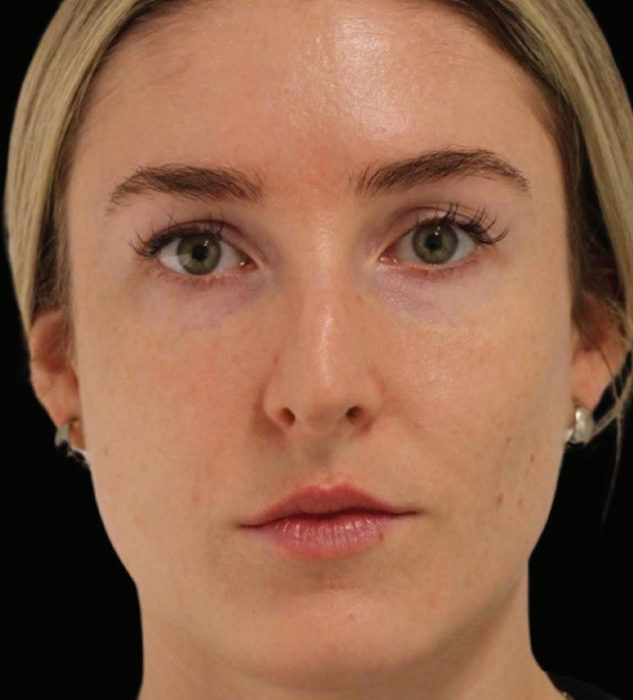
AGE-RELATED FACIAL CHANGES THE COMMON SIGNS
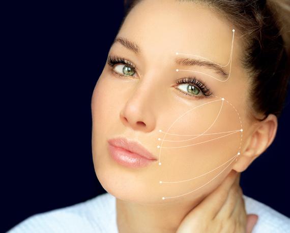
THREAD LIFTING FOR MIDFACE TISSUE SUSPENSION
TREATING CELLULITE A GAME-CHANGING APPROACH


AMWC Monaco takes place between March 27–29th, and I’m sure, like me, many of you are looking forward to three days of lively debates, excellent scientific content and world-class products on show.
Turning to this issue, which benefits from extensive distribution at AMWC Monaco, we begin with a celebration of the advancements shaping the future of the industry with our coverage of the highly anticipated 10th edition of the AMWC Aesthetic Awards, set to take place on March 27th. Turn to page 12 to view this year’s finalists, representing the industry’s finest and celebrate their contributions to the field of aesthetic and anti-ageing medicine.
Injectables remain a cornerstone of aesthetic medicine and continue to revolutionise the industry, offering patients transformative results with minimal downtime. We sat down with a panel of experts to explore the latest trends and innovations shaping the landscape. The feature is accompanied by our 2024 injectables survey, highlighting what our readers and AMWC delegates think about the future of injectables. Among the results, over 80% of respondents believe more training needs to be provided for new injectors. Additionally, with the advent of biostimulators and the move toward regeneration rather than volumising, over 87% of respondents say they already use biostimulators or plan to in the next 12 months. A revelation surely of where the injectables market is heading. You can read the full feature from page 18.
Non-surgical rhinoplasty is on the rise, offering patients a minimally invasive alternative to traditional surgery. Arash Jalali explores the risks and benefits of this increasingly popular procedure, shedding light on its growing appeal among patients seeking subtle yet transformative changes.
Finally, Jesper Thulesen explores the most common signs of age-related facial changes, offering valuable insights into the factors that drive patients to seek cosmetic consultation. From fine lines and wrinkles to volume loss and sagging skin, we explore the predictable signs of ageing and the strategies for effective intervention.

If you would like to attend any of the AMWC events in person, and I highly recommend you do, be sure to book your place at www.im-aesthetics.com. In addition, Aesthetic Multispecialty Society Premium Members will receive 20% off their delegate pass to all shows paid for during their membership, as well as further enhanced benefits, and you can sign up here: multispecialtysociety.com.
Balraj Juttla Editor, PRIME balraj.juttla@informa.com


 AMWC MONACO

NEWS

7 AAFPRS unveils aesthetic statistics from annual facial plastic surgery survey

8 The FACE Conference Gets a Facelift for the 2024 edition
AMWC AESTHETIC AWARDS 2024
12 Celebrating the industry finalists at the 10th edition of the AMWC Aesthetic Awards
INDUSTRY INSIDER
18 The state of injectables: stimulating further evolution
With injectables continuing to dominate the landscape of aesthetic medicine, we talk to a panel of experts about what they see on the horizon
CASE REPORT
26 Nordlys™ Nd:YAG treatment of periorbital veins
Hady Hamade shares the transformation of periorbital veins with two successful case studies using the Nordlys Nd:YAG 1046 nm laser applicator
28 A game-changing approach to cellulite using Sunekos Cell15
Andreea Boca explains how she uses the combined benefit of hyaluronic acid and six essential amino acids present in Sunekos Cell15 to successfully treat cellulite
AESTHETIC FEATURES
32 A balancing act: navigating the risks and benefits of non-surgical rhinoplasty
Arash Jalali delves into the growing popularity of nonsurgical rhinoplasty with hyaluronic acid fillers
40 The efficacy and safety of Duraform® dermal filler Jamel Fares, Carlos Vivas, Yaribel Chan, and Juan Contreras share the results of their clinical study on a new long-lasting PCL dermal filler

44 The Excellence line models from Aptos: principles of operation
George Sulamanidze, Lamzira Ebralidze, and Tamara Beshidze, describe the lasting effects of the Visage Excellence Method in midface tissue suspension
50 Age-related facial changes: the most common signs Jesper Thulesen explores the predictable signs of facial ageing that drive patients to seek cosmetic consultation
56
An in-depth look at hypochlorous acid
Humzah Dalvi explores the potential and clinical evidence for hypochlorous acid use in skincare, from wound care to disinfection, offering a deeper understanding of its physiological role and regulatory landscape
COMMENTARY
62 The transformative role of point of care ultrasound in aesthetic medicine
Jack Kolenda and Alfredo Ferreyra explain how ultrasound’s potential in aesthetic medicine, from filler identification to vascular complication prevention, can revolutionise patient care
64 Growth factors in the treatment of androgenetic alopecia
66 Full-face regeneration of soft tissue with Juvelook: the PDLLA + HA hybrid bio-stimulator
68 A comprehensive comparative analysis of Sunekos 1200 and Sunekos Performa in aesthetic medicine
72 P0105 Erbium-glass laser monotherapy in treatment of inflammatory acne vulgaris in adolescents
EVENTS
74 A round-up of the major industry events happening around the world over the next 12 months
The American Academy of Facial Plastic and Reconstructive Surgery (AAFPRS), the world’s largest association of facial plastic and reconstructive surgeons, released its 2023 member survey outcomes. With life finally returning to prepandemic levels of activity, facial plastic surgery and non-invasive treatments continue to boom in demand. Facelifts are back, and with the advancement of technology, facial ‘tweakments’ are as popular as ever because of accessibility and appeal to all ages.
Modern techniques like subcutaneous and deep plane facelifts are breathing new life into this age-old enhancement and drawing a younger crowd to the procedure. Since 2019, a striking 90% or more of AAFPRS surgeons have performed facelifts each year. On average, members performed 48 facelifts or partial facelifts in 2023, demonstrating a 60% increase since 2017. Over the past seven years, the number of facelifts performed has steadily increased year over year. The survey also noted a directional increase among patients ages 35–55, suggesting that facelift patients are getting younger.
77% of AAFPRS members believe there will be greater emphasis on early maintenance and prevention starting in the 20s and 30s.
‘At some point in the ageing process and with a certain degree of laxity and sagging, you will get diminishing returns on your non-invasive procedures,’ shares Sherard A. Tatum, President of the AAFPRS. ‘At this point, it’s best to opt for a facelift or partial facelift to get the desired effect. Facelifts also soared in popularity this year due to the “Ozempic Effect” where patients lost a large amount of weight in a condensed period of time, resulting in sagging skin.’
Consistent with years past, the top three surgical procedures were rhinoplasty (performed by 83% of surgeons), blepharoplasties (49%),

facelifts and partial facelifts (48%) across all genders.
Gen Z has entered the chat
From the ‘Sephora Tween’ phenomenon to TikTok’s wildly popular ‘Get Ready with Me’ videos, Gen Z (ages 11–26) is coming into their own purchasing power and prioritising aesthetics. This year’s survey supports this, showing that 77% of AAFPRS members believe there will be a greater emphasis on earlier maintenance and prevention starting in the twenties and thirties to forestall signs of ageing.
‘This generation is growing up with a greater awareness of what is possible when it comes to aesthetic treatments thanks to the normalisation online,’ says Dr. Tatum. ‘Rapid advances in non-invasive treatments and technologies allow younger patients entry into aesthetics with very little pain and downtime, making it more attractive to a larger patient pool.’
The new data points to this, with 83% of the total number of procedures performed in 2023 being minimally invasive. The remaining 17% were surgical. Of minimally invasive procedures, the three most common treatments were neurotoxins, fillers, and topical treatments (micro-needling and chemical peels). Rhinoplasty remains the single most requested surgery among patients under 34 years old.
It is still no surprise that women continue to reign when it comes to undergoing facial plastic surgery. However, this year’s results reveal that 44% of AAFPRS surgeons expect more men to have treatments and surgeries in the coming years. AAFPRS members also noted that they are seeing more men age under 35 seeking surgical and non-surgical enhancements.
‘As minimally invasive technology continues to advance, this opens the door for more men to get discreet, quick-to-heal cosmetic treatments,’ shares Dr. Tatum. ‘From non-invasive neck lifts to needle-free enhancements, there are more options than ever for men to keep looking as vital and youthful as they feel. The rise of minimally invasive options seems to be slowly closing the gender gap when it comes to facial plastic surgery.’
‘Our field is growing at such a fast pace. It’s an exciting time to be in facial plastic and reconstructive surgery,’ states Dr. Tatum. ‘Over the next year, we will certainly see the rise of AI in aesthetic medicine as surgeons integrate this technology to better analyse facial features, guide both their cosmetic and reconstructive surgery work, and predict outcomes of interventions over time. Things like 3D imaging allow us to simulate potential outcomes for more precise treatment planning, injectable placement and more. Custom computer-generated implants are available to better enhance or reconstruct facial contour problems.’
‘We may also see a reduction in the demand for injectables since a top concern for patients is appearing “overdone”,’ shares Tatum. According to 24% of survey respondents, looking unnatural is their patients’ biggest fear when considering a facial procedure. ‘I predict we’ll see a greater focus on more natural outcomes and graceful ageing. Some outward appearance of maturity can be taken as a sign of experience and wisdom. Although I am speaking as a boomer.’


THE REVAMP WILL INTRODUCE SEVERAL NEW DEVELOPMENTS
For over two decades, FACE Conference has been uniting international physicians and aesthetic professionals in London with its premier scientific programme dedicated to the in-depth exploration of non-surgical aesthetics and anti-ageing medicine. In 2024, the congress is evolving in many exciting ways.
A brand-new venue in the heart of London FACE Conference is moving the prestigious Business Design Centre, located in the heart of London. This setting will be home to a thoughtfully redesigned programme that remains unparalleled in the UK market.
Featuring four main agendas — Advanced Injectables, Multispecialty Aesthetics, Industry Workshops, and Aesthetics Open Stage — the full programme encompasses eight core industry segments: injectables, skin, regenerative medicine, business and practice management, genital treatments, hair, threads, and medical devices. Delegates can expect a programme that they know, value, and trust which has evolved in a very positive way.
The new Aesthetics Open Stage agenda has been specially curated for all medical and non-medical certified practitioners, including nurses, aestheticians, cosmeticians, beauty therapists,
and skin specialists. Accessing this educational agenda is free.
This year, we are introducing a Free Pass where all aesthetic professional profiles (at any experience level) can engage with the programme by attending the Aesthetics Open Stage, Industry Workshops, and commercial exhibition.
The Full Pass and VIP Pass grants entry to our Advanced Injectables and Multispecialty Aesthetics agendas. These tracks comprise high-level sessions for physicians, dentists, and nurses to enhance their skillset and keep themselves at the forefront of the industry. Preferential rates are available for nurses and UK-residents.
This year, we are introducing a Free Pass where all aesthetic professional profiles (at any experience level) can engage with the programme by attending the Aesthetics Open Stage, Industry Workshops, and commercial exhibition.
All FACE sessions and live demonstrations are backed by evidence-based research and presented by leading speakers, all of whom are highly esteemed in their respective fields.
At FACE, attendees have the opportunity to discover a 2,000 sqm international exhibition of over 100 trusted companies. It is the ideal place to mingle with brands, attend practical workshops, connect with peers, and foster valuable relationships between scientific sessions.

The FACE Team looks forward to welcoming you to London’s Business Design Centre on 12–13 July for an enlightening 2-day event where you will refine your practice, put yourself ahead of competitors,
In the EU, Merz Aesthetics® announced today the approval for Radiesse® to treat the décolleté area. Radiesse® is a regenerative biostimulator capable of regenerating multiple components of the skin tissue, resulting in healthier-looking skin, and is now approved for use in the décolletage, adding to the product’s versatility.
This new regulatory milestone directly answers patient demand, given there is a significant shift toward regenerative biostimulation as a preferred method of aesthetic treatment for patients. Google Trends Data further support this showcasing a continual surge in searches for ‘collagen’ between 2016–2022, confirming a shift in aesthetic preferences for patients.
‘Despite being one of the most visible areas of our bodies, the décolleté area is often overlooked when it comes to aesthetics,’ says Dr. Samantha Kerr, Chief Scientific Officer, Merz Aesthetics®.
‘Radiesse® presents a unique solution to improving décolleté wrinkles and provides our customers and their patients a non-surgical, effective option to address ageing décolleté skin.’
Recent data from the ‘International Society of Aesthetic Plastic Surgery’ reveal a 171.8% increase in worldwide non-surgical procedures minimally invasive treatments between 2018 and 2022 involving calcium hydroxylapatite further showcasing the patient need.
A number of studies, including this large, multicentre clinical trial have supported the décolleté approval and has provided substantial evidence of Radiesse®’s effectiveness, demonstrating a significant improvement in the appearance of décolleté wrinkles. The various studies have found that up to 73.5% of patients experienced improved décolleté wrinkles, 16 weeks after their last treatment. Furthermore, 76.1% of patients demonstrated an enhancement in skin quality over the same period.
Notably, physicians reported a high level of satisfaction among 77% of patients 16 weeks after their last treatment.


Celebrating the industry finalists at the 10th edition of the AMWC Aesthetic Awards, which takes place in Monaco on March 27th 2024
THE AMWC AESTHETIC Medicine Awards 2024 stand as a beacon of excellence, bringing together the brightest minds and most cutting-edge products in the field of Aesthetic and Anti-Aging Medicine. With a star-studded panel of 77 judges and input from attendees, these awards recognize the game-changers and trend-setters who are shaping the future of beauty.
Each nominee undergoes rigorous evaluation based on specific criteria, including application quality, clinical evaluation, market access approval, innovation and advancement, clinical efficacy, professional endorsement, and priceperformance ratio. These stringent standards ensure that only the most deserving entries make it to the final round.
The award categories span a wide
spectrum, encompassing various aspects of aesthetic medicine. Notable categories include Best energy-based treatment, Best non-surgical body shaping, Best complication management, Best suspension thread, and Best injectable dermal filler. These categories ensure that all areas of aesthetics, from innovative treatments to effective complication management, receive the recognition they deserve.
The finalists, narrowed down through peer and jury voting, represent the epitome of excellence in aesthetic medicine. Their contributions, whether through groundbreaking clinical cases or innovative products and devices, showcase the transformative potential of aesthetic procedures and the positive impact they have on patients’ lives.
And the best part? The winners get to take home the ultimate prize: recognition and
validation for their hard work and dedication to advancing the field of aesthetic medicine.
The AMWC Aesthetic Awards Ceremony, held at AMWC Monaco, serves as a platform to unveil the winners and celebrate their achievements. From pioneering practitioners to innovative companies, each winner receives acknowledgment for their dedication to advancing the field of aesthetic medicine.
In an ever-evolving industry where innovation and artistry redefine beauty, the AMWC Aesthetic Awards stand as a testament to the tireless efforts of individuals and establishments pushing the boundaries of aesthetic medicine. Through their commitment to excellence, these awards inspire and set new standards for the future of the field.
Aesthetic devices
U225 EVO


Lasers, light and energy based devices


Regenerative aesthetic medicine


Discovery PICO Variopulse Technology®


Cosmetics
Alastin Skin Nectar Melan-Ox Serum


Dermastir Dropper Serum Oil


MelaQuest Cysteamine Serum


Dermastir Post-Op


EXO Balm


Definisse [KP1] Regenerating Serum


Pro Restore


Biojuve


Aesthetic products and devices integration
640 C


Anti-aging supplements
Skin Regen MD Radiance


Neuromodulators
AGE Breaker Alluzience® Activ’ anti-rides ultimate Nuceiva®


Injectable
Restylane®
Revanesse Shape

Ellansé


Injectables for skin revitalization
Dermastir H13


Purasomes NC150+

Restylane® Skinboosters™

Hyal-System DUO


Juvelook
Non-invasive


Stimulate


Hyal-System ACP

Wonder Technology for Muscle Creation & Fat Burning


With injectables continuing to dominate the landscape of aesthetic medicine, we talk to a panel of experts about what they see on the horizon
AESTHETIC MEDICINE WAS ONCE perceived as facelifts for celebrities and the wealthy on the one side, and spa treatments with questionable, or at least unproven, efficacy on the other. Nowadays the picture is quite different. While injectables unquestionably dominate the landscape, the terrain has evolved significantly since the heady days of Alistair and Jean Carruthers.
Injectables are safe, effective, and accessible to an extraordinarily wide range of patients—and practitioners, which experts believe can be a double-edged sword. As public awareness increases, demand increases, and the buying public has always wanted natural-looking results, according to Nashville-based dermatologist Dr. Michael H. Gold. ‘People are not trying to look weird or “done.” What they want is to look better, younger, or even a little bit different, with an effect that’s subtle but profound. The spectrum of injectables available today, used individually or adjunctively, do just that, which is why injectables took off and have continued to hold so much importance in the aesthetic marketplace. We still have a lot of work to do maintaining industry-wide injector quality, and patients should take care choosing where they are treated if they want the best results, safely.’
The landscape of fillers has undergone a significant transformation. ‘In the 1990s and early 2000s they were considered the ultimate solution for anti-ageing, while biocollagen stimulators lingered in the background,’ said Ft. Lauderdale, Florida-based dermatologist Dr. Shino Bay Aguilera. ‘However, with advancements in our understanding at a cellular level and from gene expression studies, these stimulators, along with fat matrix stimulators, have now taken centre stage as the most coveted treatments for anti-ageing today.’
Dermatologist Sue Ellen Cox is based in Chapel Hill, North Carolina. ‘As an industry, we’ve been gravitating toward regenerative medicine for some time. More than ever we’re not just putting product into a face to reshape it, we’re trying to induce the body to produce new collagen and whatever else will create healthier tissue and a more natural effect, which hopefully persists,’ she said. ‘Using subcutaneous platelet-rich plasma (PRP) to improve skin quality or remodel acne scars are good examples. We want to induce regenerative processes. Exosomes are going to be huge for that but at this point they are absolutely not for injection.’
FDA clearance,’ Dr. Cox continued. ‘It creates improvement of skin hydration and appearance that lasts up to six months.’ This was shown in a randomised, evaluatorblind study assessing safety and effectiveness of intradermal VYC-12L treatment for improving fine lines, hydration, and cheek skin smoothness1
According to anatomist and aesthetic physician Dr. Kyu-Ho Yi, based out of Seoul, Korea, ‘Of particular interest in contemporary aesthetics is the exploration of polynucleotide (PN) as a skin booster or potential alternative to traditional fillers, contingent upon its concentration.’ Injectable PN involves the use of DNA fragments as biostimulators, which may have a variety of applications. ‘It has garnered attention for its regenerative properties, which may offer notable benefits in improving skin quality and addressing age-related concerns. As such, its potential applications in aesthetic medicine warrant further investigation and consideration for integration into clinical practice,’ he continued. The use of polynucleotide has become prevalent in Asia. ‘Polynucleotide is highly regarded for its minimal side effects and its ability to promote hydration due to its DNA particle structure.’ Patients often notice immediate radiance and a transparent appearance post-injection, contributing to demand, he added.

As an industry, we’ve been gravitating toward regenerative medicine for some time. More than ever, we’re not just putting product into a face to reshape it, we’re trying to induce the body to produce new collagen.
Recent trials led to the rollout of injections of hyaluronic acid-based gel products to freshen and revitalise skin. ‘This is something that has been going on for a while around the world but has only now received
Experts seem to agree that the premier development in injectables is the ongoing shift in the paradigm of how we use them and view them in the context of the armamentarium — now as a vital component to the current toolkit in regenerative aesthetics (RA). Foremost in this is the evolving role of biostimulatory fillers such as poly-L lactic acid (PLLA) and calcium hydroxylapatite (CaHA). The claim was made by two separate 2023 review articles, one by Goldie2 and the other by Aguilera and colleagues3, which make clear cases for this, especially regarding CaHA. The Goldie review provides a key definition of RA as focusing chiefly ‘on the regeneration of soft tissue lost or damaged due to ageing processes which consequently impacts on the aesthetics of the individual.’2 It suggests the importance of teasing out RA as a distinct sub-field of regenerative medicine as a whole deserving of special attention to firm up definitions and goals. Because the ageing process compromises both the structure and function of tissue and taking into account the delicate balance of the complex environment of senescent tissue, there are countless considerations and roadblocks to consider. Dysfunction at the cellular level, the extracellular matrix, and everything upward, which reduces overall skin quality and barrier integrity due to senescence, may have a variety of possible counteractive pathways, but which ones are nearer to ideal? The Goldie paper further advanced the idea that regenerative scaffolds, such as those employed with the use of


biostimulatory fillers, are one of three key vectors through which RA is performed. ‘Biostimulatory agents play a crucial role in stimulating collagen production and promoting tissue rejuvenation,’ Dr. Yi said. ‘By offering durable volumisation and structural support, they have revolutionised the approach to facial rejuvenation and contouring. Furthermore, these agents are utilised for reconditioning the dermal layers, contributing to overall skin health and vitality. It’s about improving structure, function and overall quality.’
Among biostimulatory fillers, the focus is primarily on CaHA because it has been shown to induce a more favourable inflammatory response than PLLA when directly compared4. After 24 hours incubation within two dilutions each (1:50 and 1:100) of commercially available CaHA and PLLA, the expression of 40 cytokines in tested primary human macrophages was measured. The cytokine profile of PLLA was shown to initiate significant expression of cytokines known to play a role in inflammation, while CaHA induced a non-inflammatory response. This immunological perspective was further illuminated in a review by Corduff5 explaining how scaffold formation and healing after injury is affected by inflammation. With the initiation of the wound healing cascade, inflammation and the foreign body response will determine the course between two healing pathways: one leading to reduced inflammation resulting in healthier new tissue, the other promoting inflammation leading to the formation of less healthy tissue (scars). The review states, ‘Anti-ageing aesthetic medicine uses interventions like biomaterial-based fillers to influence these immunological responses and renew aged tissue structure and function.’5 With its non-inflammatory response, CaHA promotes the genesis of healthier tissue.
Another review study (Amiri 2023)6 delineated that CaHA appeared to induce cell proliferation with synthesis of collagen types I and III, increase elastin (with a possible time-dependency) and stimulate angiogenesis, which is vital to microcirculation, all of which are essential building blocks of a solid regenerative aesthetic treatment. ‘The important reality is that there is more to this than simply stimulating fibroblasts,’ said Munich-based dermatologist Dr. Tatjana Pavicic. ‘Inducing the kind of regenerative response requires a delicate balance that works with, rather than against, the microenvironment and promotes the expression of more regenerative pathways. In the past, it was about neocollagenesis; now, we know more about collagen production and rations of collagen types. These studies are an application of and expansion of that knowledge.’
Merz Aesthetics’ regenerative biostimulator Radiesse® is comprised of CaHA microspheres suspended in a carboxymethylcellulose (CMC) gel. The CMC gel is largely responsible for the immediate enhancement effect; the


CaHA microspheres get to work in direct contact with the fibroblasts resulting in the long-term genesis of healthy skin tissue that supplants the CMC gel over time. Radiesse® is backed by 20+ years of scientific and clinical experience and has shown a favourable safety profile in various clinical studies, and thus has been a mainstay among biostimulatory treatments for some time. Regeneration of collagens I and III, as well as elastin and proteoglycans, according to the Aguilera review article3, helps to restore youthful skin quality as well as tissue structure with its bioregenerative scaffold.
Homogeneous particle size and distribution, as well as the particle physiology itself (smoothness, free of defects), are mentioned as playing key roles in the regenerative process and immune response. ‘This is very important to the overall mechanism of action,’ explained Dr. Gabriela Casabona from Marbella, Spain. ‘When trying to induce the most therapeutically relevant signaling we are also trying to reduce signaling that might be counterproductive. Whenever we inject something into the body, there will be a reaction. We want to produce a “less angry” reaction from the body and communicate with the fibroblasts in a way that influences the more advantageous responses. Radiesse® is just such a product, inducing a cascade that will produce a more balanced extracellular matrix leading to healthier-looking skin*.’
‘What we’re also looking at,’ Dr. Pavicic added, ‘is improvement in elastin and angiogenesis, which support healthier new tissue formation, which we’ve seen with Radiesse®. You’re not just creating a scaffold, you’re improving the structure and function of tissue.’
Unsurprisingly, many of these studies were backed by Merz Aesthetics, whose commitment to science is a major component to the substantial footprint Merz Aesthetics enjoys in the marketplace, and the confidence physicians place in their products. ‘Not many companies continue to develop products with the rigour and enthusiasm that Merz does,’ Dr. Casabona said. ‘They continue to explore new indications and if they can do so, they back them with science which helps accountability throughout the industry when their products are used. Consider that Radiesse® has been around for two decades, we have a new dilute indication in the EU, and the regenerative aesthetics development is groundbreaking. They back this science with training, which keeps injectors informed and creates a unified message, which positively affects the industry overall.’
People want to look as healthy and beautiful as possible, and treating the décolleté is part of that because if you only treat the face, the healthier and younger look will clash with the neck and/or chest because people notice the incongruity.
In the EU, Radiesse® is now approved for the treatment of décolleté wrinkles using a 1:2 dilution; study publication is forthcoming. Dr. Pavicic is the lead author, and Dr. Casabona is a co-author. ‘We use a cannula to implant the product in many small boluses along specific lines with minimal insertion points,’ explained Dr. Pavicic, ‘which, between the dilution and the multiple boluses, maximises the surface area of the injected product in contact with tissue. The goal is to harness the regenerative effect to improve tissue quality without compromising safety.’
‘People want to look as healthy and beautiful as possible,
and treating the décolleté is part of that because if you only treat the face, the healthier and younger look will clash with the neck and/or chest because people notice the incongruity,’ Dr. Casabona added. ‘It becomes distracting instead of aesthetically pleasing otherwise. A relatively young face with an old chest doesn’t match.’
Another big plus for injectables in general is that they work well as an adjunct to countless therapies, but especially with other injectables. Today, this goes beyond the original one-two punch of neurotoxin with fillers, according to Dr. Aguilera. ‘Injectable fillers, when combined with PRP, PRF, liquid fat, and skin boosters, have shown remarkable potential in enhancing the effects of biocollagen stimulators,’ Dr. Aguilera stated. ‘This underscores the significance of cellular-level nutrition in fostering the thriving and transformation of ageing cells into more active and functional versions of themselves, thereby yielding superior outcomes. For instance, a recent publication by Elina Theodorakopoulou et al.7 (Dr. Aguilera is a co-author) highlights the efficacy of blending Radiesse® with NCTF 135 HA. This study emphasises the importance of prepping the skin with essential vitamins, coenzymes, hyaluronic acid, and cofactors present in a polyrevitalising micronutrient blend. Remarkable improvements in skin tone, texture, volume, and retraction were observed at 3and 6-weeks post-treatment.’
Subsequent research (some still unpublished) is still moving the ball forward. ‘Authors like Dr. Alessio Redaelli, who combined PLLA with NCTF 135 HA, has yielded similar enhancements in volume and skin quality. These synergistic blends offer a co-effective approach, reducing the number of sessions required for optimal results. Moreover, Dr. Hassan Galadari has demonstrated the efficacy of mixing neurotoxins with NCTF HA 135 to address concerns such as pore size, redness, and overall skin tone and texture. Additionally, Dr. Alessandro Gennai and colleagues published findings on combining Superficial Enhanced Fluid Fat Injection (guided SEFFI) with calcium hydroxylapatite (CaHA)8. ‘This innovative approach effectively counters volume loss and skin ageing in both facial and bodily regions, representing a promising avenue for regenerative aesthetic treatments,’ Dr. Aguilera said. ‘The emergence of these blends underscores the importance of providing the skin with optimal nutrition at a cellular level, marking a new era in aesthetic medicine characterised not only by regeneration but also functional enhancement.’
‘With further research we are growing our understanding of the signaling, the microenvironment, and how we can interact with it to induce therapeutic regenerative processes,’ Dr. Casabona said. ‘Within five years we will know so much more.’
What used to be one toxin and a handful of fillers is now a much more crowded-looking space, although with growth there has been room, hence the proliferation of successful products and greater variety. Among toxins the global list
of toxin offerings continues to proliferate but the big five are onabotulinumtoxinA, abobotulinumtoxinA, incobotulinumtoxinA, prabotulinumtoxinA-xvfs(cosmt), and the more recent daxibotulinumtoxinA-Ianm. ‘The big five are definitely good products and have earned their positions,’ Dr. Gold said. ‘The questions we keep hearing are “How long does it last?” and “How fast is the onset time?” which, of course, does not crowd out the importance of safety. The newer of the five, daxibotulinumtoxinA-Ianm, has the six months’ approval and the general consensus is that it performs in many patients, more or less in some, but it’s a good toxin and comparable to its predecessors.’
Injectable fillers, when combined with PRP, PRF, liquid fat, and skin boosters, have shown remarkable potential in enhancing the effects of biocollagen stimulators.
Gold added that there are also new toxins in the FDA approval pipeline, most of which are available elsewhere in the world, including the EU. ‘There are liquid toxins pending FDA approval from well-known companies, and one was showing rapid onset in the one-day range, with good numbers out to six months.’ He explained that the key advantage to ‘liquid’ neurotoxins is that it requires no reconstitution. ‘You get a vial, you take out the units you want, you don’t have to mix it, there’s no variation. This is probably the immediate future of neurotoxin and is going to change things for a lot of injectors because of that. The purists who’ve been around a long time will probably stick with touting the old standbys because they have understandably high levels of confidence in their skills at injecting and reconstituting. And none of these products is going away anytime soon.’
So, do we need another neurotoxin? It is easy to say ‘no’ but the experts look at this from a different angle. ‘After years working with various neurotoxins, it becomes evident that each one imparts a distinct aesthetic effect. While subtle to some, those with a discerning eye will

notice these differences,’ said Dr. Aguilera. ‘I remain receptive to exploring new neurotoxins that may excel in specific areas. Moreover, healthy competition within any market fosters progress, driving innovation and ultimately benefiting patients and practitioners alike.’
Toronto-based dermatologist Dr. Vincent Bertucci agreed that neurotoxin development hasn’t reached its zenith.
‘The development of liquid toxin that doesn’t need to be reconstituted, and has both a good shelf life and solid science behind it is changing things and as with many developments, we’re still learning what the impact will be. More than that, people are always looking for a better, longer-lasting result, so the obvious next move was larger doses, but any toxin scientifically proven to be longer lasting and/or with faster onset will make people stand up and take notice.’

Developments in other markets may bring impact to the injectables market. ‘It’s worth mentioning that while facial rejuvenation treatment was previously primarily associated with muscle paralysis, there is now increasing recognition for treatment through muscle strengthening,’ said Dr. Yi. ‘When considering ageing, muscles weaken, and contractures develop. To strengthen them, devices such as electrical stimulation therapy are available in the market. This aspect, too, is an area that doctors should pay attention to in the future.’
Throughout medicine, changes in the doctor-patient relationship over the decades have impacted and reshaped the injectables market profoundly as well. The treatment decision is a totally different animal than it was in the twentieth century. Evolution in the way we look at medicine and the doctor-patient relationship has more recently been accompanied by the rise of social media, which has tremendous influence, according to Dr. Bertucci. ‘Generally, I’ve seen two situations develop. The first is the patient who seeks and sees a doctor because they’ve got, or want, a positive relationship of trust. The doctor is experienced, aware of and understanding of forefront developments, reads the literature, is reasonable and rational, and can help guide the patient. After a healthy conversation they’ll take the doctor’s advice on what solution is most appropriate. The other type involves a new doctor-patient interaction. The patient has just Google searched and found this person, but there’s no context; is
this doctor amazing or is this doctor average? Experienced and credentialed, or new to practice? And in those circumstances, if the doctor doesn’t have the confidence, knowledge, and ability, their decisions will be more patient-driven. That’s not good or bad, more an observation that we should be aware of. And patients should continually be encouraged to seek safety and competence.’
The influence of social media
Social media has also impacted the balance between marketing and scientific rigour, he continued. ‘Social media drives demand, and new fads come and go even more rapidly than they used to.
Elevated patient demand influences marketing, and I’ve seen science being pushed aside somewhat, which concerns me. From the industry perspective I think aesthetic medical companies and the FDA have done a good job of keeping regulation relevant, but human beings want what they want, so science and even safety are sometimes made secondary to the patient by social media influence.’
We see this with the most reputable products: science drives success. If we align ourselves accordingly, we serve the industry and our patient base best.
‘In the best-case scenario we’re talking about products that we’re enthusiastic for because of the science,’ Dr. Cox added. ‘We see this with the most reputable products: science drives success. If we align ourselves accordingly, we serve the industry and our patient base best.’
* ‘Healthy-looking skin’ is defined by regeneration of collagen I and III, elastin, proteoglycans, and angiogenesis.
1. Alexiades M, Palm M, KaufmannJanette J, et al. A randomized, multicenter, evaluator-blind study to evaluate the safety and effectiveness of VYC-12L treatment for skin quality improvements. Dermatologic Surgery 2023 Jul;49(7):p 682-688.
2. Goldie K. The evolving field of regenerative aesthetics. J Cosmet Dermatol 2023;22(Suppl 1):1-7.
3. Aguilera SB, McCarthy A, Khalifian S, Lorenc ZP, Goldie K, Chernoff WG. The role of calcium hydroxylapatite (Radiesse) as a regenerative aesthetic treatment: A narrative review. Aesthet Surg J 2023;43(10):1063–1090.
4. Nowag B, Schäfer D, Hengl T, Corduff
N, Goldie K. Biostimulating fillers and induction of inflammatory pathways: A preclinical investigation of macrophage response to calcium hydroxylapatite and poly-L lactic acid. J Cosmet Dermatol 2023;00:1-8.
5. Corduff N. Introducing aesthetic
regenerative scaffolds: An immunological perspective. J Cosmet Dermatol 2023;22(Suppl1):8-14.
6. Amiri M, Meçani R, Niehot CD, et al. Skin regeneration-related mechanisms of Calcium Hydroxylapatite (CaHA): a systematic review . Frontiers in Medicine 2023;10:1-16.
7. Theodorakopoulou E, McCarthy A, Perico V, Aguilera SB. Optimizing skin regenerative response to calcium hydroxylapatite microspheres via poly-micronutrient priming. J Drugs Dermatol 2023 Sep 1;22(9):925-934.
8. Gennai A, Baldessin M, Melfa F, et al. Guided superficial enhanced fluid fat injection (SEFFI) procedures for facial rejuvenation: an italian multicenter retrospective case report. Clin Pract 2023 Aug;13(4):924-943.
Curious about a smoother, more youthful looking chest area?
Provides collagen stimulation together with long-term renewal of healthy-looking skin and enhanced skin quality1–3
Improves collagen and elastin production which help to provide immediate lift to smooth moderate and severe lines and wrinkles in the upper chest area (décolleté)4–6


Patient
ARE YOU CURRENTLY USING BIOSTIMULATORS OR PLAN TO IN THE NEXT 12 MONTHS ?
WHAT DO YOU CONSIDER THE BEST INVESTMENT STRATEGY FOR NEUROMODULATORS/ FILLER MANUFACTURERS?
■ MARKETING 3.03%
■ RESEARCH AND DEVELOPMENT 34.75%
■ PROFESSIONAL EDUCATION 50.91%
■ PATIENT EDUCATION 11.31%

ARE THERE NEEDS THAT CURRENT SOFT TISSUE FILLERS DO NOT MEET OR AREAS WHERE THEY COULD BE IMPROVED?
DO YOU USE INJECTABLES IN COMBINATION WITH OTHER TREATMENTS?
I think sales should be more limited to professionals who are actually trained to use them.
WITH FACIAL AGEING, DO YOU PREFER TO
DO PHYSICIANS NEED MORE BOTULINUM TOXIN PRODUCTS FOR AESTHETIC USE?
More training and more research to level up the quality of treatment for the patient.

Quality is slowly becoming more important than marketing. Price is not the most important criterion but the quality of the product, its safety based on research and clinical work.
INSIDER
Emphasis on ethics and on natural results!
WHAT ARE YOUR NON-NEGOTIABLES FOR NEUROMODULATOR PRODUCTS – THE CHARACTERISTICS THEY ABSOLUTELY MUST HAVE?
SELECTED IN ORDER OF PREFERENCE
1ST = MOST IMPORTANT
6TH = LEAST IMPORTANT
1 ST SAFETY
2 ND DURATION OF EFFECT
3 RD
PREDICTABLE RESULTS
4 TH BIOCOMPATIBLE
5 TH EASY TO USE
6 TH AFFORDABLE
DO YOU COMPLY WITH PATIENTS’ REQUESTS FOR A SPECIFIC BRAND OF TOXIN?
■ YES, USUALLY 21.37%
■ YES, IF I HAVE IT AVAILABLE 30.44%
■ NO, I MAKE THE DECISION 48.19%

Increase the scope of medical education in terms of injection techniques, make it more reachable to users. We need to develop new agents that can melt granuloma, nodules and fibrotic tissues.
WHAT ARE YOUR NON-NEGOTIABLES FOR DERMAL FILLER PRODUCTS – THE CHARACTERISTICS THEY ABSOLUTELY MUST HAVE?
SELECTED IN ORDER OF PREFERENCE
1ST = MOST IMPORTANT
6TH = LEAST IMPORTANT
1 ST SAFETY
2 ND PREDICTABLE RESULTS
3 RD DURATION OF EFFECT
4 TH BIOCOMPATIBLE
5 TH EASY TO USE
6 TH AFFORDABLE
DO YOU THINK MORE NEEDS TO BE DONE TO PROVIDE TRAINING AND EDUCATION FOR NEW INJECTORS ?
NO, THERE ARE A LOT OF OPTIONS ALREADY AVAILABLE
HAS MARKETING OVERTAKEN SCIENTIFIC RIGOUR IN THE USE OF INJECTABLES?

Hady Hamade shares the transformation of periorbital veins with two successful case studies using the Nordlys Nd:YAG

HADY
HAMADE, RPH DIRECTOR, is a highly experienced Cosmetic Laser Specialist, Laserologist, Skin Care & Laser Clinic, Hamilton, Ontario, CanadaKEYWORDS
Candela, Nordlys™, periorbital veins, Nd:YAG
THIS CASE STUDY SHOWCASES THE EFFECTIVENESS OF utilising the Candela® Nordlys™ multi-application platform with the long-pulsed Nd:YAG 1064 nm laser applicator to treat prominent periorbital veins (POVs). The Nordlys platform is a multi-modality system offering an Nd:YAG 1064 nm laser, non-ablative fractional lasers (1550 nm and 1940 nm), as well as a range of Selective Waveband Technology (SWT®) narrowband IPL applicators to address a variety of skin conditions*. Unlike invasive surgical therapies for prominent POVs that can leave unsightly scars, the Nd:YAG vascular laser with its long 1064 nm wavelength can penetrate deep into the skin to heat the entire vessel wall and coagulate visible POVs with minimal post-treatment care1,2
We report here on two case studies of POVs successfully treated with the Nd:YAG 1064 nm laser. Patients were instructed to avoid excessive sun exposure for at least one month prior to treatment and to avoid exfoliants, alpha hydroxy acid (AHA), beta hydroxy acid (BHA), or retinoids (vitamin A derivatives) for two weeks pre-procedure. Post-treatment care consisted of applying a hydrating cream (La Roche-Posay® Cicaplast) for seven days followed by sunscreen. Under-eye puffiness subsided within hours to 1 week post-treatment.
A 25-year-old female with brown hair and blue eyes presented with a network of POVs that, due to her fair skin (Fitzpatrick Skin Type II), were quite visible and perceived as dark circles under the eyes. She received two Nd:YAG treatments with a 6-week interval for the optimal cosmetic outcome. The treatment was performed directly over the vessels, with no overlap, to the clinical endpoint of vessel disappearance.
The second patient was a 35-year-old female with brown hair and brown eyes. The patient had Fitzpatrick Skin Type III, a prominent blue POV that appeared raised and several smaller vessels. A single Nd:YAG treatment significantly minimised the prominent vessel.
The two case studies presented here demonstrate a successful treatment technique using the Nordlys Nd:YAG applicator to treat or minimise prominent POVs quickly and non-invasively. The long-pulsed Nd:YAG 1064 nm laser targets haemoglobin in the blood vessels, causing coagulation and vessel closure without damaging the surrounding skin. The treatment involves precise application along the periorbital vein,
The two case studies presented here demonstrate a successful treatment technique using the Nordlys Nd:YAG applicator to treat or minimise prominent POVs quickly and noninvasively.
inducing rapid disappearance of the characteristic blue hue because of efficient haemoglobin absorption and subsequent coagulation. There were no treatment complications, and downtime was minimal for the patients.
Treatment of periorbital veins is an advanced treatment and should be performed by experienced treatment providers. Always perform test spots and observe treatment response prior to performing the full treatment. Ocular shields should be used when treating near the eye. The number of treatments can be customised according to the patient’s cosmetic concerns and the vessel size.
Declaration of interest Funding for submission of this paper was provided by Candela
* Nordlys and Frax Pro Systems, CE Mark
Figures 1–2 © Hady Hamade


Andreea Boca, MD, explains how she uses the combined benefit of hyaluronic acid and six essential amino acids present in Sunekos Cell15 to successfully treat cellulite
 ANDREEA BOCA, MD PHD, is a Dermatologist, Cluj-Napoca Romania
fibrosclerotic panniculopathy, what we commonly refer to as cellulite is a primarily aesthetic skin condition that is constantly in the spotlight.
In years prior, exposing a celebrity photograph depicting cellulite was considered a major blow to one’s career, as society was aspiring to promote the perfect body: the perfect height, weight, body proportions, skin texture, and demeanour.
In response to the never-ending chase of perfection, a movement towards accepting human variation and celebrating beauty in all its forms was born. The scientific consensus is that cellulite occurs in over 80% of women1,2. Cindy Crawford’s ‘leaked’ candid images, showcasing the tell-tale dimples of cellulite, went viral as an empowering memento to women to have realistic expectations.
While society tries to lean towards accepting this seemingly benign condition, at an individual level, people constantly seek solutions. With media giants such as Cosmopolitan posting guides to say goodbye to cellulite in 2024 and Elle providing reviews of the best cellulite-fighting creams, it seems like the war on cellulite has its wellestablished place in the aesthetic market.
The significance of cellulite is striking when taking into account quality of life studies: 78% of patients with cellulite report seeking treatment for their condition, while over 40% of patients report receiving embarrassing remarks regarding their skin condition. This considerable social impact is amplified when considering another study that reports that 60% of responders believe that having cellulite is mainly their fault3
This explains partially why in only one year, over 86,000 minimally invasive cellulite treatments are reported to be performed by physicians solely in US2
KEYWORDS
Cellulite, Sunekos Cell15, edematous fibrosclerotic panniculopathy
The colloquial term ‘cellulite’ is known medically under a plethora of names: gynoid lipodystrophy, nodular liposclerosis, edematofibrosclerotic panniculopathy, adiposis edematosa, dermopanniculosis deformans, status protrusus cutis and several others. This has less to do with overzealous polyonymy and more to do with the ongoing discussion regarding the multifactorial pathogenesis of cellulite: initially considered a mechanical response of adipose septa to the strain of adipocytes, further research has uncovered a series of other key players, such as inflammation and vascular compromise.

The topographic feature of skin with cellulite is the ‘orange peel’ dimpling caused by adipose tissue protruding into the dermal tissue by way of herniation. The supportive septa undergo fibrosis, followed by shortening and retraction, forming the depressions that sum up the visual appearance of cellulitis. While it would be reasonable to assume that an increase in adipocyte volume would be directly correlated with septum fibrosis and cellulite, there seems to be no statistically significant correlation between cellulite severity and adipose layer thickness1
Cellulite occurs almost exclusively in the buttocks, abdomen and upper thigh. While over 80% of female patients suffer from it, male patients are affected as an exception, with 2% reporting cellulite lesions in the context of androgen deficiency, either as a consequence of a
systemic condition (hypogonadism, Klinefelter’s syndrome) or as a response to antiandrogen therapy2 These demographic features indicate that mere mechanics are insufficient to explain the pathogenesis of cellulite. Endocrine factors were then taken under consideration to explain cellulite: oestrogen increases vascular wall permeability, leading to oedema of surrounding tissues and impairing regional microcirculation. Meanwhile, the lack of progesterone and hyperestrogenism influence metabolic function, impairing adipocyte function and favouring fibrosis and nodule formation4
A low-grade inflammatory state has been correlated with dermal atrophy, as well as with the development of the fibrotic septae. This type of low-grade inflammation, frequently associated with obesity and insulin
The topographic feature of skin with cellulite is the ‘orange peel’ dimpling caused by adipose tissue protruding into the dermal tissue by way of herniation.

ATL-C technique with cannula
● Cannula 27G and 40-50mm or 22G and 50-70mm
● 0.8ml per injection 12cm 12cm
● Deep dermis injection
● Fan technique


resistance, contributes to endothelial and soft tissue dysfunction.
Adipose tissue is a metabolically active organ, secreting a number of peptide and steroid hormones, of which several have been correlated with endothelial damage: interleukin-6, tumour necrosis factor-alpha and others of the kind2. The synergy of mechanical and biological stimuli produced by hypertrophic adipose cells entertains a steady context of low-grade inflammation coupled with vascular dysfunction.
All the above lead to a chronic, ongoing condition that, if left untreated, goes through four stages of evolution:
■ The starting point of cellulite is represented by adipose tissue dystrophy, accompanied by initial oedema and microcirculation impairment. Macroscopic changes may include pale skin which loses its elasticity; however, it is mostly asymptomatic
■ The second stage refers to lobule dystrophy, stromal hypertrophy, oxidative stress and further vascular impairment: this is where local oedema influences collagen fibres by way of hypertrophy and hyperplasia, leading to pale, cold skin with potential sensitivity alterations
■ The third stage revolves around the hallmarks of inflammation, decreased pH and fibrosis: micronodules encapsulated by collagen further alter microcirculation and metabolic exchange in the skin. There is an important loss of extracellular matrix homeostasis. Visually, this stage is easily identified by the well-known ‘orange skin’ appearance
■ The final stage of cellulite is marked by irreversible sclerosis, with well-formed nodules, thick adherent septa and vascular dilatation.
The medical and cosmetic industries alike have developed a formidably diverse armamentarium to address cellulite, from marketing-driven thermogenic gels and creams all the way to invasive procedures.
The science behind anti-cellulite creams is underwhelming in comparison to their variety: topical formulations have been reported to offer modest improvement with consequent use. However, the durability of the results is unproven1
As a direct consequence of the presumed mechanical theory of cellulite etiopathogenesis, massage therapy was seen as an obvious answer to the orange-peel appearance. However, improvements following this procedure are similarly short-lived1
More advanced physical manipulation options include extracorporeal or acoustic shock-wave therapy and, notably, radiofrequency.
Moving towards the invasive: subcision, either manual
In our experience, the full course of four treatment sessions yields excellent results. However, patients report improvements to us even after the first session.
or vacuum-assisted, is an invasive option for smoothing skin topography, with the latter option showcasing scientific-backed durability. Liposuction is a controversial procedure, with literature suggesting that subcutaneous fibrosis and consequent lipoatrophy may aggravate existing cellulite lesions or cause secondary cellulite. All these procedures have the drawback of increasing an already present state of inflammation and not addressing the main etiopathogenetic factors of cellulite.
In light of our current understanding of the underlying key factors driving cellulite, aesthetic medicine strives towards regenerative, optimised, efficient approaches to treating this chronic condition.
Sunekos Cell15, a patented formula consisting of premixed low molecular weight hyaluronic acid with six essential amino acids (HY6AA), buffered with a vial of carbonate and bicarbonate salts, was designed to address the main mechanisms involved in cellulite. This unique, patented formulation acts through several different
Figure 1 The Across Tension Lines (ATL) technique Uppermechanisms, aimed specifically at the main pathogenic checkpoints, such as neo-synthesis of collagen and elastin, control of local inflammation, the balance of altered pH, and a decrease of fibrosis and neoangiogenesis. The efficiency of Sunekos Cell15 is also supported by a recent clinical study which, considering a group of 20 female patients, reported a 2-point improvement on the Global Aesthetic Improvement Scale at 12 weeks after four treatment sessions.
In our experience, the full course of four treatment sessions yields excellent results. However, patients report improvements to us even after the first session. Sunekos Cell15 is delivered by way of needle or cannula, applying the ATL (Across Tension Lines) technique (Figure 1), with injections being made perpendicular to tension lines. The treatment areas are marked with the patient standing, after which the 15 ml resulting product per ampoule is divided, yielding 7.5 ml for each side (Figure 2).
As a visual example, meet our patient M.C., a 30-yearold female with a history of weight fluctuation due to lifestyle factors, with weight maintenance for the past 2 years. In her case, we decided to focus on the upper thigh and trochanteric area, as retractions were noticeable up to 10 cm above the popliteal fossa. Marking from below the gluteal crease and down towards the last lesion visible while standing, we obtained an area of 10 cm by 18 cm, which we divided in three equal parts. Each section received 2.5 ml of reconstituted solution by way of a 25G cannula. The patient reported minimal discomfort and improvement visible to her and others from the first session. The four-session protocol yielded the beforeand-after images visible in Figure 3
While the results obtained by way of Sunekos Cell15 are remarkable, a holistic approach is key to maintaining durable results. One of the few points of consensus in the scientific literature regarding cellulite is that significant improvement is noted when intrinsic and extrinsic factors are addressed strategically5: this is where lifestyle modifications such as diet come into play.

A B


Diets aimed at reducing cellulite, unlike conventional
1. Bass LS, Kaminer MS. Insights Into the Pathophysiology of Cellulite: A Review. Dermatol Surg. 2020 Oct;46 Suppl 1(1):S77-S85. doi: 10.1097/ DSS.0000000000002388. PMID: 32976174; PMCID: PMC7515470
2. Gabriel A, Chan V, Caldarella M,
Wayne T, O’Rorke E. Cellulite: Current Understanding and Treatment. Aesthet Surg J Open Forum. 2023 Jun 21;5:ojad050. doi: 10.1093/asjof/ ojad050. PMID: 37424836; PMCID: PMC10324940.
3. Bass LS, Hibler BP, Khalifian S,
Shridharani SM, Klibanov OM, Moradi A. Cellulite Pathophysiology and Psychosocial Implications. Dermatol Surg. 2023 Apr 1;49(4S):S2-S7. doi: 10.1097/DSS.0000000000003745.
Epub 2023 Mar 13. PMID: 37000912
4. Tokarska K, Tokarski S, Woźniacka
weight loss diets, focus on managing inflammation and improving insulin sensitivity to maintain healthy lipolysis and metabolic health. This involves promoting a healthy gut-skin axis and gut microbiota, which are essential for regulating energy metabolism and fat storage and preventing excessive fat breakdown that leads to metabolic complications.
Cellulite is a natural occurrence, but our patient’s desire to improve their self-confidence is just as natural. Thanks to a better understanding of this chronic condition and advancements in technology, such as Sunekos Cell15, we find ourselves in a privileged position to offer optimised, regenerative solutions to this age-old concern.
Declaration of interest None
Figures 1–3 © Andreea Boca
A, Sysa-Jędrzejowska A, Bogaczewicz
J. Cellulite: a cosmetic or systemic issue? Contemporary views on the etiopathogenesis of cellulite.
 ARASH JALALI, MD, One Clinic MD, Vancouver, Canada
email: arash.jalali@oneclinicmd.com
ABSTRACT
Non-surgical rhinoplasty with hyaluronic acid fillers is increasingly popular particularly following the advent of novel products with increased cohesivity and elastic modulus. Many patients also prefer the convenience and reduced downtime of filler treatment compared with surgery. However, it comes with associated risks. In particular, rare cases of vascular compromise can result in necrosis and even vision loss. The consequences can be devastating for patients, but it is important to put this risk into perspective. Various systematic reviews have estimated the rate of these events to be
BALANCING RISK AND BENEFIT IS A daily challenge for all healthcare professionals— and that includes aesthetic practitioners. Non-surgical rhinoplasty based on hyaluronic acid (HA) fillers is a prime example. It is an increasingly popular procedure and the results can be impressive, but when things go wrong the consequences may be catastrophic. So how can injectors strike a rational balance between the potential benefits and the possible risks? In this commentary, I will review the current evidence and provide some practical tips.
KEYWORDS
When using fillers in the nose, rare instances of vascular compromise — resulting from injection-related obstruction, compression or damage to key blood vessels within the ophthalmic artery system — can result in significant localised tissue necrosis and even vision loss1,2. Several reviews have collated and analysed reports in the literature documenting such events2–4 To pick one case that illustrates the danger, a report was recently published concerning a 32-year-old woman
less than 0.1% in clinical studies. Large, single-injector case series also suggest that the risk of major complications is low. Nonetheless, non-surgical rhinoplasty is an advanced indication and should only be attempted by experienced and highly skilled practitioners with detailed knowledge of nasal anatomy. Technical optimisation is also essential. Thorough consideration should be given to pretreatment preparation, choice of product and device, injection technique, emergency workflow, and post-treatment follow-up. In this way, the benefits of non-surgical rhinoplasty can be fully realised while mitigating the associated risks.
who received treatment with 0.5 mL of HA filler to correct a nasal hump5. She experienced sudden, acute, unilateral blindness in her left eye while still in the treatment room, accompanied by orbital pain, total ophthalmoplegia, and anterior and posterior segment ischaemia. Urgent interventions were made by the treating physician and in the Emergency Department, including repeated hyaluronidase injections, vigorous massage, and systemic steroid treatment. The localised skin necrosis did resolve (with residual scarring), and extraocular mobility was recovered over time, but the patient did not regain her vision5
Looking more broadly, cases of partial or complete vision loss following HA filler injection are typically unilateral and immediate, and can result even from small treatment volumes, as low as 0.2 mL3. These cases are often accompanied by severe periocular pain, ptosis, ophthalmoplegia, headache and nausea, and many patients are left with permanent facial scarring as a result of accompanying skin necrosis3. Eyesight is often not fully recovered.
However, a recent analysis of cases at a Chinese tertiary referral centre noted improvements in visual
Blindness, filler, hyaluronic acid, necrosis, non-surgical rhinoplasty, VYC-25L
When using fillers in the nose, rare instances of vascular compromise — resulting from injection-related obstruction, compression or damage to key blood vessels within the ophthalmic artery system — can result in significant localised tissue necrosis and even vision loss.
In general terms, the use of injectables can have important advantages for patients compared with surgery. These include the convenience of an outpatient procedure, greatly reduced downtime, freedom from anaesthesia, decreased overall safety risk, reversibility (with hyaluronidase), and lower upfront costs.
acuity in 36% of patients following intra-arterial thrombolytic treatment (IATT)6. Furthermore, the median time interval from vision loss to IATT was 23.5 hours6 , suggesting that the ischaemic survival time of the retina may be longer than previously thought7. Further studies are needed to assess the relationship between treatment time interval and patient outcomes with IATT. Nonetheless, these results provide a sense of direction and fresh hope that solutions can be found to the issue of central retinal artery occlusion caused by HA fillers.


Although the consequences of these events can be highly significant for patients and their families, it is important to put the size of the risk into perspective. Several recent literature reviews have attempted to collate disparate reports of vision loss with dermal fillers2–4. Clearly, not all cases will have been captured in the literature, but a review published in 2022 found a total of 233 reported instances of compromised vision following filler injection, of which 105 were related to HA products2. Around 40% of cases result from injection of the nose only, with many of the remainder derived from the treatment of the glabella or forehead (Figure 1)2,3
There have also been several attempts to quantify the size of the risk using clinical study data4,8–10. For example, a recent high-quality, PROSPERO-registered systematic review by Mortada and colleagues identified 23 cohort studies and case series reporting outcomes following non-surgical rhinoplasty with HA fillers — enrolling a total of 3928 patients10. Only eight of these individuals developed major complications, none of which were related to vision loss. Other systematic reviews have also found very low rates of vision-related complications, including those by Williams (0%), Kumar (0%), and DeVictor (0.09%)4,8,9. Thus, estimates are uniformly less than 1 in 1000 across published cohorts. Similarly, with regard to necrosis, systematic reviews put the reported rates of such events below 0.1% among available studies4,8–10
Large, single-injector case series also suggest that the risk of major complications is low in the hands of experienced and highly skilled practitioners. For example, a retrospective review by Rivkin of 2488 nonsurgical rhinoplasty procedures over a 10-year period identified five patients (0.20%) who experienced a serious adverse event — all of which related to ischemia and necrosis with no cases of vision problems11. This aligns with my own personal experience; in a recently published case series, I had no cases of vision loss or necrosis in almost 500 non-surgical rhinoplasty patients12
Having considered the risk, it is, of course, essential to weigh this against the potential benefits of non-surgical rhinoplasty. In general terms, the use of injectables can have important advantages for patients compared with surgery. These include the convenience of an outpatient procedure, greatly reduced downtime, freedom from
Figure 2 This was a 27-year-old female who presented requiring correction of a deviated septum. She was initially treated using 0.45 mL of VYC-25L, with a touch-up based on 0.15 mL. We see her before treatment (A) and 6 weeks after treatment (B). She was satisfied with the result: FACE-Q Satisfaction with Nose, 39/40 (Rasch-transformed 90/100); Satisfaction with Outcome, 24/24 (Rasch-transformed 100/100).anaesthesia, decreased overall safety risk, reversibility (with hyaluronidase), and lower upfront costs13–15
Furthermore, although surgery has long been the gold standard for treating the nose, the advent of novel HA fillers with increased cohesivity and elastic modulus (G′) is altering this balance. Such products provide enhanced lift capacity, greater resistance to deformation, and reduced lateral spread — making them ideal for treatment of the nose, where elevated skin tension over bone and cartilage can lead to rapid loss of projection with less cohesive fillers16,17. In the vanguard of this product development process is VYC-25L (Juvéderm Volux, Allergan Aesthetics, an AbbVie Company, Madison, NJ), which has the rheologic and physicochemical properties required for use in the nose. Other products with potentially appropriate rheology include Teosyal Puresense Ultra Deep (Teoxane, Geneva, Switzerland).
Results with VYC-25L are proven to be highly durable. Indeed, in a prospective clinical trial (albeit in the chin and jaw rather than the nose), it was associated with maintained efficacy through 18 months18. For treatment of the nose, a number of real-world studies have demonstrated the safety and effectiveness of VYC25L12,19,20. Moreover, patient-reported outcomes were highly positive. For example, in my own retrospective analysis of 492 patients undergoing non-surgical rhinoplasty, mean Rasch-transformed FACE-Q scores were 90.2% for Satisfaction with Nose and 99.2% for Satisfaction with Outcome — equating to untransformed scores of 39/40 and just under 24/24, respectively12 Similarly, in an Italian cohort, 97% of patients were satisfied with outcomes following treatment with VYC25L19


Looking more broadly, recent systematic reviews of clinical studies based on the treatment of the nose with HA fillers have demonstrated high levels of patient satisfaction, with rates generally greater than 90%8–10 A B A B


(Rasch-transformed 100/100).

Figures 2–4 show case studies of my patients demonstrating the benefit of non-surgical rhinoplasty. In addition, novel HA fillers may offer the possibility of treatment beyond routine aesthetic practice into more complex nasal deformities — such as cases of ‘cocaine nose’ after long-term drug use21. This warrants further investigation in the future.
Risk can never be entirely erased from any procedure. However, it can be minimised — and likely benefit maximised — through judicious technical optimisation. In this regard, it is important first to state that filler injection of the nose should only be performed by experienced and highly skilled practitioners. Before attempting non-surgical rhinoplasty, it is essential to undergo appropriate training and to gain a thorough knowledge of nasal anatomy, particularly relating to the vasculature (Table 1)12. Even with experience and knowledge, high levels of caution are always required. Appropriate patient selection is also important. Key criteria for excluding an individual from non-surgical
Figure 3 This was a 32-year-old female who presented requiring correction of dorsal hump and reshaping. She was initially treated using 0.40 mL of VYC-25L, with a touch-up based on 0.15 mL. We see her before treatment (A) and 6 weeks after treatment (B). She was satisfied with the result: FACE-Q Satisfaction with Nose, 39/40 (Rasch-transformed 90/100); Satisfaction with Outcome, 24/24


A–C show her before treatment (A), 19 months after her first treatment (B) and then 6 weeks after re-treatment (C). She was satisfied with the result: FACE-Q Satisfaction with Nose, 39/40 (Raschtransformed 90/100); Satisfaction with Outcome,
rhinoplasty include large obtuse nasofrontal angle (greater than ~130 degrees); over-projected nose; thick skin with limited pliability; bulbous tip; active skin infection; pregnancy or breastfeeding; and unrealistic expectations of treatment. In addition, the following may be considered as relative contraindications, although still feasible in some cases: previous surgical rhinoplasty, chronic smoking, chronic skin condition, immunocompromised state, and previous nasal fracture or trauma.
Before proceeding with treatment for any individual patient, a thorough diagnostic examination should be performed, personal treatment goals should be ascertained, and the benefits, risks, and limitations of non-surgical rhinoplasty discussed.
I typically perform non-surgical rhinoplasty based on five different injection areas — the radix, rhinion, columella, supratip, and tip with specific use of these points customised to individual patient needs.
Key technical considerations include the product, device, injection depth and techniques employed. As already noted, HA fillers with high cohesivity and G′ offer the required levels of lift capacity and reduced lateral spread needed in the nose. With regard to injection plane, the most important consideration is minimising the risk of vascular compromise. Anatomical layers in the nose include the skin, subcutaneous fat, superficial musculoaponeurotic system (SMAS), a sub-SMAS space, the periosteum or perichondrium, and then bone or cartilage22. Anatomical studies have shown that the arterial vasculature is usually located superficially
(subdermal)23,24. Hence, injection into the periosteum/ perichondrium is widely recommended as a relatively avascular plane12,19,25,26
I typically perform non-surgical rhinoplasty based on five different injection areas – the radix, rhinion, columella, supratip, and tip – with specific use of these points customised to individual patient needs.
The tip is often the best place to start but there are exceptions. For example, when treating a moderate to severe nasal hump, it is advisable to begin at the radix to reduce hump severity, then treat other areas, and finally return to the radix to refine the outcome. As far as possible, it is important to stay in the midline throughout the treatment procedure, given that the arterial vasculature is usually (although not always) lateral or paramedial27–29
With regard to the device, I use a 30G needle when injecting the radix, rhinion and supratip, and a 25G cannula for the columella and tip12. Nonetheless, this choice remains an area of active debate within the aesthetic medicine community, and I would advise all injectors to read widely on this topic and seek guidance from experienced practitioners. The bluntness of cannulas means that they may naturally end up in the ‘plane of least resistance’, which is typically more superficial (and therefore more dangerous), whereas a sharp needle might be easier to direct to a safer deep plane; on the flip side, because cannulas are blunt, they could be less likely than needles to penetrate a blood vessel26,29,30, On this latter point, a cadaver study found that cannulas typically required greater force to penetrate arteries as compared with correspondingly sized needles31. The required penetration force also rose as the device diameter increased31. However, even larger-gauge devices have been associated with significant complications in the nose, particularly in relatively unskilled hands, and it is important to avoid a false sense of security30
Other measures that may help to improve safety and optimise effectiveness include the use of small product volumes, slow administration, and general alertness for signs and symptoms of intravascular placement (e.g. pain or blanching)32. I also favour the use of aspiration. Although controversial in some quarters33, and certainly not a foolproof method for guaranteeing safety, many injectors find it helpful. Practitioners should ensure adequate aspiration duration (5–7 seconds), take heed that waiting times for visualising flashback may be affected by the properties of the filler, and consider that not all injectors will be able to maintain a steady needle position for sufficient time32
In the unlikely event that a major complication like vision loss occurs during treatment, it is essential that emergency plans and workflows are already in place so that no time is lost unnecessarily (Table 2). Central to these plans should be the initiation of an immediate response based on the administration of hyaluronidase using a high-dose pulsed protocol34
Training and knowledge
Pre-treatment preparation
Product and equipment
Injection plane and position
Technical specifications
● Build experience with fillers and a thorough knowledge of nasal anatomy
● Seek training on treatment methods for the nose
● Perform a thorough diagnostic examination
● Understand the patient’s personal treatment goals
● Educate on the benefits, risks and limitations of treatment
● Choose an appropriate HA filler with high cohesivity and elastic modulus
● Carefully consider the advantages and disadvantages of using a needle versus a cannula
● Inject onto the periosteum / perichondrium to minimise the risk of vasculature compromise
● Stay in the midline as far as possible
● Use small volumes of filler
● Administer the product slowly
● Consider the use of aspiration
● Remain watchful for signs and symptoms of intravascular placement
Emergency plans
Post-treatment considerations
● Establish preset emergency plans and workflows in case of major complications during the procedure
● Brief patients on vigilance for complications and general aftercare requirements
● Ensure appropriate follow-up on the telephone and/or in person
1. Recognition of complication
2. Immediate response
3. Patient assessment
4. Documentation
5. Patient reassurance
6. Transfer to emergency care
7. Follow-up
8. Review and improve
● Recognise any potential complication during or immediately after the nonsurgical rhinoplasty procedure
● In particular, remain vigilant for signs such as severe pain, excessive swelling, discolouration, etc
● In the event of a suspected major complication, immediately administer a high-dose pulsed hyaluronidase protocol34
● If there is a risk of anaphylaxis, administer epinephrine if available, and call for emergency medical assistance
● Evaluate the patient’s vital signs, airway, breathing and circulation
● Document the nature and onset of the complication
● Maintain detailed records of the procedure, substances used, and the observed complication
● Take photographs if this is within ethical considerations
● Reassure the patient and keep them informed about the actions being taken
● Encourage them to report any sensations or symptoms promptly
● Transfer when necessary
● Ensure that appropriate post-procedural care and monitoring are provided
● Schedule follow-up appointments to assess recovery and address any ongoing concerns
● Conduct a thorough review of the incident, focusing on the cause and response
● Implement any necessary improvements to procedural protocols and emergency plans
Non-surgical rhinoplasty with hyaluronic acid fillers is increasingly popular but comes with associated risks – including rare but potentially devastating cases of necrosis and vision loss resulting from vascular compromise
It is important to put this risk into perspective, with systematic reviews estimating the rate of such events at < 0.1% in clinical studies
Nonetheless, non-surgical rhinoplasty is an advanced indication and should only be attempted by experienced and highly skilled practitioners with detailed knowledge of nasal anatomy
Furthermore, technical optimisation is essential, including pretreatment preparation, choice of product and device, injection technique, emergency workflow, and post-treatment follow-up
In this way, the benefits can be fully realised while mitigating the associated risks
1. ISAPS, 2023. ISAPS Global Survey 201. Jung DH, Tran KNT, Li H, Hyun SM. Symptoms and treatment of necrosis of the nose after filler injection. Rhinoplasty Archive 2020. Available at: https://www. rhinoplastyarchive.com/articles/ rhinoplasty-special-topics/symptoms-andtreatment-of-necrosis-of-the-nose-afterfiller-injection [Last accessed 15 January 2024].
2. Kato JM, Matayoshi S. Visual loss after aesthetic facial filler injection: a literature review on an ophthalmologic issue. Arq Bras Oftalmol 2022;85(3):309-19.
3. Kapoor KM, Kapoor P, Heydenrych I, Bertossi D. Vision loss associated with hyaluronic acid fillers: A systematic review of literature. Aesthetic Plast Surg 2020;44(3):929-44.
4. DeVictor S, Ong AA, Sherris DA. Complications secondary to nonsurgical rhinoplasty: A systematic review and meta-analysis. Otolaryngol Head Neck Surg 2021;165(5):611-6.
5. Eldweik L. Orbital infarction syndrome following hyaluronic acid filler rhinoplasty. Am J Ophthalmol Case Rep 2021;22:101063.
6. Wang Y, Li Q, Ye Y, et al. Intraarterial thrombolytic treatment for visual deficits caused by hyaluronic acid filler: Efficacy, safety, and prognostic factors. Plast Reconstr Surg 2023;152(6):1226-33.
7. Walker L, Convery C, Davies E, Murray G, Croasdell B. Consensus opinion for the
High levels of overall satisfaction and durable outcomes can be achieved with minimal downtime and improved patient convenience compared with surgery.
In the absence of any immediate safety concerns, once the procedure is complete, patients should be briefed on vigilance for complications and general aftercare requirements (e.g. avoidance of icing, refraining from alcohol and foodstuffs associated with histamine release, abstention from wearing make-up, and not wearing glasses for a few weeks)12

rhinoplasty can be realised while simultaneously mitigating the related risks.
Non-surgical rhinoplasty using HA fillers offers a valuable alternative to surgical approaches in appropriately selected patients. High levels of overall satisfaction and durable outcomes can be achieved with minimal downtime and improved patient convenience compared with surgery. However, treatment of the nose must be considered as an advanced indication. It is absolutely essential that practitioners first gain the required experience and training. Injectors should also give meticulous consideration to their pretreatment preparation, choice of product and device, injection technique, emergency workflow, and post-treatment follow-up. In this way, the full benefits of non-surgical
Declaration of interest Dr Jalali has been a speaker and sat on advisory boards for Allergan and has been a speaker for Teoxane. The author received no outside funding for this work.
Acknowledgements Editorial assistance was provided by Timothy Ryder, DPhil, of Biological Communications Limited (London, United Kingdom).
Figures 1-4 & tables 1-2 © Dr Jalali
management of soft tissue filler induced vision loss. J Clin Aesthet Dermatol 2021;14(12):E84-94
8. Williams LC, Kidwai SM, Mehta K, et al. Nonsurgical rhinoplasty: A systematic review of technique, outcomes, and complications. Plast Reconstr Surg 2020;146(1):41-51.
9. Kumar V, Jain A, Atre S, et al. Non-surgical rhinoplasty using hyaluronic acid dermal fillers: A systematic review. J Cosmet Dermatol 2021;20(8):2414-24.
10. Mortada H, Korb A, Mawdsley E, et al. The use of hyaluronic acid in non-surgical rhinoplasty: A systematic review of complications, clinical, and patientreported outcomes. Aesthetic Plast Surg 2023; published online 22 May 2023. doi: 10.1007/s00266-023-03386-z.
11. Rivkin A. nonsurgical rhinoplasty using injectable fillers: a safety review of 2488 procedures. Facial Plast Surg Aesthet Med 2021;23(1):6-11.
12. Jalali A. Nonsurgical rhinoplasty using the hyaluronic acid filler VYC-25L: Safety and patient satisfaction in a retrospective analysis of 492 patients. J Cosmet Dermatol 2023; published online 22 September 2023. doi: 10.1111/jocd.15997.
13. Philipp-Dormston WG, Bergfeld D, Sommer BM, et al. Consensus statement on prevention and management of adverse effects following rejuvenation procedures with hyaluronic acid-based fillers. J Eur
Acad Dermatol Venereol 2017;31(7):1088-
95.
14. Alam M, Tung R. Injection technique in neurotoxins and fillers: Planning and basic technique. J Am Acad Dermatol 2018;79:407-19.
15. Josipovic LN, Sattler S, Schenck TL, Sattler G. Five-point liquid rhinoplasty: Results from a retrospective analysis of a novel standardized technique and considerations on safety. J Cosmet Dermatol 2022;21(11):5614-20.
16. Pierre S, Liew S, Bernardin A. Basics of dermal filler rheology. Dermatol Surg 2015:41(Suppl 1):S120-6.
17. de la Guardia C, Virno A, Musumeci M, Bernardin A, Silberberg MB. Rheologic and physicochemical characteristics of hyaluronic acid fillers: Overview and relationship to product performance. Facial Plast Surg 2022;38(2):116-23.
18. Ogilvie P, Benouaiche L, PhilippDormston WG, et al. VYC-25L hyaluronic acid injectable gel is safe and effective for long-term restoration and creation of volume of the lower face. Aesthet Surg J 2020;40(9):NP499-510.
19. Bertossi D, Malchiodi L, Albanese M, Nocini R, Nocini P. Nonsurgical rhinoplasty with the novel hyaluronic acid filler VYC-25L: Results using a nasal grid approach. Aesthet Surg J 2021;41(6):NP51220.
20. Calvisi L, Palazzolo D, Triolo A.
Nonsurgical reshaping of the nose, chin, and jawline: A retrospective analysis using experience-based eligibility criteria. J Cosmet Dermatol 2022;21:1436-44.
21. Jalali A. Cocaine nose correction: A nonsurgical approach using a novel hyaluronic acid filler. Plast Reconstr Surg Glob Open 2023;11(10):e5329.
22. Bertossi D, Giampaoli G, Verner I, Pirayesh A, Nocini R, Nocini P. Complications and management after a nonsurgical rhinoplasty: A literature review. Dermatol Ther 2019;32(4):e12978.
23. Cotofana S, Lachman N. Arteries of the face and their relevance for minimally invasive facial procedures: an anatomical review. Plast Reconstr Surg 2019;143(2):416-
26.
24. Alfertshofer MG, Frank K, Ehrl D, et al. The layered anatomy of the nose: An ultrasound-based investigation. Aesthet Surg J 2022;42:349-57.
25. de Maio M. MD codes™: A methodological approach to facial aesthetic treatment with injectable hyaluronic acid fillers. Aesthetic Plast Surg 2021;45:838-43.
26. Swift A, Remington K, Frank K, Cotofana S. Superficial nasal filler injections – how I do it. Facial Plast Surg Clin North Am 2022;30(2):183-91.
27. Tansatit T, Apinuntrum P, Phetudom T. Facing the worst risk: Confronting the dorsal nasal artery, implication for
non-surgical procedures of nasal augmentation. Aesthetic Plast Surg 2017;41:191-8.
28. Lee W, Kim JS, Oh W, Koh IS, Yang EY. Nasal dorsum augmentation using soft tissue filler injection. J Cosmet Dermatol 2019;18:1254-60.
29. Wu WTL. Commentary on: Three-dimensional arterial distribution over the midline of the nasal bone. Aesthet Surg J 2022;42(7):791-4.
30. Magacho-Vieira FN, Santana AP. Are large diameter cannulas safe in nonsurgical rhinoplasty? J Cosmet Dermatol 2023;22(8):2174-7.
31. Pavicic T, Webb KL, Frank K, Gotkin RH, Tamura B, Cotofana S. Arterial wall penetration forces in needles versus cannulas. Plast Reconstr Surg 2019;143(3):504e-12e.
32. Heydenrych I, De Boulle K, Kapoor KM, Bertossi D. The 10-point plan 2021: Updated concepts for improved procedural safety during facial filler treatments. Clin Cosmet Investig Dermatol 2021:14:779-814.
33. Goodman GJ, Magnusson MR, Callan P, et al. Aspiration before tissue filler-an exercise in futility and unsafe practice. Aesthet Surg J 2022;42:89-101.
34. DeLorenzi C. New high dose pulsed hyaluronidase protocol for hyaluronic acid filler vascular adverse events. Aesthet Surg J 2017;3:814-25.


Many individuals desire a more youthful appearance, leading to a global surge in the popularity of dermal fillers.


Drs Jamel Fares, Carlos Vivas, Yaribel Chan, and Juan Contreras share the results of their clinical study on a new long-lasting PCL dermal filler
ABSTRACT
The demand for non-surgical procedures is on the rise as more people seek to maintain a youthful appearance. Patients are looking for safe and effective options that provide longlasting results. Soft-tissue dermal fillers are
Aable to meet these needs, and a new trend in collagen stimulators is showing promise in restoring and preserving facial contours. The use of long-lasting dermal fillers based on polycaprolactone (PCL) compounds is increasing among both patients and physicians. These collagen stimulators offer long-lasting effects, safety, and high patient satisfaction. Their use is expanding beyond just the face, with indications for volume loss, skin laxity, and lipoatrophy in HIV patients, as well as body areas such as striae and cellulite
S LIFE EXPECTANCY CONTINUES TO rise, there has been a consistent increase in the desire for non-surgical facial and body rejuvenation. Many individuals desire a more youthful appearance, leading to a global surge in the popularity of dermal fillers. These fillers effectively address concerns such as lost volume, wrinkles, and the enhancement of facial contours.
DURAFORM® is the first collagen stimulator with longlasting effects using a new technology developed by Laboratoire GlobalSkin France called ICH (INNOVATION CONCEPT HYBRID). This technology combines microspheres ranging from 25 to 40 microns with a gel of hyaluronic acid, which acts as a transport to the skin using 25 gauge needles or cannulas. DURAFORM® is composed of polycaprolactone (PCL) and hyaluronic acid (HA), with PCL accounting for 30% and HA for 70%. Polycaprolactone is an FDA-approved, hydrophobic, biocompatible, and biodegradable polymer with good mechanical properties; therefore, it is used for a variety of applications1. A recently published study has shown that the PCL-based dermal filler induces neocollagenesis, a process associated with improvement in the appearance of the skin2. Its specific physicochemical and mechanical properties, viscoelasticity and ease of shaping led to the production of PCL-based products with various shapes and durations dependent on its biodegradation kinetics. PCL has been safely used in the biomedical field for more than 70 years, from sutures to tissue and organ replacement via 3D printing3. Based on
the evidence, it can be inferred that PCL is a material that can be considered safe.
A clinical study was conducted at multiple centres to observe and describe the safety, effectiveness, and longterm effects of treatment on male and female subjects aged 30 to 55 years. The study involved 200 patients, 90% of whom were women and 10% men, who received treatments at different clinics and countries. The study followed good clinical practice guidelines, and all patients gave their informed consent.
Indications for the use of DURAFORM include skin laxity, nasolabial folds, facial contour, chin augmentation, nose, hands, depressed scars, and lipoatrophy in patients with HIV.


JAMEL FARES, MD, Plastic Surgeon, Academia
Internacional Dermo Cosmiatria, Brazil; CARLOS VIVAS, MD, Plastic Surgeon, Aesthetic
Doctor, Plasticenter Clinic, Dominican Republic; JUAN CONTRERAS, MD, Academia
Internacional Dermo Cosmiatria
Venezuela; YARIBEL CHAN, MD, Medical Esthetic Center, Panama
Sample
Ethnicity
200 patients
Hispanic and non hispanic
Control 720 days
Age 30-55 years
Gender
180 women, 20 men
Fitspatrick skin types II, III & IV
Control days 0, 30, 90, 180, 360 & 720
Glasgou Wrinkle Scale® Score 3: 60%, score 4: 40%
KEYWORDS
Polycaprolactone, hyaluronic acid, dermal filler, collagen stimulator, face rejuvenation, skin laxity and body area
Table 1 Inclusion criteriaOut of the 200 patients, 160 patients (80%) expressed complete satisfaction with the Duraform implantation, citing long-lasting results and improved skin quality as the reasons for their satisfaction.

Exclusion criteria comprised skin disease, medical history (hypersensitivity to any ingredient in the study, including anaesthesia), history of allergies and scars, previous treatment with permanent dermal fillers or collagen stimulators, a non-permanent filler such as hyaluronic acid in the last 2 years, laser treatment or chemical peeling in the last year, pregnancy or any other facial treatment in the last year.


The physician utilises clinical evaluation and photographic evidence to manage and monitor the patient’s progress.
Follow-up was conducted over 720 days by the clinical practice with patients’ visits to the physician’s practice at 30, 90, 180, 360, and 720 days, clinically and with photos. Prior to the initial interview, the physicians assessed all the patients and gathered comprehensive information, including photographs. During the first interview, they evaluated the wrinkles, scales and laxity of the skin, as well as measured the severity of the wrinkles.
Different techniques have been described for injecting dermal fillers, such as linear threading, fanning, serial puncture, and bolus injections. The volume of the
Figure 1 (A) Before and (B) 90 days after applying 2 ml Duraform for the nasolabial foldsinjection varies depending on the specific indication for the filler and can range from the subdermal (subcutaneous) to supraperiosteal layers. While dermal fillers were initially approved for use with needles, the use of cannulas is now considered the volume technique. The choice between injection techniques on cannula versus needle use is typically the injector’s personal preference and experience.
The cannula technique minimises trauma and prevents bruising and intradermal ridges, especially in nasolabial folds. It requires only a single cannula penetration through a tiny hole made by a needle, in this case, 23 or 25 gauge.
The touch-up procedure can be done between 3 to 6 months after the initial treatment. Clinical practice has shown that approximately 30% of patients may require a second treatment in order to achieve full correction, especially those with lipoatrophy and scars.
Collagen stimulators are widely acknowledged as both safe and efficient. All patients received treatment from skilled and experienced physicians across multiple facilities. Although some side effects may occur with dermal filler injections, they typically resolve within a few days. These include:
■ swelling
■ local pain
■ bruising
■ redness
■ oedema
■ discolouration.
All the patients completed the trial without any treatment or post-treatment-related adverse effects.
Out of the 200 patients, 160 patients (80%) expressed complete satisfaction with the Duraform implantation, citing long-lasting results and improved skin quality as the reasons for their satisfaction. On the other hand, 20% of the patients reported that the treatment did not meet their expectations.
Collagen stimulators combined with PCL, a widely recognised polymer in the field of aesthetics, present a new perspective on skin rejuvenation using injection techniques. This unique method delivers lasting results for patients and physicians, as evidenced by the successful clinical trial of Duraform, which demonstrates exceptional outcomes even after more than 2 years.
Declaration of interest The authors have consulted for Duraform
Figures 1-4 & tables 1-2 © Dr Fares


Polycaprolactone is a FDA approved material
By promoting neocollagenesis, DURAFORM dermal filler made from PCL, improves the overall appearance of the skin
The trial revealed remarkable results that continue to be evident even after a period of more than 2 years


George Sulamanidze, Lamzira Ebralidze, and Tamara Beshidze
ABSTRACT
Thread lifting technique, the most popular minimally invasive cosmetic procedure for facial rejuvenation, makes ground for competitive products in the market. The Visage Excellence Method (EV) has emerged as a thriving method for facial rejuvenation. It employs sterile synthetic absorbable monofilament surgical sutures composed

Aof Poly-L-lactide-co-ε-caprolactone (P(LA/ CL)). Detailed mechanisms of action and technological characteristics underlying the efficacy of EV in midface tissue suspension and elevation come to the cardinal focus of attention for practitioners (plastic and aesthetic surgeons, cosmetologists and dermatologists) and individuals demanding the procedure. This article analyses the key
GEING RESULTS IN FACIAL FAT LOSS, notably in the cheeks, eyes, jowls, and neck, accompanied by skin ageing marked by thinning elastic fibres and reduced facial elasticity1,2 These processes lead to facial elongation and wrinkle formation due to diminished support. Thread lifts, using surgical-grade threads or sutures, tighten and lift sagging facial areas, combat gravity and ageing effects, and provide facial rejuvenation3
Polypropylene (PP) was among the first materials utilised in face-lifting sutures, known for its nonabsorbable characteristics, requiring more than a year to degrade fully4. This procedure is favoured by individuals seeking to address signs of ageing without undergoing traditional surgical face-lifts, as it typically involves less downtime and fewer risks. Aptos threads were one of the first threads used in aesthetic medicine1
aspects of the Aptos thread configuration and specialised barb design, along with insights into the lasting effects of the suture material. With its enhanced tissue integration and prolonged degradation profile, EV stands as a notable advancement in facial rejuvenation techniques, offering patients and practitioners an effective and reliable solution for addressing age-related changes in facial contours.
In the 1980s, Sulamanidze introduced the first barbed thread for noninvasive aesthetic techniques2. Since then, absorbable alternatives such as polycaprolactone (PCL), polylactic acid (PLA), polyglycolic acid (PGA), and polydioxanone (PDO) have gained attraction in this field5
Poly(L-lactide-co-ε-caprolactone) — P(LA/CL) — threads, composed of 75% polylactic acid and 25% polycaprolactone, incorporate bidirectional barbs to enhance tissue fixation and integration, resulting in a long-lasting effect6. P(LA/CL) threads can attribute their collagen-stimulating properties and superior strength to earlier generations7
This article gives an overview of the principles of operation for the Aptos absorbable barbed threads — Visage Excellence. The Excellence line of Aptos threads includes six models: Visage Excellence Method (EV), Visage Excellence Method Soft, Nano Excellence Method, Visage Excellence Method with hyaluronic acid (HA), Visage Excellence Method Soft

GEORGE SULAMANIDZE, MD, PHD, Plastic Surgeon, Tbilisi, Georgia; LAMZIRA EBRALIDZE, PHARMD, PHD, Tbilisi,
email: aptos@aptos.ge
KEYWORDS
Aptos threads, Barbed threads, Visage Excellence Method, Facial rejuvenation, Poly-Llactide-co-ε-caprolactone, thread degradation profile
Georgia; TAMARA BESHIDZE, PHD, Tbilisi, Georgia
Thread lifts, using surgical-grade threads or sutures, tighten and lift sagging facial areas, combat gravity and ageing effects, and provide facial rejuvenation.
NAME

DESCRIPTION
Visage Excellence Absorbable P(LA/CL) / P(LA/CL) with HA coating;
Method/Visage Excellence multidirectional barbs thread 19cm. Thread USP 2/0; Method HA round tip cannula; cannula size 15cm; cannula diameter 20G; 10 pieces in pack
Visage Excellence Method S/ Absorbable P(LA/CL) / P(LA/CL) with HA coating;
Visage Excellence Method S multidirectional barbs thread 10cm. Thread USP 2/0; HA round tip cannula; cannula size 10cm; cannula diameter 21G; 10 pieces in pack
Nano Excellence Method Absorbable P(LA/CL) / P(LA/CL) with HA coating;
Nano Excellence Method HA multidirectional barbs thread 8cm. Thread USP 2/0; round tip cannula; cannula size 7cm; cannula diameter 21G; 10 pieces in pack for threads with HA


with HA, Nano Excellence Method with HA, and the Body Excellence Method. The models present three threads of different lengths, but all models have the same application layer, technique, and intended use (Table 1). Threads of different lengths are important to allow the most appropriate product to be used based on the size of the treated area, as wider faces would require longer threads. The different needles’ lengths and diameters were also adjusted to thread length. In addition, in the Excellence line, three models are coated with HA. HA is a biodegradable, non-immunogenic, and non-toxic glycosaminoglycan, an essential component of the extracellular matrix8. HA stimulates elastin and collagen, speeds up
The Aptos barb is one of the principal technological features of the device that achieves adequate mechanical fixation of the threads in subcutaneous tissues.
wound healing, and also reduces inflammation8. Below are the described principles of operation of the Visage Excellence Method — one of the widely known and used examples of Aptos barbed threads.
The Visage Excellence Method (EV) utilises multiple pairs of opposing direction barbs to achieve repositioning and compression of subcutaneous tissue by grabbing and holding/fixing it in a repositioned site. Below is a description of the specific technological characteristics of the Visage Excellence Method.
The Aptos barb is one of the principal technological features of the device that achieves adequate mechanical fixation of the threads in subcutaneous tissues. Variables such as the geometric shape, frequency, alignment, and sequence of Aptos barbs are optimised for use with subcutaneous fat tissue. The 19 cm thread contains a total of 56 single barbs, evenly distributed and organised into pairs through a specific arrangement.
The barb geometry, designed with the optimal dimensions (Figure 1), facilitates secure hooking onto the subcutaneous tissue, ensuring stable fixation. The barbs are strategically spaced at 2.5–5.0 mm on either side of a short retainer-free segment. The optimised cut depth of the Aptos barb primarily controls the base length and the bending rigidity of the barbs. Within the space formed between the barb and thread base, subcutaneous fat tissue is compressed, enhancing the thread’s holding strength at that precise point.
The Visage Excellence Method thread features 28 pairs of Aptos barbs that are evenly distributed along its entire length (Figure 2). Each pair comprises both the proximal and distal ends of a single barb, creating a mirror array of barbs in the opposite direction.
Upon insertion, the P(LA/CL) thread embeds subcutaneous fat tissue between 28 pairs of barbs distributed along its entire length. The proximal barbs reposit subcutaneous fat tissue in one direction (Figure 3), but distal barbs offer resistance when attempting to revert to their original position (Figure 4A). Being bidirectionally oriented, each pair of barbs effectively hooks onto the tissues, creating multiple fixating points within the treated area (Figure 4B).
Besides considering the optimal dimensions of Aptos barbs, the density of their placement along the thread’s length is crucial too. Threads with low barb density do not lift the same amount of tissue as those with a higher barb density. A greater contact area between the barbs and facial tissue demonstrated a more secure tissue hold. The proposed device boasts a carefully designed barb density, which maximises the contact surface area, ultimately promoting superior tissue integration. Table 2 below illustrates the total surface area of the barbs.
Figure 1 Aptos barbed threads Figure 2 Aptos barb dimensionsThe Visage Excellence Method utilises 28 pairs of oppositely oriented Aptos barbs along the entire 190 mm length of each thread, totalling 56 single barbs. During the treatment procedure, one package, including 10 threads, is applied per patient, with five threads on each side of the midface. Consequently, a total of 560 single barbs (10 threads x 56 single barbs, equating to 280 pairs) reposit and hold the sagged tissue, covering an impressive surface area of 403.2 mm2
The substantial surface area of the fixational units (barbs) ensures a more uniform distribution of loading forces over fixational points (pairs of barbs) along the thread, which provides the thread with an optimal holding capacity and minimises thread breakage.
Prolonged degradation profile of thread material: Poly-L-lactide-co-ε-caprolactone
EV offers an approach to suspend the midface subcutaneous tissue, temporarily holding it in an elevated position. The tissue holding capacity of the P(LA/CL) polymeric suture material is strongly determined by its degradation duration. To investigate the degradation profile of the Aptos thread, an in-vivo model has been utilised, and the results are visually depicted in the accompanying diagram (Figure 5)9,10 Notably, after 6 months of implantation, approximately 70% of the thread diameter remains intact in the implanted area. This finding further supports the expectation that the thread material maintains its integrity, providing sustained support to the tissue.
EV achieves facial subcutaneous tissue elevation by effectively gripping and temporarily maintaining the tissue in an elevated position, employing multiple pairs/ sets of opposing-direction barbs. Each pair of barbs involves bidirectionally/oppositely oriented single barbs, allowing the thread to maintain tissue fixation through the following principles:
■ Upon insertion, the proximal barb of each paired barb repositions descended (mobile) soft tissue, while the distal barb provides fixation and resists moving back to the original position
■ The insertion of multi-pair barbed thread ensures thread engagement with the tissue by bidirectional traction, reinforcing the thread-repositioning capability of the superficial fat compartment.
The Visage Excellence Method of utilising multiple pairs of bidirectionally/oppositely oriented barbs creates antagonistic traction between the barbs in two opposing directions. This balanced tension counteracts restoring forces and effectively keeps the thread elevated.
The degradation profile of the P(LA/CL) thread material ensures that both the thread itself and the barb retain their structural integrity to provide sustained support to the tissue.
The Visage Excellence Method (EV) utilising Poly-L-


The
56
Evaluation of full absorption of Aptos absorbable thread poly L-lactide-ε-Caprolactone P(LA/CL)
The Visage Excellence Method achieves facial subcutaneous tissue elevation by effectively gripping and temporarily maintaining the tissue in an elevated position, employing multiple pairs/sets of opposing-direction barbs.
Figure 3 The direction of the force as the thread is repositioning the tissue Figure 4 (A) Restoring force exerted on the proximal part of the paired barbs by repositioned tissue trying to return to its original position. (B) The distal part of the paired barbs facilitates and stops the repositioned tissue from returning to its original position
Key points
A detailed analysis of Aptos thread configuration reveals bidirectional barbs designed for enhanced tissue fixation and integration, ensuring a prolonged and effective lifting effect
Composed of 75% polylactic acid and 25% polycaprolactone, P(LA/ CL) threads exhibit collagen-stimulating properties and superior strength, ensuring optimal tissue support and integration over time
The Visage Excellence Method offers practitioners and patients a reliable solution for addressing age-related changes in facial contours, with its advanced thread configuration and prolonged degradation profile, marking a significant advancement in facial rejuvenation techniques
lactide-co-ε-caprolactone (P(LA/CL)) threads presents a successful approach for facial rejuvenation. Through meticulous analysis of its technological features and mechanisms, it becomes evident that EV with P(LA/CL) threads offer several advantages over traditional materials. The specialised design of Aptos barbs ensures effective tissue fixation, while the prolonged
The Visage Excellence Method (EV) utilising Poly-L-lactide-co- εcaprolactone (P(LA/CL)) threads presents a successful approach for facial rejuvenation.
degradation profile of P(LA/CL) threads provides sustained support to the tissue over time. With their optimised thread configuration and superior strength, EV demonstrates enhanced tissue integration and longlasting results, making it a preferred choice in midface tissue suspension procedures. Overall, the Visage Excellence Method is a notable advancement in facial
1. Ilankovan V. Anatomy of ageing face. British Journal of Oral and Maxillofacial Surgery 2014;52(3):195-202.
2. Sulamanidze MA, Paikidze TG, Sulamanidze GM, Neigel JM. Facial Lifting with “APTOS” Threads: Featherlift. Otolaryngologic Clinics of North America 2005;38(5):1109-1117.
3. Garvey PB, Ricciardelli EJ, Gampper T. Outcomes in Threadlift for Facial Rejuvenation. Annals of Plastic Surgery 2009;62(5):482-485.
4. Atiyeh BS, Dibo SA, Costagliola M, Hayek SN. Barbed sutures “lunch time” lifting: evidence-based efficacy. Journal of Cosmetic Dermatology 2010;9(2):132-141.
5. Aitzetmueller MM, Cerdas CC, Nessbach P, et al. Polydioxanone threads for facial rejuvenation: analysis of quality variation in the market. Plast Reconstr Surg 2019; 144(6): 1002e-1009e.
rejuvenation techniques, offering patients and practitioners an effective and reliable solution for addressing age-related changes in facial contours.
Declaration of interest George Sulamanidze has ownership interest in medical products and techniques of Aptos LLC. Lamzira Ebralidze and Tamara Beshidze are employed at Aptos. The employment and ownership relationships have neither any interference with the activities and the results of this study nor the ability of the authors to process the reported material fairly, rationally and without any kind of bias.
Ethical Statement Study protocol and consent form used for the study were approved by the Ethics committee of the Clinic of Plastic and Aesthetic Surgery and Cosmetology “Total Charm Vake”, Tbilisi, Georgia.
Figures 1-5 © George Sulamanidze
6. Zhukova O, Dydykin S, Kubíková E, Markova N, Vasil’ev Y, Kapitonova M. A New Complex Minimally Invasive Thread Lift Method for One-Time Three-Step Fixation of the Face and Neck Soft Tissues. Archives of Plastic Surgery 2022;49(3):296-303.;49:296–303.
7. Wong V. The Science of Absorbable Poly(L-LactideCo-ε-Caprolactone) Threads for Soft Tissue Repositioning of the Face: An Evidence-Based Evaluation of Their Physical Properties and Clinical Application. Clinical, Cosmetic and Investigational Dermatology 2021;14:45-54.
8. Ziade G, Daou D, Karam D, Tsintsadze M. The third generation barbed lifting threads: Added value of hyaluronic acid. Journal of Cosmetic Dermatology 2023;23(1):186-192.
9. Sulamanidze GM, Sulamanidze MA, Sulamanidze KM, Kajaia AA, Giorgadze SG. The Subcutaneous Tissue Reaction on Poly (L-lactide-co-caprolactone)
Threads. International Journal of Clinical & Experimental Dermatology 2018;3(2):1-9.
10. Beshidze T, Otkhozoria N. Physical properties of biodegradables and biologically compatibles copolymers after implantation until its complete resorption. Foundations and Trends in Modern Learning. 2023(2):235-244.
11. Degradation and Local Tissue Response of Sutures in a Subcutaneous Rabbit Implant Model (GLP), Report Number: 98887, Performing Laboratory: Wuxi AppTec, USA
12. Sulamanidze GM*. Sulamanidze MA. Sulamanidze KM. Kaidia AA. Gioraadze SG: ISSN: 24/6 - 2415
International Journal of Clinical & Experimental Dermatoloav 2018. The Subcutaneous issue Reactior on Polv (L-lactide-co-caprolactone) based Threads.Case Report
based


It is fascinating that facial changes, to a great extent, can be predicted due to the intrinsic ageing processes that are pre-programmed in all of us.
ABSTRACT
The article aims to provide a general overview of the ‘predictable’ ageing changes in
Hthe facial surface. These signs are the ones that patients react to and present for cosmetic consultation. Clinicians should
UMANS IN SOCIETIES AROUND THE world discriminate between potential mates based on attractiveness. Facial attractiveness can activate the reward neural system and, therefore, has reward value, but it isn’t easy to define unambiguously. In simple terms, it can be regarded as a ‘health certificate’, and attractiveness at any age potentially predicts ‘health’ at later ages and influences self-esteem significantly. In this aspect, visual changes due to ageing are regarded negatively, and the visible surface changes in the face bring patients to the physician for cosmetic consultation.
In most cultures, the face is exposed, and changes become visible objectively, whereas clothes can shroud somatic ageing. It is fascinating that facial changes, to a great extent, can be predicted due to the intrinsic ageing processes that are pre-programmed in all of us. Of course, significant individual differences exist due to genetic dispositions and extrinsic ageing factors that are individual, such as sun exposure, smoking, and air pollution. This article will focus on the facial surface changes in relation to the ageing face and its aetiology. For the physician, it will provide a rationale for choosing the optimal rejuvenating treatment modalities, offer significant tools to diagnose these changes, and assess patients for optimal treatment planning and prevention of future changes.
Skin
The skin, being the most abundant facial tissue, rarely gets a rest from harmful environmental effects. The most apparent clinical cutaneous ageing changes include
understand and be aware of the anatomical alterations that underlie the changing appearance of specific facial
areas to assess patients properly and to choose the optimal rejuvenating treatment modalities.
increased skin roughness with dryness, surface changes due to chronic muscle contractions, dyschromia, and loss of elasticity and thinning due to dermal atrophy. The surface changes consist of:
■ Wrinkles/rhytides, which are generally limited to superficial creasing. They can be of partial thickness, as ‘lines’ or full thickness, as ‘furrows’
■ ‘Folds’, which are contour changes in the surface topography associated with different thicknesses of adjacent compartments, nasolabial folds are one such example
■ Contour changes between two superficial compartments with different thicknesses, such as infraorbital or submental creases.
The forehead and brow
Photodamage, with its resultant skin changes, as well as tissue ptosis, and gravity, all contribute to the changes in the forehead and brow (Figure 1). Continual flexing of the frontalis muscles creates horizontal forehead lines. Coupled with the atrophic changes in the brow fat and the upper periorbital complex in combination with the bone resorption that leads to flattening of the forehead as the glabellar angle decreases, these changes cause the brows to descend, often manifested as lateral hooding or generalized ptosis which gives the appearance of smaller eyes. The continual flexing of the glabellar muscles leads to the frown rhytides between the eyebrows.
The upper eyelids are inseparable from brow and

JESPER THULESEN, MD, PHD, DR.
FEBO is an Oculoplastic Surgeon at Hovedstadens Eye Clinic (www.eye5.dk), and an Aesthetic Medical Physician at Clinic Aesthetica Kongens Nytorv (www.clinicaesthetica.dk). Store Kongensgade 36, 4., 1264 Copenhagen K, Denmark, email: J.Thulesen@dadlnet.dk
KEYWORDS
Facial ageing, facial anatomy, facial rejuvenation, injectables
MED. SCI.,forehead changes because the ptotic brow enhances upper lid skin redundancy. Superomedial and inferolateral portions of the orbital rim undergo significant bone resorption, manifesting in increased prominence of the medial brow. Weakening of the orbital septum causes the upper periorbital fat to protrude, leading to more voluminous changes. Laxity in the suspensory apparatus of the periorbital structures (Figure 2) provokes a ptotic appearance that can also involve the lacrimal gland in the lateral section of the lid and lead to a downward slant of the eyes. Smaller and rounder eyes appear as senile enophthalmos (sunken eyes). The continual flexing of the orbicular muscle causes crow’s feet wrinkles.


The lower eyelid region is one of the first to show signs of facial ageing, predominantly due to atrophy of the infraorbital fat pads. The region undergoes the same skin, bone, and deep fat changes as the upper lid region. The lower lids’ suspensory apparatus, consisting of the medial and lateral canthal tendons, weaken and contribute to the general laxity of the tissues (Figures 1 and 2). Often this will result in increased scleral show and lateral canthal rounding. The weakening in the orbital supporting structures causes insufficient pumping of lymphatic fluid, which can lead to ‘malar oedema’, oedema with chronic tissue swelling as ‘malar mound’ or chronic swelling with lax skin and orbicularis muscle tissue as ‘malar festoon’. The result of these changes, coupled with fat protrusion, gives patients a tired and aged appearance.
The tear trough groove (the nasojugal groove), which is the depression that represents the transition between the lower lid, malar fat pad, and upper lateral aspect of the nose, appears due to the lower lid changes, maxillary bone changes and the descent/deflation of the malar fat pad. The malar fat pad comprises the nasolabial and superficial medial cheek fat pads. These changes lead to exposing the inferior orbital rim and, by this, an accentuation of the nasojugal groove, the so-called ‘tear trough deformity’ (Figures 1 and 2).
elastosis, sarcopenia, and
indentations
to attachments of the ‘true’ osteocutanneous ligaments. Permission for use of full-face photo granted
The cheeks represent the foundation of the midface and predominantly consist of the malar fat pads, the middle cheek fat pad, the deep medial cheek fat, and the buccal fat pad. In youth, these fat pads are voluminous and in a superior position, but in ageing the fat undergoes atrophy, and the malar fat pads become ptotic and deflated (Figure 1). The dentoalveolar regression and maxillary retrusion contribute to the flattening and hollowing of the cheeks. The suspensory apparatus of the midface supports the tissues against gravity and the weight of the lower face and consists of both ‘true osteocutaneous ligaments’ and the so-called ‘tethering retaining structures’ (Figure 3). By this arrangement, facial soft tissue and skin are held in place by the osteocutaneous ligaments, which run from the underlying fixed structures through facial fat and are inserted into the dermis. Effectively, these ligaments attach the superficial fascia and the overlying skin to the underlying deep fascia and bone and, by this arrangement, fixate superficial tissue layers by multiple fibrous septa to deeper structures, forming the superficial musculoaponeurotic system (the ‘SMAS’). In the posterior zone, the SMAS and the overlying skin are tethered to such a degree that soft tissue in this area does not become ptotic with age, the ‘fixed SMAS’. A vertical column of attachments fixes the anterior face; these ‘true osteocutaneous ligaments’ are the orbital retaining ligament, lateral orbital thickening (superficial canthal tendon), zygomatic cutaneous ligament, masseteric cutaneous ligament and in the lower face, the mandibular cutaneous ligament (Figure 3).
Figure 1 The ageing face with characteristic surface changes due to anatomical alterations with bone resorption, fat volume reduction, secondary surface dueThe lower eyelid region is one of the first to show signs of facial ageing, predominantly due to atrophy of the infraorbital fat pads.


In the mid-cheek, there is some mobility of these ligaments, which leads to a ‘mobile’ SMAS in this part of the face. Weakening of the suspensory apparatus in this part (the ‘mobile SMAS’) predominantly contributes to tissue descent in the ageing midface. Owing to the combination of photodamage, protrusion of lower lid fat, tissue laxity, cheek ptosis, loss of dental support, and bone resorption, this culminates in surface indentations due to the downward pulling effects of the true osteocutaneous ligaments on the skin. These indentations manifest as visible grooves in the midface, such as the palpebromalar groove and mid-cheek groove (Figure 1).
The dermal muscle insertions of levator labii superioris, zygomatic major/minor, and the risorius cross the ‘SMAS’ to form the nasolabial fold. The aforementioned midfacial ageing changes with loss of both bone and retaining fibrous support, together with the related tissue descent, will lead to a deepening of the nasolabial folds (Figure 4).
An inferior volume shift in the deep medial cheek and buccal fat pads, maxillary skeletal regression, and loss of dental vertical dimension contribute significantly to this process. In addition, the loss of muscle tone of the modiolus and the ‘SMAS’ with the resultant ptosis contribute further to the deepening of the nasolabial folds. With the ongoing weakening of the suspensory apparatus of the cheeks, the malar tissues become ptotic but will not descend beyond the level of the nasolabial fold.
In addition to photodamage, sebaceous gland hyperplasia or swelling that develops in the nasal skin, the ageing nose undergoes a gradual process of ptosis
3
its ‘true’ and tethering retaining structures. The orbital retaining ligament (with the superficial lateral canthal tendon), zygomatic cutaneous ligament, masseteric cutaneous ligament and the mandibular cutaneous ligament represent a vertical column that fixes the SMAS and divides into a posterior ‘fixed’ part and an anterior ‘mobile’ part.
Permission for use of full-face photo granted
that is multifactorial but to a great extent due to loss of bone support by the pyriform and maxillary recession, and simultaneously by the enlargement of the pyriform aperture with retraction of the columella and alar base widening (Figure 1). A weakening of the nasal bone and cartilage interface junction, laxity in the supporting intercartilaginous fibres, fat atrophy in the upper lip, and alveolar loss contribute further to nasal ptosis. The ptosis of the nasal tip causes nasal lengthening. Overgrowth of the nasal tip cartilage or skin may contribute to a bulbous nasal tip development with a downward ‘ptotic nasal tip’ appearance over time.
Vertical upper and lower lip rhytides in the skin develop, especially in females. The corners of the mouth turn downward from cheek ptosis (Figure 1). Loss of support
 Figure
The facial suspensory apparatus with
Figure
The facial suspensory apparatus with

with resorption of the maxillary and mandibular bones, alveolar resorption and loss of dental structures contribute to this change. In addition, loss of muscle tone and atrophy of the lip elevators, depressors, and modiolus, as well as soft tissue changes in the lips, lead to elongated, thin, and flat lips with rolling in of the vermilion, losing the upper lip pout, together with the development of more prominent labiomandibular folds (‘marionette lines’) lateral to the oral commissures. The latter becomes more prominent, and an individual may appear sad. This change is also influenced by the hyperactivity of the depressor muscles and volume loss in the chin zone.
The upper face frequently presents with ageing changes before the lower face, and it is not uncommon for younger patients to present for cosmetic consultations for their ‘tired’ look due to changes around their eyes.
With ageing, the combination of photodamage of the skin, ligamentous laxity in the masseteric and mandibular cutaneous ligaments, midface deflation and descent, often together with weight gain in the lower facial region, obscures mandibular definition (Figures 1-3). The development of jowling itself is a result of changes in the jowl fat compartments, the mandibular septum and submandibular fat compartments, and the overlying skin. Volume shift inferiorly in these fat compartments is very likely to contribute, and dehiscence of the mandibular
septum allows fat to flow across the mandibular border into the submandibular fat compartment. In addition, the dermal muscular attachments become weak, and the jawline is less defined.
The aesthetic clinician should be familiar with the general ageing changes in the facial surface, since these are the ones that patients typically react to
One of the most important tools for successful treatment results is the facial assessment and treatment plan made by the clinician which should be based on a solid knowledge on the facial ageing processes and their aetiologies
The predominant facial ageing changes are predictable due to pre-programmed physiological and anatomical tissue changes and should be well known for the clinician to ensure aesthetic treatment modalities that improve the natural beauty.
The skin undergoes significant changes with enlargement of pores, ‘peau d’orange’ (dimpling rhytides), fine lines, and a horizontal labiomental sulcus (‘mental crease’). Loss of subcutaneous fat, loss of tone of the mentalis dermal insertions, muscle atrophy, mandibular resorption with rotation and dental changes contribute to chin ageing, which can manifest as an altered projection or in females as a ‘witch’s chin’.
The youthful face is smooth and convex with homogenous reflections of light. In contrast, the ageing face appears with surface irregularities, broken curves, and demarcations due to gradual processes of structural weakening and disorganization of tissues, which then lead to ptotic changes by gravitational force. These changes begin as early as the late second or early third decade of life, and biological ageing can often exceed chronological ageing. Lifestyle and heredity are significant contributors to the ageing equation, but the intrinsic ageing processes are predictable. The upper face frequently presents with ageing changes before the lower face, and it is not uncommon for younger patients to present for cosmetic consultations for their ‘tired’ look due to changes around their eyes. Therefore, physicians should understand and be aware of the anatomical alterations that underlie the changing appearance of the specific facial areas due to the ageing processes. With this knowledge, the physician can assess patients properly and choose the optimal rejuvenating treatment modalities. The latter can be planned and executed to beautify the natural appearance of our patients.
Declaration of interest None
Figures 1-4 © Jesper Thulesen
1. Swift A, Liew S, Weinkle S et al. The facial aging process from the ‘inside out’. Facial Surg 2020;16:1107-1119
2. Kapoor KM, Saputra DI, Porter CE et al. Treating aging changes of facial anatomical layers with hyaluronic acid filler. Clin Cosm Invest Dermatol 2021;14:1105-1118
3. Fundaro S, Mauro G, Blasio AD et al. Anatomy and aging of cheek fat compartments. Med Dental Res 2018;1:1-7
4. Honigman R, Castle DJ. Aging and cosmetic enhancement. Clin Invent Aging 2006;1:115-119
5. Fitzegeral R, Graivier MH, Kane M et al. Update on facial aging. Aesth Surg 2010;30:11S-24S
6. Mendelson B. Anatomic study of retaining ligaments of the face and applications for facial rejuvenation. Aesth Plast Surg 2013;37:513-515
7. Mendelsen B, Wong C. Changes in the facial skeleton with aging: Implications and clinical applications in facial rejuvenation. Aesthe Plast Surg 2012;36:753-760
8. Akinbiyi T, Othman S, Familusi O et al. Better results in facial rejuvenation with fillers. Plast Reconstr Surg Glob Open 2020;8:e
9. Wollina U, Wetzker R Abdel-Naser MB et al. Role of adiposet issue in facial aging. Clin Invest Aging 2017;7:20692076
10. Sui J, Hong Lu C. Can beauty be ignored? Effects of facial attractiveness on covert attention. Psychonomic Bull Rev 2009;16:276-281
Figure 4 Midfacial ageing includes an inferior volume shift in the deep medial cheek and buccal fat pads, maxillary skeletal regression, and loss of dental vertical dimension, which contribute to the development of the nasolabial folds. Permission for use of full-face photo granted.


In clinical use, HOCl has been shown to have a high degree of antimicrobial activity against all common bacteria and yeasts/fungi that infect wounds and has been used to enhance the rate of healing and reduce wound care costs.

HYPOCHLOROUS ACID (HOCL) HAS RECENTLY BEEN USED WIDELY in aesthetics as a skin cleanser, pre-treatment, peri-treatment and posttreatment product. The use of this compound has a long history both in wound care and in physiological processes. Clinically, Dakin’s Solution, a hypochlorite solution, has been extensively used in wound care since its initial production in 19151. This solution was effective in treating wounds that were contaminated or infected. However, in a wound healing model it is cytotoxic to normal cells and was involved in producing pain, inflammation and erythema to the surrounding tissues with prolonged use and fell out of mainstream use for wounds2.
Hypochlorous acid was investigated as an agent to neutralise bacterial biofilm. In chronic wounds, it was found to be effective and non-cytotoxic and have a physiological pH that did not produce any inflammatory or erythematous response3,4. A further small study evaluated the use of hypochlorous acid on chronically infected wounds in an attempt to prepare the wound bed and continued treatment following skin grafting. All the patients studied had healed skin grafts by 2 weeks with no graft loss or bacterial infection5 Despite several in vitro and in-vivo studies demonstrating the clinical effectiveness of hypochlorous as a potent biocidal agent, use in the medical field has been low.
The role of hypochlorous acid in the immune system has been physiologically determined4. Physiologically, hypochlorous acid is produced during the oxidative burst process through the production of hydrogen peroxide which is converted by a myeloperoxidase enzyme to hypochlorous acid6. This represents the final common pathway for the microbicidal agent inside these cells7, chemically represented:

email: pdsurgery@live.co.uk
ABSTRACT
Hypochlorous acid is an extremely powerful yet safe antiseptic agent that has a physiological role; recently this has been stabilised and is being used extensively in aesthetics as a skin cleanser and antiseptic agent. The clinical evidence is examined and the current european and UK legislation regarding hypochlorous is presented.
■ H2O2 + Cl- + H+ → HOCl + H2O
HOCl disrupts the cellular integrity of the bacterial cell wall by binding to the lipid layer7; it destroys viruses by forming chloramines and nitrogen-centred radicles, which cause single and double-stranded breaks in the viral nucleic acid and inactivate the virus8
In clinical use, HOCl has been shown to have a high degree of antimicrobial activity against all common bacteria and yeasts/fungi that infect wounds9 and has been used to enhance the rate of healing and reduce wound care costs10. It has also been shown to disinfect prions in tissues and stainless steel wire particles11,12. Other documented uses have been described where disinfection is required, including farming, hospitality and healthcare sectors (Table 1).
KEYWORDS
Hypochlorous acid, skin antiseptic, electrolysed saline water, biocidal products
DALVI HUMZAH, BSC(HONS), MBBS (HONS) ,AKC, FRCS, FRCS (PLAST), MBA, Consultant Plastic Surgeon Beechwood House Clinic, UKDisinfection Use References
General surface disinfectant (Hakin, et al., 2016)13, (Hao, et al., 2013)14, (Rasmussen E.D. & Williams J.F., 2017)15, (Block & Rowan, 2020)16
Aerosol spray for spaces / COVID-19 / GI Endoscopy Rooms (Galvin, et al., 2012)17 Australian TGA, (Overholt, et al., 2018)18
Wounds and topical treatment (Wang, et al., 2007)7 (Rasmussen E.D. & Williams J.F., 2017)15 (Del Rosso & Bhatia, 2018)19
Eyelid skin disinfection (Stroman, et al., 2017)20
Farming / Poultry (Hakin, et al., 2016)13
Oral hygiene / hand sanitiser (Hao, et al., 2013)14
Dental water lines (Martin & Gallagher, 2005)21
100% HOCl 4.0 - 5.33 pH
10% Bleach 6.45 pH
20% Bleach 6.80 pH
30% Bleach 7.00 pH
40% Bleach 7.20 pH
50% Bleach 7.40 pH
60% Bleach 7.60 pH
70% Bleach 7.80 pH
80% Bleach 8.00 pH
90% Bleach 8.30 pH
Antoine Jerome Balard described the production of hypochlorous acid in 1834 when he added a dilute mix of mercury (II) oxide in water to chlorine gas. Additionally, he discovered that HOCl was an effective and safe disinfectant solution.
Michael Faraday subsequently developed a successful technique that generated HOCl from saline. The process was called electrochemical activation and is often referred to as electrolysed water. Chemically represented:
■ 2Cl- + 2e- → Cl2
■ Cl2 + H2O → HOCL + H+ + Cl-
Another method of production is the acidification of a hypochlorite solution, which is more convenient and is also used by some commercial firms to produce HOCl. Represented chemically:
■ OCl- + H+ → HOCl
One of the main problems initially was controlling the concentration of the solution and maintaining the stabilisation of HOCl as it deteriorates with exposure to air and light22,23. It is, therefore, important that HOCl is kept stored in light-proof (black) containers. More contemporary production facilities are able to produce stable HOCl with a shelf-life of 18 months.
The quantification of HOCl based on parts per million (ppm) is a measure of the concentration of the -OCl, which is the active moiety and is referred to as the available free chlorine (AFC). Aqueous solutions of HOCl approved for disinfection in the EU may contain up to 200 ppm of oxidative titratable chlorine (Cl). Wound care solutions cleared by the FDA range from 100–200 ppm Cl.
A preparation of HOCl approved as a Class III medical product for wound care in the EU contains no more than 200 ppm Cl. The potency, stability and safety of the HOCl depend on the purity of the solution and avoiding contamination with other molecular species of aqueous chlorine (such as hypochlorite, chlorate, chlorite,
HOCl is extremely active and shortlived when exposed to microbes and degrades within minutes to NaCl and H2O, becoming a benign and nonreactive salt water25. As a consequence of the reactivity, pure HOCl at a concentration of 180 ppm poses no risk of environmental contamination and has many benefits.
perchlorates, and elemental chlorine)24,25. The main issue is that hypochlorous acid behaves completely differently to hypochlorite (bleach).
HOCl has been shown to inactivate a variety of viruses, including coronaviruses, in less than 1 minute26. At a concentration of 200 ppm, it is effective in decontaminating inert surfaces carrying noroviruses and other enteric viruses in a 1-minute contact time. When diluted at 20 ppm, it was still effective in decontaminating environmental surfaces carrying viruses in a 10-minute contact time27. The -OCl chlorine species decreases when the pH is less than 3.5, and the bactericidal effect decreases; therefore, the acidity as well as the concentration must be considered in clinical use.
Adverse events from using pure HOCl (present at a pH between 4.0 and 5.33) have not been recorded. However, there are reports following exposure to relatively high pH formulations (>6.5) containing mixed-oxidants, including hypochlorite. Similar problems may arise when formulations are pH-adjusted into the neutral or higher zone. Eye and skin inflammation and respiratory irritation are common with hypochlorite (bleach), which can be present at levels of 30% or more in ‘HOCl’ solutions made or adjusted to pH 7 or in swimming pools that are improperly managed, allowing pH to rise into the alkaline range (Figure 2).

HOCl is extremely active and short-lived when exposed to microbes and degrades within minutes to NaCl and H2O, becoming a benign and non-reactive salt water25. As a consequence of the reactivity, pure HOCl at a concentration of 180 ppm poses no risk of environmental contamination and has many benefits (Figure 3).
Impure HOCl/hypochlorite solutions, like hypochlorite (bleach) itself, require PPE, hazardous material storage, and must be disposed of as both a toxic material risk and an environmental hazard. Those same hazard considerations are also present for other classes of antisepsis and disinfection agents (Figure 4).
In 2012, Thorn et al.28 stated: ‘Electrochemically activated solutions (HOCl) have been shown to have broadspectrum antimicrobial activity and have the potential to be widely adopted within the health care environment due to low-cost raw material requirements and ease of production (either remotely or in situ). Numerous studies have found HOCl to be highly efficacious as both a novel environmental decontaminant and a topical treatment agent (with low accompanying toxicity). However, they are still not in widespread use, particularly within the health care environment.’
This review so far has highlighted the potential broad spectrum of applications for HOCl within this field. Recent interest in HOCl has seen an increase in the number of HOCl-based products available on the market. The variability in the efficacy and quality of these products is vast. All HOCl products are, however, not equal, and the variability is largely down to the range of methods used to produce it and their effects on the purity/stability of the final solution.
The cruder products are made through chemical blending — this process is essentially acidifying bleach (see above). This manufacturing method produces very unstable HOCl, and laboratory tests have demonstrated
✔ ✘ ✘ ✘ ✘ ✘
Safe on skin ✔ ✘ ✘ ✘ ✔ ✘
Safe for eyes ✔ ✘ ✘ ✘ ✘ ✘
Safe for mucosa ✔ ✘ ✘ ✘ ✘ ✘
Safe inhalation ✔ ✘ ✘ ✘ ✘ ✘
Hypoallergenic ✔ ✘ ✘ ✔ ✘ ✘
No PPE needed ✔ ✘ ✘ ✘ ✘ ✘
Safe on food ✔ ✘ ✘ ✘ ✘ ✘
Rinse-free ✔ ✔ ✘ ✘ ✘ ✘
Environmentally-safe ✔ ✘ ✘ ✘ ✘ ✘
Biodegradable ✔ ✔ ✔ ✘ ✘ ✘
Non-explosive ✔ ✘ ✘ ✔ ✔ ✔
Non-flammable ✔ ✘ ✔ ✘ ✔ ✔
Non-staining ✔ ✔ ✘ ✔ ✘ ✔
No resistant organisms ✔ ✘ ✔ ✘ ✔ ✔ Biofilm disruption ✔ ✘ ✘ ✘ ✘ ✘ Odour control ✔ ✘ ✔ ✘ ✘ ✘
Product-type Description
Main group 1: Disinfectants
These product types exclude cleaning products that are not intended to have a biocidal effect, including washing liquids, powders and similar products.
PT 1 Human hygiene
PT 2 Disinfectants and algaecides not intended for direct application to humans or animals
PT 3 Veterinary hygiene
PT 4 Food and feed area
● Products in this group are biocidal products used for human hygiene purposes, applied on or in contact with human skin or scalps for the primary purpose of disinfecting the skin or scalp.
● Used for the disinfection of surfaces, materials, equipment and furniture which are not used for direct contact with food or feeding stuffs. Usage areas include, inter alia, swimming pools, aquariums, bathing and other waters; air conditioning systems; and walls and floors in private, public, and industrial areas and in other areas for professional activities.
● Used for disinfection of air, water not used for human or animal consumption, chemical toilets, waste water, hospital waste and soil.
● Used as algaecides for treatment of swimming pools, aquariums and other waters and for remedial treatment of construction materials.
● Used to be incorporated in textiles, tissues, masks, paints and other articles or materials with the purpose of producing treated articles with disinfecting properties.
● Used for veterinary hygiene purposes such as disinfectants, disinfecting soaps, oral or corporal hygiene products or with anti-microbial function.
● Used to disinfect the materials and surfaces associated with the housing or transportation of animals.
● Used for the disinfection of equipment, containers, consumption utensils, surfaces or pipework associated with the production, transport, storage or consumption of food or feed (including drinking water) for humans and animals.
● Used to impregnate materials which may enter into contact with food.
● Used for the disinfection of drinking water for both humans and animals.
poor shelf life, with products typically completely degrading within 6 months of manufacture. These products should not be labelled as HOCl and should instead be labelled as bleach. However, the HSE has been overloaded since Brexit, and many of these types of products are still being sold illegally as HOCl.
The most refined method of producing HOCl is through the electrolysis of a saline solution. H and NaCl are broken down through electrolysis and then, using membrane technology, recombined to make HOCl and NaOH. Again, there is a large variation in quality with this method as electrolysis and membrane efficiency varies between manufacturers.

It is possible to produce HOCl that is 99.6% pure (tested products are 50–75% pure).
It is possible to produce HOCl that is 99.6% pure (tested products are 50–75% pure). This very high level of purity ensures that these high-quality products have a long and reliable shelf life. It also means that those products do not contain other chlorine species (some of which are ineffective, others of which are harmful).
So why has HOCl not been fully integrated into healthcare in the UK? In Europe and the UK, claims of biocidal activity in humans can only be made if the product is compliant with the European Biocide Regulations (EU 528/2012), and the companies should have achieved BPR EU 528, Article 95, compliance. Article 95 is the scheme under the Biocidal Products Regulation (BPR) for establishing a list of approved sources of biocidal products.
The Biocidal Products Regulation (BPR) concerns the placing on the market and use of biocidal products, which are used to protect humans, animals, materials or articles against harmful organisms like pests or bacteria, by the action of the active substances contained in the biocidal
1. Dakin , H. D., 1915. On the use of cerrtain antiseptic substances in the treatment of infected wounds.. BMJ, Volume 2, pp. 318- 320.
2. Levine, J., 2003. Dakin’s solution: Past, present, and future. Adv. Skin Wound Care, Volume 26, pp. 410-414.
3. Niezgoda, J. A., Sordi, P. J. & Hermans M.H., 2010. Evaluation of Vashe Wound Therapy in the clinical management of patients with chronic wounds. Adv. Skin Wound Care, Volume 23, pp. 352-357.
4. Day, A. et al., 2017. Disruption of biofilms and neutralization of bacteria using hypochlorous acid solution. An in vivo and in vitro evaluation.. Adv Skin Wound Care, Volume 30, pp. 543-551.
5. Odum, E., Mundschenk, M.-B., Kathryn, H. & Ward III, D. W., 2019. The Utility of Hypochlorous Acid Wound Therapy in wound bed preparation and skin graft salvage. PRS, 143(3), pp. 677e- 678e.
6. Kettle, A. J. & Winterbourn, C., 1997. Myeloperoxidase: A key regulator of neutrophil oxidant production.. Redox Rep, 3(1), pp. 3-15.
7. Wang, L., Bassiri, M., Najafi, R. & et.al., 2007. Hypochlorous Acid as a Potential Wound Care Agent Part I. Stabilized Hypochlorous Acid: A Component of the Inorganic Armamentarium if Inanate Immunity. J. Burns and Wounds, Volume 6, pp. 65-79.
8. Winter, J. et al., 2008. Bleach Activates a Redox-Regulated Chaperone by Oxidative Protein Unfolding. Cell, 135(4), pp. 691-701.
9. Linden, B., 2013. Hypochlorous acid: Its Multiple Uses for Wound Care. Ostomy/wound management, Volume 59, pp. 8-10.
10. Sen, C. K. et al., 2002. Oxygen, Oxidants and Antioxidants in Wound Healing. Annals of the New York Academy of Sciences, 957(1), pp. 239-49.
11. Hughson, A., Race, B., Fraus, A. & et.al, 2016. Inactivation of Prions and Amyloid Seeds with Hypochlorous Acid. PLOS Pathogens, 12(9), pp.
product. This regulation aims to improve the functioning of the biocidal products market while ensuring a high level of protection for humans and the environment.
A biocidal product can be sold on the EU market only if the manufacturer or supplier of the relevant substance in the biocidal product, or the manufacturer or supplier of the biocidal product for the applicable product type, is included on the Article 95 list of the Biocidal Products Regulation. The European Chemicals Agency (ECHA) is responsible for the publication of this list.
Hypochlorous acid (HOCl) demonstrates potent antimicrobial activity, effectively neutralizing bacterial biofilms in chronic wounds without cytotoxicity or inflammatory responses.
HOCl, produced during the oxidative burst process, disrupts bacterial cell walls and inactivates viruses, supporting wound healing and reducing infection rates.
While HOCl offers promising benefits in wound care and disinfection, regulatory hurdles in the UK limit its use for human biocidal activity, necessitating awareness among practitioners regarding legal requirements and permissible claims.
Following Brexit — EU regulations do not apply, and in the UK, HOCl is not licensed for PT1 Human biocidal activity. Nevertheless, HOCL manufacturers in the UK must file their BPR Active Substance Dossiers under the GB Biocidal Products Regulation (GB BPR), and if they are a legal manufacturer and supplier of hypochlorous acid in the UK, they will require a Letter Of Access to comply with the legal requirements to sell their products in the UK; The UK HSE oversees the GB BPR (Executive, 2022). Claims for biocidal activity in human use are currently not accepted within the regulations of the MHRA/GB BPR.
Physicians using HOCl in their practice must know the clinical uses and current research as well as the legal requirements of the HSE UK in relation to the product they are using. Currently, HOCl is not recognised in the UK for PT1 use, and such claims of biocidal activity may be open to investigation by the UK regulatory bodies; the use as a cosmetic cleanser with no clinical claims for biocidal action is, however, permissible, and the producer must have filed their production dossiers under the UK BPR.
Declaration of interest None
Table 1 & figures 2-5 © Mr Humzah
e1005914 1-27.
12. Robins, L. et al., 2018. Chemical conversion of electrolytically generated pure HOCL to HOBr:analytical characterization of the reaction product and its efficacy against scrapie prions and resistant microbes. International Journal of Infectious Diseases, Volume 73S, p. 96.
13. Hakin, H., Alam, M. S., Sangsriratanakul, N. & et al., 2016. Inactivation of bacteria on surfacesby sprayed slightly acidic hypochlorous acid water: in vitro experiments. Vet Med Sci, 78(7), pp. 1123-1128.
14. Hao, X., Li, B. M., Zhang, Q. & et al., 2013. Disinfection effectiveness of slightly acidic electrolysed water in swine barns. J Appl Microbiol, 115(3), pp. 703-710.
15. Rasmussen E.D. & Williams J.F., 2017. Stabilized hypochlorous acid disinfection for highly vulnerable populations: brio HOCl wound disinfection and area decontamination. s.l., s.n.
16. Block, M. & Rowan, B., 2020. Hypochlorous Acid: a review. J Oral Maxillofac Surgery, 78(9), pp. 1461-1466.
17. Galvin, S., Boyle, M., Russell, R. & et al, 2012. Evaluation of vaporised hydrogen peroxide, Citrox and pH neutral Ecasol fo rdecontamination of an enclosed area: a pilot study. J Hosp Infect, Volume 80, pp. 67-7-.
18. Overholt, B., Reynolds, K., Wheeler, D. & et al., 2018. More effective method for cleaning and disinfectiong GI endoscopic procedure rooms. Open FOrum Infect Dis, 5(1), p. s346.
19. Del Rosso, J. & Bhatia, N., 2018. Status report on topical hypochlorous acid: clinical relevance of specific formulations, potential modes of action and study outcomes. J Clin Aesthet Dermatol, 11(11), pp. 36-39.
20. Stroman, D., Mintun, K., Epstein, A. B. & et al., 2017. Reduction in bacterial load using hypochlorous acid hygiene soluton on ocular skin.
Clinical Ophthalmology, Volume 11, pp. 707 - 714
21. Martin, M. & Gallagher, M., 2005. An investigation of the efficacy of super-oxidised (Optident/Sterilox) water for the disinfection of dental unit water lines. BDJ Open, 198(6), pp. 353-354.
22. Nagamatsu, Y. et al., 2002. Durability of Bactericidal Activity in Electrolyszed Neutral Water by Storage. Dental Materials Journal, 21(2), pp. 93-104.
23. Rossi-Fedele, G., Gogramaci, E., Steier, L. & et al., 2011. Some factors influencing the stability of Sterilox, a super-oxidised water. Br Dent J, Volume 210, p. E23.
24. Cherney, D. P., Duirk, S. E., Tarr, J. C. & et al., 2006. Monitoring the Speciation of Aqueous Free Chlorine from pH 1 to 12 with Raman Spectroscopy to Determine the identity of the Potent Low pH Oxidant. Applied Spectroscopy, 60(7), pp. 764-772.
25. Luke, A., Fabian, I., Suzuki, B. & et al., 1992. Hypochlorous Acid Decomposition in the pH 5-8 Region. Inorganic Chemistry, 31(17), pp. 3534-41.
26. Kampf, G., Todt, D., Pfaender, S. & et al., 2020. Persistence of coronaviruses on inanimate surfaces and their inactivation with biocidal agents. J Hosp Inf, 104(3), pp. 246-251.
27. Park, G. W. et al., 2007. Evaluation of liquid- and fog-based application of Sterilox hypochlorous acid solution for Surface Inactivation of Human Norovirus for. Appl Environ Microbiol, 73(14), pp. 4463-4468.
28. Thorn, R., Lee, S. H. W., Robinson , G. M. & et al., 2012. Electrochemically activated solutions: evidence for antimicrobial efficacy and applications in healthcare environments. Eur J Clin Microbiol Infect Dis, 31(5), pp. 641-653
Joint meeting with


11 th congress Asian Society for Pigment Cell Research
24-25-26 May 2024


 JACK KOLENDA, MD, FRCSC, Facial plastic surgeon, Canada; ALFREDO FERREYRA, RDMS, RMSKS, ASAR, CEO of MSKORTHO, Miami, FL, USA
email: jackkolenda@gmail.com
POINT-OF-CARE ULTRASOUND (POCUS) has transformed healthcare by enabling immediate patient-care decisions. Its adoption has grown significantly across various medical specialties in the past decade, thanks to the increasing portability and compactness of ultrasound machines. POCUS has reshaped the way medical care is delivered, from emergency departments to intensive care units. However, one emerging frontier where POCUS is gaining prominence is in the field of aesthetic medicine, particularly concerning facial and body fillers and the diagnosis of associated adverse events.

Some critics will often argue against adopting ultrasound in aesthetic medicine, citing several valid concerns. These include the potential difficulty in interpreting ultrasound images, the risk of inexperienced practitioners misinterpreting images, and the possibility of practitioners becoming overconfident in their skills, leading to riskier procedures. Cost considerations and potential procedure lengthening are also cited as drawbacks. Nonetheless, it’s essential to remember that we are navigating complex vascular networks, where individual variations can occasionally lead to adverse vascular occlusions.
KEYWORDS Ultrasound, POCUS
Imagine if our clients had the transparency of glass frogs, where every internal detail is visible, and injections are virtually risk-free. The adoption of ultrasound effectively transforms our clients into metaphorical ‘glass frogs.’ Ultrasound offers real-time visualisation of facial and superficial anatomy, enhancing safety and reducing the risk of vascular complications. It instils confidence in practitioners, especially when injecting in sensitive areas. Furthermore, ultrasound aids in the diagnosis and treatment of nodules and granulomas. It is a valuable tool in identifying the types of fillers used by clients, which is particularly important in today’s globalised world. Most critically, ultrasound plays a pivotal role in preventing vascular occlusions through vascular mapping and real-time injection monitoring. It becomes indispensable in the
Imagine if our clients had the transparency of glass frogs, where every internal detail is visible, and injections are virtually risk-free.
algorithmic approach to treating vascular occlusions. The work of Dr Leonie Schelke and others has proven the effectiveness of ultrasound in targeted, real-time reversal of vascular occlusions caused by fillers. Overall, point-ofcare ultrasound provides an increased level of safety for our patients.
Ultrasound can reliably identify and detect different types of cosmetic fillers, with hyaluronic acid fillers being the most common. Their characteristic hypoechoic appearance on ultrasound aids in identification. However, the accuracy of filler identification depends on factors like the amount of filler present and the quality of the ultrasound image. Here, we introduce the ‘Fremitus Filler Identifying Technique (FFIT),’ a method that utilises power Doppler settings in conjunction with patient vocalisation to enhance filler detection. This technique is particularly useful for novice ultrasound users, improving the detection rate.

subtle changes in blood flow, which affects FFIT. While handheld devices at present offer convenience and accessibility, as well as lower cost, they may sacrifice some advanced imaging capabilities in exchange for portability.
The future adoption of AI into ultrasound systems will also help propel the understanding of ultrasound imaging.
The FFIT technique was developed on the Mindray TE5 system. The TE5 Ultrasound System is designed to maximise imaging quality in point-of-care environments. Regular ultrasound machines, such as Mindray TE5, are larger, more complex, and often more specialised for various medical applications. They are equipped with advanced hardware and software components, including powerful processors and high-quality transducers, which allow for more precise and detailed Doppler imaging. In the aesthetic medicine space, greater reliance is placed on handheld ultrasound devices. Handheld ultrasound devices are typically designed for portability and ease of use in point-of-care settings. Due to their smaller size and cost-effective design, they have less sophisticated processing capabilities and lower sensitivity in detecting
Even though point-of-care ultrasound in aesthetic medicine is undoubtedly growing as a reliable tool in daily clinical practice, potential barriers exist. The learning curve is substantial but not insurmountable; after all, the effectiveness of the ultrasound is operator-dependent. One would argue that the learning curve is significantly dependent on the ultrasound system purchased. Systems that have more precise imaging allow for faster learning, as anatomical knowledge can be translated faster by a novice user. The future adoption of AI into ultrasound systems will also help propel the understanding of ultrasound imaging.
In conclusion, the future of ultrasound in aesthetic medicine is promising. While the exact timeline for its standardisation remains uncertain, recent developments, such as Florida’s mandatory ultrasound use in Brazilian buttock lift surgeries, hint at its growing significance. Investing in POCUS education and integration should be seen as a strategic choice, driven not solely by mandates but by the commitment to providing the best care for our clients.
Declaration of interest None


AZIMOVA
ONE OF THE MOST common causes of hair loss is androgenic alopecia, which sharply reduces an individual’s quality of life since appearance can have a significant bearing on self-esteem and confidence. Two factors are vital in the pathogenesis of androgenetic alopecia - genetic and androgenic1–4. One new and promising drug for the treatment of androgenetic alopecia is vitten Hair RX (JETEMA), which consists of essential fibroblast growth factor, vascular endothelial growth factor, keratinocyte growth factor-2, stem cell factor, insulinlike growth factor-1, fibroblast growth factor 9, and noggin peptide.
This study aims to improve the pathogenetic method of treating androgenetic alopecia in females.
We observed 52 female patients with androgenetic alopecia, most of whom had Ludwig stage II-III (38 patients, 73.1%) and clinically significant thinning hair in the frontoparietal region of the scalp, and 14 (26.9%) had stage I of the disease, in which a small (0.7–1 cm wide) thinning of hair in the crown was observed.
In patients with androgenic hair loss, hormonal status was determined (LH, FSH, DHEA, SHBG, 17-OH). These hormone levels in females with androgenetic alopecia were higher than normal in 16.3% of
No Stages of androgenetic alopecia
1 I degree (n=14)
2 II-III degree (n=38)
6 patients
cases, and the patients were referred for consultation with an endocrinologist. The provoking factors in patients with androgenetic alopecia were postpartum period (29.4%), adolescence (16.7%), menopause (37.5%), and stress (16.4%). All patients with androgenetic alopecia underwent video trichodermatoscopy, which showed a significant decrease in the number of anagen phase hairs and an increase in telogen phase hairs both in the parietal region of the scalp and in the occipital region.
For all patients with androgenetic alopecia, skin fragments were taken from the scalp in the form of biopsies with the patients’ informed consent to perform immunohistochemical analyses. Sections were scored according to the intensity of plasma cell staining as follows: 0 for no positive cells, 1 for 1% to 20% positive cells, 2 for 21% to 50%, 3 for 51% to 80%, and 4 for 81% to 100% positive cells.
Hair follicle growth with the help of growth factors and their regression
3 patients
19 patients
11 patients
13 patients
is a carefully regulated process of cell proliferation followed by massive cell death. It is accompanied by a cyclic expression of a pair of genes regulating apoptosis, Bcl-2, and Bax.
Apoptosis, a morphologically and biochemically defined cell suicide programme by which excess or damaged cells are eliminated from the body, plays a critical role not only in the process of embryonic tissue morphogenesis but also in tissue homeostasis in adults.
Therefore, using growth factors in the treatment of androgenetic alopecia, it is possible to increase the activity of the anti-apoptotic protein Bcl-2, and conversely, a decrease in the activity of the latter indicates a decrease in growth factors and regeneration of the hair follicle.
All patients (52) received monotherapy using mesotherapy vitten Hair RX (JETEMA, Korea) in an amount of 2.5 ml (the dry mixture was diluted with isotonic sodium chloride solution) intradermally to a depth of 0.5 mm every two weeks for three months. FGF-9 is a growth factor that helps hair follicle
regeneration by activating Wnt signalling necessary for hair follicle development. The noggin peptide inhibits BMP4, activating Wnts and differentiation of hair follicle stem cells, increasing proliferation. The control group consisted of 20 patients with androgenetic alopecia who received mesotherapeutic preparations based on amino acids and biotin.
An immunohistochemical study of the anti-apoptotic protein Bcl-2 in patients with androgenetic alopecia after mesotherapy with vitten Hair RX (JETEMA, Korea) showed high levels of average reaction (from 21% to 50%) in three patients with stage I and a high reaction (from 51% to 80%) in 11 patients. With II–III degrees of androgenetic alopecia in women, a low reaction (from 1% to 20%) was observed in six patients, an average reaction (from 21% to 50%) in 19 patients, and a high reaction (from 51% to 80%) in 13 female patients. (Table 1).
The hair restoration in women with androgenetic alopecia is shown in Table 2. In patients receiving vitten hair RX mesotherapy, growth of vellus hair was noted on 15.4±1.26 (p<0.01) days of therapy, terminal 19 .1±2.01 (p<0.01) days, a decrease in the amount of hair loss— 13.8±1.17 (p<0.01) days and an increase in hair diameter by 6.4%. Based on the better results of hair growth in women when using vitten Hair RX mesotherapy, it is possible to consider this drug highly effective in treating androgenetic alopecia.
The use of vitten Hair RX peptides FGF-9, FGF-5s, and noggin peptide (JETEMA, Korea) in the treatment of androgenetic
“One new and promising drug for the treatment of androgenetic alopecia is vitten Hair RX, which consists of essential fibroblast growth factor, vascular endothelial growth factor, keratinocyte growth factor-2, stem cell factor, insulin-like growth factor-1, fibroblast growth factor 9, and noggin peptide.”
alopecia showed high effectiveness, which was expressed in active hair growth in the parietal region of the patient’s head at earlier stages of treatment (p<0.001) and a five times increase in hair diameter compared to individuals in the control group.
For more information, contact global@jetema.com
1. Muratova R.R., Emirsanova S.O., Prokhorov D.V., Kuznetsova M.Yu., Ispiryan M.B. Current possibilities for the treatment of androgenetic alopecia in men and women // Crimean Therapeutic Journal. 2022; 3: 54-58
2. Davis D., Callender V. Review of quality of life studies in women with alopecia // Int. J. Women’s Dermatol. 2018; 4: 18-22
3. Kabir Y., Goh C. Androgenetic alopecia: update on epidemiology, pathophysiology, and treatment // J. Egypt Women’s Dermatologic Soc. 2013; 10: 107-116.
4. Martinez Jacobo L., Villarreal Villarreal C.D., Ortiz López R., Ocampo Candiani J., Rojas Martínez A. Genetic and molecular aspects of androgenetic alopecia // Indian J. Dermatol. Venereol. Leprol. 2018; 84 (3): 263-268.

Symptoms Control Group (amino acids and biotin) (n=20) 24 hours
Experimental Group (vitten Hair RX mesotherapy) (n=52) 24 hours
Hair loss reduction 20.5±0.2 days 13.8±1.17 days **
Vellus hair growth 26.7±0.14 days 15.4±1.26 days ***
Terminal hair growth 39.6±1.8 days 19.1±2.01 days ***
Thickening of hair shafts Increase in hair diameter by 1.2% Increase in hair diameter by 6.4%
Note: * differences relative to the control group data are significant (** - p<0.01, *** - p<0.001)


JUVELOOK STANDS OUT AS ONE of the few PLA (Poly-Lactic Acid) fillers capable of providing comprehensive skin rejuvenation for the entire face. Notably, Juvelook can be administered into the dermis with needle injection methods or with different delivery tools, such as a multi-needle injector, extending the treatment to include the forehead.
JUVELOOK not only provides a prolonged effect of skin rejuvenation but also addresses specific signs of ageing, including periorbital
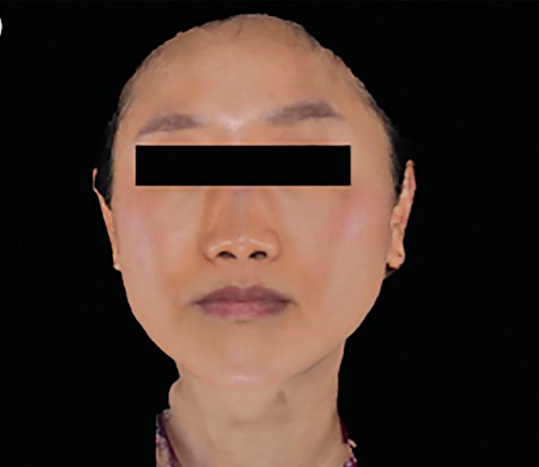

wrinkles, tear trough volume loss, neck wrinkles, and irregular pigmentation. Additionally, it can target specific skin concerns such as stretch marks and acne scars.
There was no study on injectable PDLLA targeting the entire face. Recently, the manufacturer of this innovative hybrid bio-stimulator, VAIM Global, has published clinical research performing a full-face dermis injection, observed full-face skin rejuvenation, and assessed the efficacy and safety of injectable PDLLA in participants with skin ageing changes.


The procedure was performed on 16 patients (All Asian, average age 50.6 years, 15 women and 1 man). The injecting solution was prepared by resolving PDLLA/HA (PDLLA 42.5 mg + HA 7.5 mg; Juvelook, VAIM co. LTD.) in a 10 mL mixture of non-cross-linked HA (2.5 ml), 1% lidocaine hydrochloride hydrate (2.5 mL), and normal saline (5 mL) mixture at a 1:1:2 ratio. All 16 patients received 2–3 sessions with 4-week intervals between sessions.
Therapeutic effects were evaluated by measuring five ageing skin signs using a 10-point scale by two dermatologists who did not participate in the treatment before the first treatment session and at follow-up visits (12 weeks or 16 weeks after the final treatment). At follow-up visits, patients were also asked about their overall improvement based on the following scoring method.
■ No improvement
■ 1%–24% improvement
■ 25%–49% improvement
■ 50%–74% improvement
■ 75%–100% improvement.
Overall, statistically significant differences were observed in all signs of skin, such as fine wrinkles, skin texture, irregular pigmentation, telangiectasia, and facial erythema before and after the treatment.
There were relatively minimal differences in treatment outcomes of facial erythema and telangiectasia. However, the participants in the following picture showed remarkable improvement in facial erythema in both cheeks (Figure 1) and skin contouring effect (Figure 2), especially in both mandible areas.
In some patients, remarkable improvement in skin texture and irregular pigmentation was observed in the forehead, cheeks, and chin. In
the context of improving uneven pigmentation, there are conditions, such as lentigines or melasma, which are associated with photoaging. The alteration of the dermal matrix due to photoaging plays a significant role, and improving the dermal environment through PDLLA injection contributed to the amelioration of irregular pigmentation.
No serious adverse events were identified throughout the experiment except temporary procedural pain and facial erythema. The duration of temporary pain reported by the patients disappeared immediately after the procedure.
Recently, VAIM Global discovered two mechanisms of Juvelook’s collagen production, and they are believed to be attributed to the effects of the abovementioned procedure. Those mechanisms make Juvelook stand out among the other bio-stimulators or polymer fillers.
VAIM Global has first shown that PDLLA, PN, and CaHA can result in angiogenesis, thereby contributing to the rejuvenation of aged skin.
Polymer fillers such as PN and CaHA achieve angiogenesis by upregulating proangiogenic factors such as HSP90, HIF-1α, and VEGF. These growth factors play a crucial role in stimulating angiogenesis and promoting rejuvenation in the skin and multiple organs. Notably, when compared to other polymer fillers like PN or CaHA, PDLLA exhibits the highest level of upregulation of these factors. This finding suggests that PDLLA may effectively contribute to the rejuvenation of aged skin by promoting angiogenesis.
Additionally, PDLLA demonstrates increased expression of type 1 collagen in the skin by reducing reactive oxidative species (ROS) and increasing TGF-β. This signifies PDLLA’s superior effectiveness in skin rejuvenation and tissue regeneration.
“PDLLA demonstrates increased expression of type 1 collagen in the skin by reducing reactive oxidative species (ROS) and increasing TGF- β . This signifies PDLLA’s superior effectiveness in skin rejuvenation and tissue regeneration.”
Furthermore, dermal thickness was reduced in aged mice and increased post-injection, with PDLLA exhibiting the most substantial effect. Skin elasticity, lower in aged mice, improved after injections, with PDLLA again demonstrating the most significant impact.
The key lies within the unique structure of PDLLA particles

The microparticles in PLA dermal filler exhibit additional biological effects that trigger collagen synthesis. Following the injection of PLA filler, immune cells, including macrophages, lymphocytes, and giant cells, envelop the particles, leading to a mild inflammatory reaction. PDLLA represents a chiral variant of PLLA, sharing the same chemical formula but generating different physical and chemical properties. With the patented technologies of VAIM Global, PDLLA microparticles have distinct shapes from usual PLA particles. PLLA particles take on irregular solid structures, while PDLLA microspheres adopt a sponge-like sphere formation with crystallization close to 0%. The interior reticular structures of PDLLA microspheres stimulate the synthesis of collagen I and III and function as threedimensional scaffolds, maximising the contact surface area involved in neo-tissue generation.
ECM materials enter the particles through the surface pores of PDLLA, binding to the reticular structure beneath. As the internal structure gradually decomposes into CO2 and

water, promoting new collagen formation in the area, the outer surface of PDLLA remains undamaged, preserving the inner structure. This protective mechanism enables the extended development of collagen and elastin fibres.
For more information, visit https://juvelook.com
 MARKO MANCE MD, UNIV. MAG. PHD, Skin Health Lab, Zagreb, Croatia
SKIN AGEING IS A complex biological process characterised by the gradual loss of structural integrity, elasticity, and hydration, leading to the formation of wrinkles, sagging, and volume depletion1. This occurs due to multiple simultaneous processes including2,3:
Oxidative stress
Reactive oxygen species (ROS), generated through normal cellular metabolism and external factors like UV radiation and pollution, contribute to oxidative stress. ROS damage cellular components, including lipids, proteins, and DNA, leading to impaired cellular function and accelerated ageing.
DNA damage and repair
UV radiation and other environmental factors induce DNA damage in skin cells. Activation of DNA repair mechanisms, such as nucleotide excision repair and base excision repair, is critical for maintaining genomic integrity and preventing mutagenesis.
Inflammation
Chronic inflammation, triggered by factors like UV exposure and environmental pollutants, contributes to skin ageing. Pro-inflammatory cytokines, such as interleukin-1 (IL-1) and tumor necrosis factor-alpha (TNF-α), activate signaling pathways like nuclear factor kappa B (NF-κB), leading to the production of matrix
metalloproteinases (MMPs) and degradation of extracellular matrix (ECM) components.
Extracellular matrix (ECM) degradation
ECM proteins, including collagen, elastin, and glycosaminoglycans (GAGs), provide structural support and elasticity to the skin. MMPs, enzymes upregulated by factors like UV radiation and inflammation, degrade ECM proteins, leading to loss of skin firmness, wrinkles, and sagging.
Decline in collagen and elastin synthesis
Fibroblasts are the primary cells responsible for producing collagen and elastin in the skin.
Ageing and external factors like UV exposure inhibit collagen and elastin synthesis while increasing their degradation, resulting in decreased skin firmness and elasticity.
Glycation
Glycation, the non-enzymatic reaction between reducing sugars and proteins, forms advanced glycation end-products (AGEs) in the skin. AGEs cross-link with collagen and elastin fibres, impairing their function and contributing to skin stiffness and loss of elasticity.
Decline in hyaluronic acid (HA) levels
HA, a glycosaminoglycan abundant in the skin, plays a crucial role in maintaining hydration and tissue integrity. Ageing and oxidative stress
lead to a decrease in HA synthesis and an increase in its degradation, resulting in reduced skin hydration and volume.
Telomere shortening
Telomeres, repetitive DNA sequences at the ends of chromosomes, protect genomic integrity by preventing chromosome degradation and fusion. Telomere shortening, occurring with each cell division, leads to cellular senescence and eventual apoptosis, contributing to skin ageing.
Dysregulation of autophagy
Autophagy, a cellular process responsible for degrading damaged organelles and proteins, declines with ageing. Impaired autophagy leads to the accumulation of damaged cellular components, oxidative stress, and inflammation, exacerbating skin ageing.
Hormonal changes
Hormonal fluctuations, such as a decline in oestrogen levels during menopause, affect skin structure and function. Oestrogen depletion leads to decreased collagen synthesis, loss of skin thickness, and increased wrinkle formation. Understanding these intricate biochemical processes involved in skin ageing is crucial for developing effective anti-ageing strategies and interventions aimed at maintaining skin health and vitality.
Repair and rebuild key skin components
Injectable treatments, such as
Sunekos 1200 and Sunekos Performa, have emerged as effective interventions for addressing ageing skin concerns by restoring dermal homeostasis leading to the repair and regeneration of key ECM and subcutaneous tissue components. While both products share similarities in composition, they exhibit distinct differences in concentration and ratios between hyaluronic acid vs amino acids, their mechanism of action, clinical indications, treatment protocols, and safety profiles4
Sunekos 1200 and Sunekos Performa are composed of hyaluronic acid, a glycosaminoglycan crucial for skin hydration and volume, alongside a patented formula combining 6 amino acids (Glycine, L-Alanine, L-Proline, L-Valine, L-leucine and L-Lysine) specifically tailored to act as antioxidants and to stimulate fibroblast activity respectively. The benefit of HA includes fibroblast activation and subsequent increased production of key ECM components including, collagen I, III, IV and VII, elastin and fibronectin synthesis. Also there is a 40% decrease in hyaluronidase degradation due to the repelling forces generated by the positively charged AA, L-lysine and the oily hydrophobic components of L-Alanine, L-Valine and L-Leucine. This means that the HA found in both Sunekos 1200 and Sunekos Performa have a longer duration of action on fibroblast receptors compared to products that are HA only or with a different mix in percentage of amino acids. In fact, the HA+6AA formula increased elastin and collagen synthesis significantly more compared to other biostimulatory

products including peptides and collagen. When compared to a control group, there was a 124% and 60% increase in elastin and fibronectin, respectively4,5
Sunekos 1200
Comprised of high molecular weight HA (1000–1500 kDa); Sunekos 1200 acts as an antioxidant and antiinflammatory when injected by protecting fibroblasts and structural elements of the dermis since it acts as a free radical ‘scavenger’ improving the subcutaneous homeostasis4
Sunekos Performa
Comprised of low molecular weight HA (up to 200kDa) to regulate ECM homeostasis, leading to more robust collagen-stimulating effects and superior improvements in skin texture, elasticity, and firmness. The precise balance of HA and 6 amino acids maximise its efficacy by hydrating/retaining water and increasing the synthesis of structural elements as collagens, elastin, fibronectin through fibroblast stimulation4

0.1 ml
0.03 ml
0.02 ml - optional
Sunekos 1200 and Sunekos Performa are indicated in young patients as a preventative treatment against the early signs of ageing as well as in older patients who present advanced signs of ageing, including deep wrinkles, severe skin laxity, photoaging, solar induced pigmentation and volume depletion. Sunekos 1200 is indicated for facial and hand treatments only, while Sunekos Performa is indicated for face, neck, decolletage and hand treatments. Treatment protocols
1 point just below the chin

Standard points
Optional points (0.3 ml each with needle)
Optional point 2 (with needle)
0.3 ml - 0.6 ml (Cannula)
0.3 ml (Needle)
Access point cannula
Direction cannula
Treatment included 3 ml of Sunekos 1200 and 3.5 ml of Sunekos Performa in injection sites including malar, mandibular, chin (Sunekos 1200 and Sunekos Performa), periocular and frontal regions (Sunekos Performa only)

for Sunekos Performa involve more sessions and higher injection volumes due to its regenerative action compared to Sunekos 1200, which is used for tissue repair and antioxidant activity in the first stage of facial treatment only4 (Figure 1).
Both Sunekos 1200 and Sunekos Performa have demonstrated excellent safety profiles in clinical studies, with minimal risk of adverse reactions or complications. Adverse events, such as transient swelling, bruising, or erythema at the injection site, are rare and typically resolve spontaneously. However, meticulous adherence to recommended
1. Trojahn C, Dobos G, Lichterfeld A, Blume-Peytavi U, Kottner J. Characterizing facial skin ageing in humans: disentangling extrinsic from intrinsic biological phenomena. Biomed Res Int. 2015;2015:318586. doi: 10.1155/2015/318586. Epub 2015 Feb 12. PMID: 25767806; PMCID: PMC4341846.

injection techniques and patient selection criteria is essential to mitigate potential risks4,6
The following case is a 35 year old female patient who presented to our clinic with complaints of looking tired and poor skin. She was treated using the high performance technique protocol4. The first treatment included 3 ml of Sunekos 1200 and 3.5 ml of Sunekos Performa in typical injection locations including malar, mandibular, chin (Sunekos 1200 and Sunekos Performa), periocular and frontal regions (Sunekos Performa only) (Figure 2). The second and third sessions were given 10 days
2. Boismal F, Serror K, Dobos G, Zuelgaray E, Bensussan A, Michel L. Vieillissement cutanéPhysiopathologie et thérapies innovantes [Skin aging: Pathophysiology and innovative therapies]. Med Sci (Paris). 2020 Dec;36(12):1163-1172. French. doi: 10.1051/medsci/2020232. Epub 2020 Dec 9. PMID: 33296633.
apart and included injections of 3.5 ml Sunekos Performa in the above mentioned locations according to the protocol suggested by Professional Dietetics.
Photographs were taken before the treatment protocol and 6 weeks after the final treatment (Figure 3 ). We see a marked clinical improvement in skin laxity, facial shape, skin hydration, periocular dis-colouration, reduced fine wrinkles, and a younger overall appearance.
Sunekos 1200 and Sunekos Performa represent valuable options in aesthetic medicine for skin rejuvenation, offering tailored approaches to address various degrees of skin ageing. While both products share a common objective of stimulating collagen and elastin production, their differences in composition, concentration, mechanism of action, clinical indications, treatment protocols, and potency enable clinicians to customise treatment plans according to individual patient needs. By leveraging the most detailed scientific evidence available, clinicians can make informed decisions to achieve optimal aesthetic outcomes in clinical practice.
For more information, visit www.professionaldietetics.com
3. Tobin DJ. Introduction to skin aging. J Tissue Viability. 2017 Feb;26(1):37-46. doi: 10.1016/j. jtv.2016.03.002. Epub 2016 Mar 14. PMID: 27020864.
4. Fasola E. 2023. Sunekos Injectable and the High Performance Technique Counteracting Skin Aging. Prime Journal: Aesthetic adn Anti-aging medicine. Accessed: 25,February,2023. [https://issuu. com/imaesthetics/docs/prime_mar_ apr_2023].
5. D’Agostino A, Stellavato A, Corsuto L, Diana P, Filosa R, La Gatta A, De Rosa M, Schiraldi C. Is molecular size a discriminating factor in hyaluronan interaction with human cells? Carbohydr Polym. 2017 Feb 10;157:21-30. doi: 10.1016/j.carbpol.2016.07.125. Epub 2016 Sep 5. PMID: 27987920.
6. Nandi R. 2023. Sunekos Performa, The Enhanced Formula for Skin Renovation. Prime Journal: Aesthetic and Anti-aging medicine. Accessed:25,February,2023. [https:// issuu.com/imaesthetics/docs/ prime_mar_apr_2023].


27–29 MARCH 2024
AMWC 2024 Monte Carlo, Monaco www.euromedicom.com
16–18 MAY 2024
EADV Symposium St Julian’s, Malta eadv.org/symposium
30 MAY – 1 JUNE 2024


2–4 JULY 2024
Annual Meeting of the British Association of Dermatologists Manchester, UK badannualmeeting.co.uk
12–13 JULY 2024
FACE 2024 London, UK www.faceconference.com
26–27 SEPTEMBER 2024
Beauty Through Science 2024 Stockholm, Sweden www.btsstockholm.com
13–16 JUNE 2024
5-CC World Congress 2024 Lisbon, Portugal www.5-cc.com
BAAPS 2024 London, UK baaps.org.uk/
25–29 SEPTEMBER 2024
EADV 2024
Amsterdam, Netherlands eadv.org
30 MAY – 1 JUNE 2024
Vegas Cosmetic Surgery Las Vegas, NV www.vegascosmeticsurgery.com
27–30 JUNE 2024
The Aesthetic Show Las Vegas, NV www.aestheticshow.com/en/home.html
1–4 AUGUST 2024
AAD Innovation Academy Seattle, Washington www.aad.org
24–26 MAY 2024
AMWC ASIA TDAC Taipei, Taiwan www.amwc-asia.com
24–25 AUGUST 2024
AMWC India 2024 Mumbai, India www.amwcindia.com/
18–20 OCTOBER 2024
AMWC China Chengdu, China en.amwcchina.com/

BUSINESS DESIGN CENTRE, LONDON, UNITED KINGDOM

1 2 - 1 3 JULY 2024
WWW.FACECONFERENCE.COM

Under the scientific supervision of the Aesthetic Multispecialty Society (AMS)

