International Journal of Science and Research (IJSR), India Online ISSN: 2319-7064
Detection of Cysts in Ultrasonic Images of Ovary Ashika Raj M. Tech, Computer Science & Engineering, Calicut University, Calicut, India
Abstract: In this paper, cysts are detected in the ultrasonic images of ovary. PCOS is an endocrine disorder affecting women of reproductive age. This syndrome is mainly seen in women whose age is in between 25 and 35. We are proposing methods for identifying whether a person is suffering from Polycystic Ovary Syndrome (PCOS) or not. Ultrasound imaging of the follicles gives important information about the size, number and mode of arrangement of follicles, position and response to hormonal stimulation. A thresholding function is applied for denoising the image in the wavelet domain. Before the segmentation process the ultrasonic image is preprocessed using contrast enhancement technique. Morphological approach is used for implementing contrast enhancement. This is performed in order to improve the clarity and quality of the image. Fuzzy c-means clustering algorithm is applied to the resultant image. Finally the cysts are detected with the help of clusters. The efficiency of the algorithm depends upon the value of Mean Square Error (MSE) and Peak Signal to Noise Ratio (PSNR). Keywords: Denoising, Contrast Enhancement, Morphological Operations, Segmentation, Fuzzy C-Means Clustering.
1. Introduction Cysts are follicles which have abnormal size. Based on the detection of follicles, the suspected patient can be treated as normal or polycystic. Ovarian follicles are roughly spherical, fluid-filled structures in which oocytes (eggs) develop. Follicles imaged in two dimensions appear as dark, roughly circular regions in ultrasound images since fluid does not reflect ultrasonographic pulses. PCO ovaries typically exhibit a larger number of smaller, possibly irregularly shaped follicles, and a peripheral distribution of follicles. The ultrasonographic morphology of a polycystic ovary (PCO) is characterized by the presence of 12 or more ovarian follicles which are 2-9 mm in size. These follicles are termed as cysts. The symptoms of PCOS are menstrual irregularity, obesity, hyperandrogenism, diabetes, acne, increased risk of cardiovascular disease, male-pattern facial and bodily hair growth and balding, excessive production of male hormones, infertility etc. An automatic detection of cysts for a PCOS patient is implemented using fuzzy c-means clustering. Before performing this algorithm we are denoising and contrast enhancing the ultrasonic image which is given as the input. Diagnostic ultrasound uses frequency between 2 and 15 MHz. Ultrasonic waves are produced from the transducer and penetrates into the body tissues and when the wave reaches an object or a surface with different texture or acoustic nature, some fraction of the energy is reflected back. The echoes so produced are received by the apparatus and changed into electric current. These signals are then amplified and processed to get displayed on CRT (Cathode Ray Tube) monitor. The image so obtained is called ultrasound scan and the process is known as ultrasonogram. This image is given as the input. Ultrasound imaging technique is inexpensive and is very effective for cyst recognition. The overall quality of the ultrasound image is the end product of a combination of many factors originating from the imaging system and the performance of the operator. Ultrasonic image may contain noises due to loss of proper contact or air gap between transducer and body part. Noises can also be formed during beam forming process or signal processing. The noises may cause the image blurred and thereby lead
to poor segmentation. Hence we are performing denoising. A soft thresholding function is proposed for the process of removing noise. Contrast enhancement increases the appearance of largescale light-dark transitions of an image. The edges of the image become clearer. This process automatically brightens images that appear dark or blurred and applies appropriate correction to deliver optimal quality and clarity. Morphological operations are performed for contrast enhancement technique. Fuzzy c-means clustering algorithm is applied for the segmentation of follicles from the ultrasonic image. The purpose of clustering is to identify natural groupings of data from a large dataset to produce a concise representation of a system’s behavior. The follicles are detected inside the ovary and the obtained results are compared with the manual results.
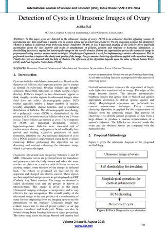
2. Proposed Methodology Figure 1 gives the schematic diagram of the proposed methodology.
Figure 1: Block Diagram for cyst detection
Volume 2 Issue 8, August 2013 www.ijsr.net
185