10 XI November 2022 https://doi.org/10.22214/ijraset.2022.47386
ISSN: 2321 9653; IC Value: 45.98; SJ Impact Factor: 7.538

Volume 10 Issue XI Nov 2022 Available at www.ijraset.com

ISSN: 2321 9653; IC Value: 45.98; SJ Impact Factor: 7.538
Volume 10 Issue XI Nov 2022 Available at www.ijraset.com
M. M. Kalpashree1 , G. P. Vyshali2 , Preetha Florina3 , Dr. K. Krishna4

1Research Scholar,2 M.SC, Student, 3 Research Scholar,4 Professor, Post Graduate Department of Botany, Yuvaraja’s College, University of Mysuru, Mysuru,570005


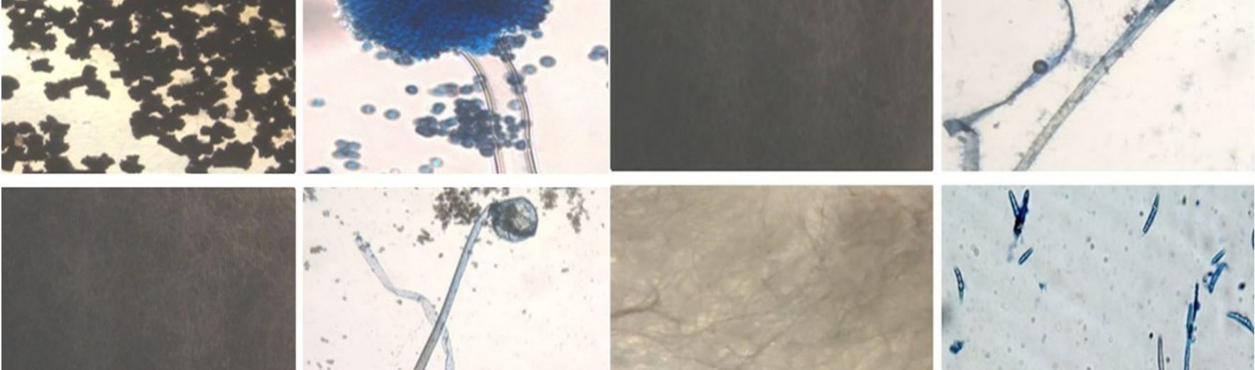
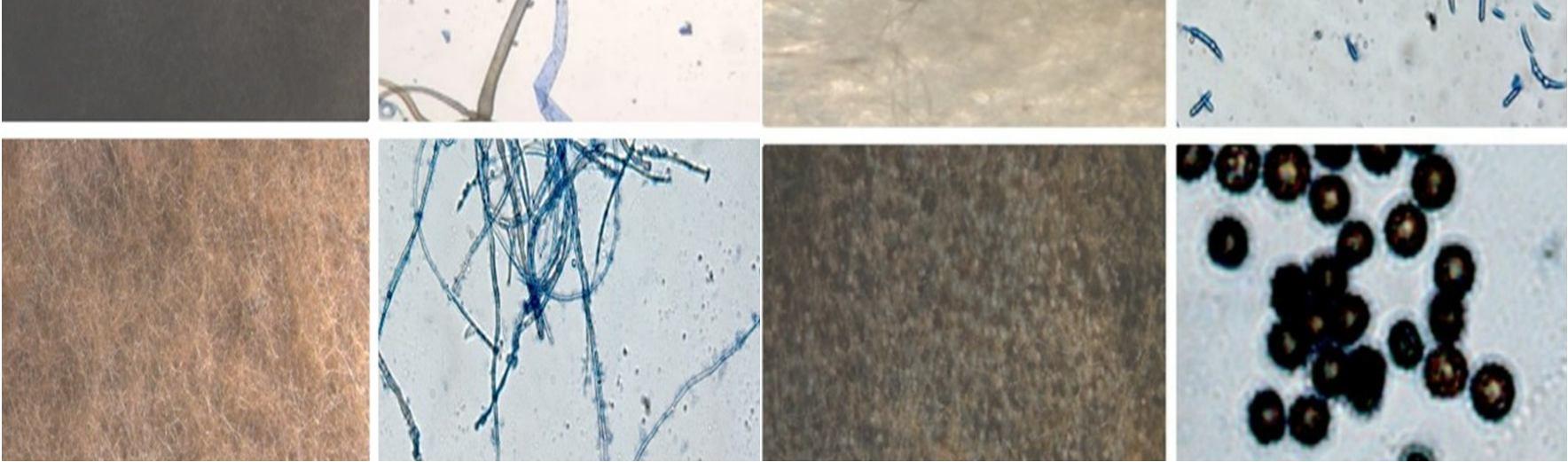
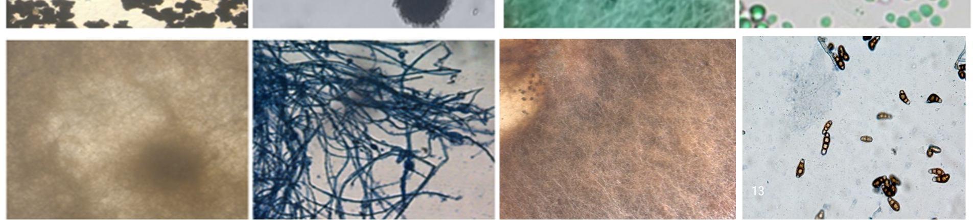
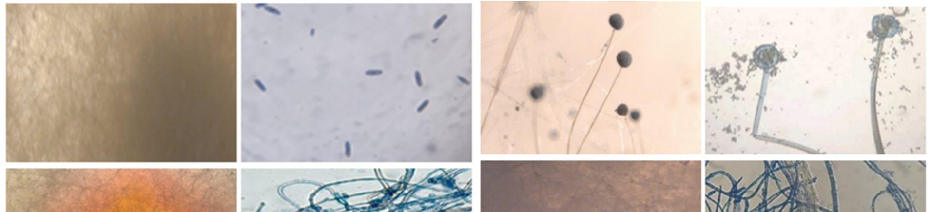
Abstract: The present study focused on the isolation and identification of fungal endophytes from Talinum fruticosum. Endophytic fungi live inside the higher plants apparently without causing harm. Talinum fruticosum has medicinal value and is a good source of vitamins A and C, an excellent source of calcium and phosphorus. Leaves and stems of Talinum fruticosum were evaluated to isolate endophytic fungi from Talinum fruticosum collected from different regions of Mysuru, Karnataka, India. All isolates of fungal species were identified based on morphological characteristics, mycelium, asexual spores, and sexual spores observe under the compound microscope. 20 species were isolated from leaves, and 5 fungal species were isolated and identify fungal species from leaves are Rhizopus sp, Aspergillus sp, Penicillium sp, Rhizopus stolonifera, Aspergillus sp, Fusarium sp, and sterile hyphae. 3 fungal species were unidentified. And 20 species were isolated from the stems and 6 fungal species were isolated and identify the fungal species from stems were Fusarium sp, Curvalaria sp, Aspergillus sp, Rhizopus stolonifera, Colletotrichum sp, Trichoderma sp, Sterile hyphae. 5 fungal species were unidentified.
Keywords: Endophytic fungi, Talinum fruticosum, Leaves, and stems, Isolation, and Identification

These endophytic fungi play an important physiological and ecological role in the life of the host The term “ENDOPHYTE” was introduced by De Bary and was initially applied to any organism found within plant tissues without any symptoms of the disease[1][2]. They have indicated the presence of one or more types of endophytes in every plant studied. Endophytic fungi are defined as fungi that complete part or all of their life cycle living naturally in the plant tissues without causing any disease symptoms. Endophytic fungi produce some of the most widely used antibiotic and anticancer drugs [3] The presence of these endophytic fungi increases the plant’s natural defense system against both biotic and abiotic stress. In the present study, Isolated and identification of endophytic fungi of Talinum fruticosum. The plant is a member of the Talinaceae family. Talinum fruticosum is an herbaceous, perennial plant that is native to Mexico, west Africa, Central America, etc. It is widely grown in tropical regions as a leaf vegetable as it is rich in vitamins (vit a and c), and minerals such as iron and calcium. This green leafy vegetable is recommended on a daily diet due to its aid in the easy digestion of food. The common name is waterleaf it is scientifically classified as a weed plant. It carried out a phytochemical analysis of Talinum triangulare in other to ascertain its medicinal properties. Endophytic fungi are present in living tissues of various parts i.e., in the root, stem, leaf, flowers, and seed [4]. The endophytic fungi complete their life cycle in this plant without causing any visible manifestation of disease.
Healthy leaves and stem segments of Talinum fruticosum were collected from different regions of Mysuru Karnataka, India. The plant material were brought to the laboratory in sterile bags and processed immediately to reduce the chances of contamination.
To enumerate and observe of sporulation of endophytic fungi. Suspend 3.00 grams of agar in 100 ml distilled water with 0.5 grams of sucrose and streptomycin 0.5 µg/ml into water agar media. Sterilize by autoclaving at 15 lbs pressure (121 ̊c) for 15 minutes.40 leaves and stem pieces[1cm] are placed in particulates of 2% water agar media. The fungal colony growth was examined under a stereo binocular microscope on the 7th day.
ISSN: 2321 9653; IC Value: 45.98; SJ Impact Factor: 7.538 Volume 10 Issue XI Nov 2022 Available at www.ijraset.com
Potato dextrose agar (PDA) is used for the cultivation of fungi or enumerate the fungi. 20 grams of potato dextrose agar was taken in 500 ml of distilled water and 2 grams of sucrose added followed by antibiotics into PDA containing media Bring to a boil to completely dissolve the medium. Sterilize by autoclaving at 15 lbs pressure (121 ̊c) for 15 minutes. In sterile condition pour the media into Petri dishes (i.e., subculture) then place to solidify. 40 leaves and stem segments approximately cut around 1 cm are placed in Petri plates under the sterilized condition. After placing the leaves and stems segments of Petri dishes. The fungal colony growth was examined under a stereo microscope and binocular microscope for 10 days.
Isolation of endophytic fungi was done [4] with slight modifications. The plant materials were taken and first rinsed in running tap water to remove the dust and the other debris present in them. Segments of approximately 1 cm were cut in sterile lancet blades and surface sterilized by agitating in 70% ethanol (5 s), followed by treatment with 0.1% sodium hypochloride (90 s)and then rinsed in sterile distilled water (10 s) 40 ( 20 leaves, 20 stems) segments from the Talinum fruticosum plant are processed for the isolation of endophytic fungi. The leaf and stem segments were placed into water agar media for the enumeration of fungi inoculated at room temperature for 7 days. leaf and stem segments were then placed onto potato dextrose agar (PDA) amended with. The one tour weeks pure cultures of the fungal isolates were identified using culture and morphological features such as a model of colony growth, morphology and pigmentation conidia , using by slide culture techniques . The Petri dishes were sealed using cling film paper and incubated at room temperature for 4 5 days. The fungi that grew out from the tissues were isolated.
The endophytic fungal isolates were identified up to the genus level based on morphological features such as colony morphology, pigmentation, growth pattern, spore structures, and other Characteristics of hyphae using the help of the standard mycological manuals. A microscopic examination was also done to study their reproductive spores [5].
To know the endophyte richness, the frequency of fungal endophytes in plant species was calculated by the number of segments colonized by endophyte species divided by the total number of segments examined into 100 [6]. Colonization frequency[CF%]:
CF%= Noofindividualfungirecorded Totalnumberofsegmentsexaminedₓ100
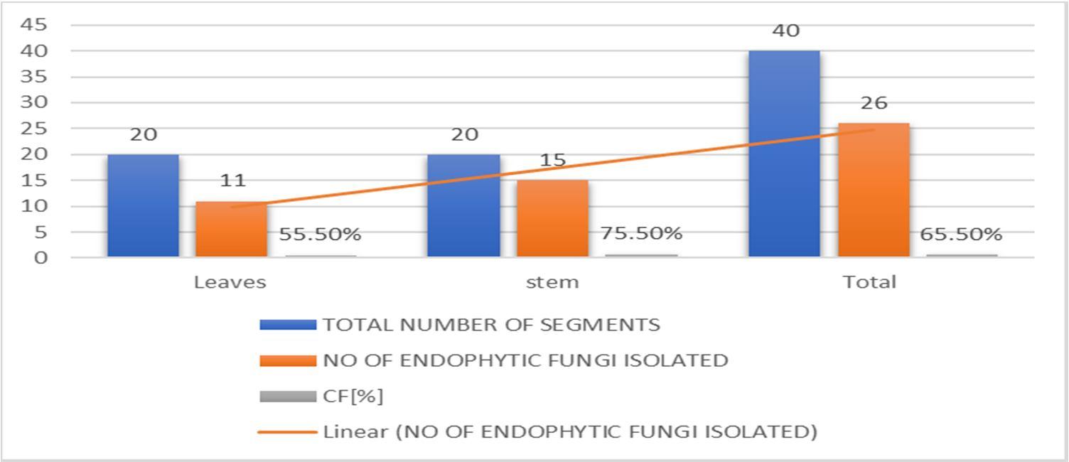
About 40 segments (20 segments of leaf and 20 segments of stem part respectively) of the plant were placed for the isolation of endophytic fungi It was observed that more endophytic fungi were from stems (75.5%) as compared to the leaves(55.5%).A total of 40 endophytic fungi were isolated and identification from the plant Talinum fruticosum (Table 1) .The leaf segments showed a maximum repository for endophytic fungi than the other segments. Among the 26 endophytic fungi, the predominant endophytic fungi isolated and identified The remaining 8 fungal species were unidentified. The predominant endophytic fungi belonged to the genera of Rhizopus sp, Aspergillus sp, Alternaria sp, Penicillium sp, Curvalaria sp, Fusarium sp, and Colletotrichum sp and Rhizopus stolonifera
Tables 1,2and 3 showed the CF value and taxonomic position of endophytic fungi.
Table 1: Endophytic fungi isolated from the plant, Talinum fruticosum (L.) Juss.
SITE OF LOCATION TOTAL NUMBER OF SEGMENTS
NO OF ENDOPHYTIC FUNGI ISOLATED
CF[%]
Leaves 20 11 55.5% stem 20 15 75.5%
Total 40 26 65.5%
ISSN: 2321 9653; IC Value: 45.98; SJ Impact Factor: 7.538 Volume 10 Issue XI Nov 2022 Available at www.ijraset.com
Graph 1:Percentage of endophytic fungi from leaves and stems of Talinum fruticosum (L.) Juss
A. Isolation and identification of endophytic fungi from leaves of Talinum fruticosum (l.) Juss.
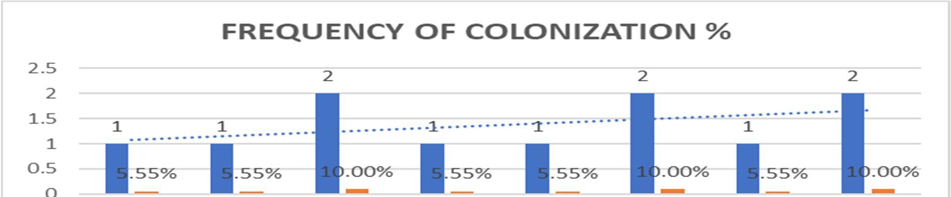
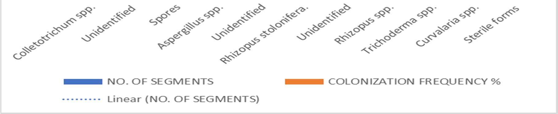
Table 2: Colonization Frequency (%) of Endophytic Fungi Isolated from Leaves of Talinum fruticosum (L.) Juss.
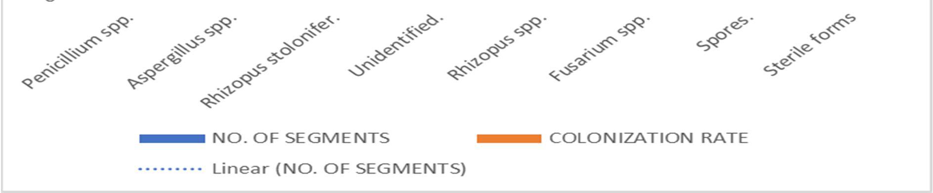
SL.NO. ISOLATED ENDOPHYTES FUNGAL CLASS NO. OF SEGMENTS COLONIZATION RATE
1. Penicillium spp Eurotiomycetes 1 5.55%
2. Aspergillus spp. Ascomycetes 1 5.55%
3. Rhizopus stolonifer. Zygomycetes 2 10.00%
4. Unidentified. 1 5.55%
5. Rhizopus spp. Zygomycetes 1 5.55%
6. Fusarium spp Hyphomycetes 2 10.00%
7. Spores. 1 5.55% 8. Sterile forms 2 10.00% TOTAL 11 55.5%
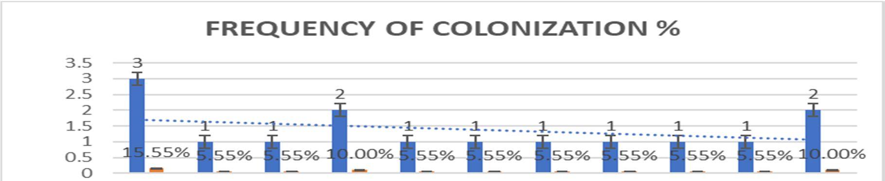
Graph 2:Colonizing frequency [%] of endophytic fungi isolated from leaves of Talinum fruticosum(L.) Juss.


ISSN: 2321 9653; IC Value: 45.98; SJ Impact Factor: 7.538 Volume 10 Issue XI Nov 2022 Available at www.ijraset.com


ISSN: 2321 9653; IC Value: 45.98; SJ Impact Factor: 7.538 Volume 10 Issue XI Nov 2022 Available at www.ijraset.com
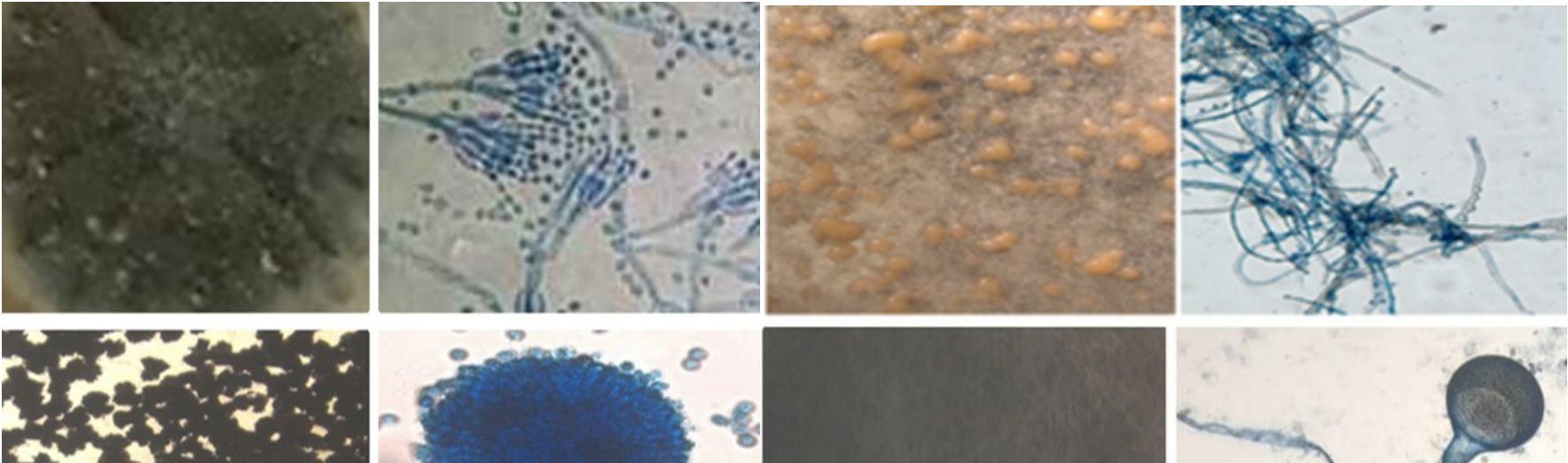
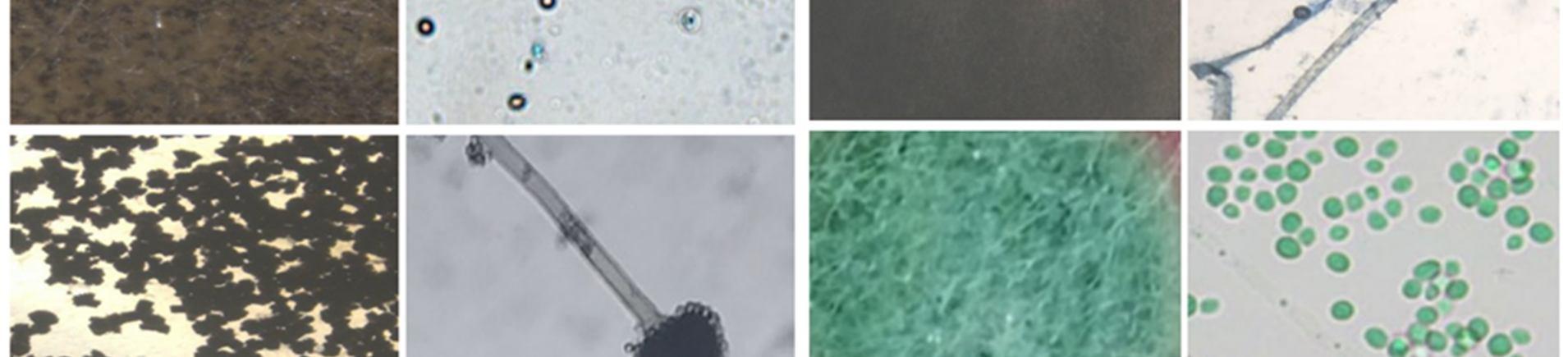
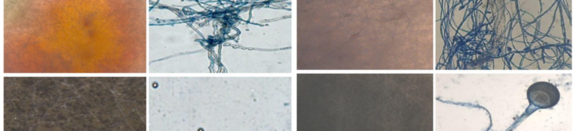
This study was carried out to isolate and identify of endophytic fungi from Talinum fruticosum collected from different regions of mysuru. Identification of fungi was done based on the morphology characteristics of the fungi growing on the culture medium (PDA). The morphology includes macroscopic and microscopic characteristics. The microscopic identification of colonies such as color, and colony growth the observation was done for seven days during the fungal culturing. The macroscopic identification of the colony such as color, diameter, colony growth, and colony reverse, observation was done for seven days during the fungal culturing. the microscopic characterization was done by observing the shape and size of conidia, and hyphae.
Observation of conidia including its arrangements (singular, in a chain, or in a cluster), the number of cells (single celled or multicellular), and conidial measurement. Observation of hyphae was also performed on the presence or absence of septa in the hypha, its shape, morphology (nodular, pectinate, spiral, rhizoid, chandler), and its modifications of hyphae (rhizoid, stolon, rhizomorph, haustorium, appressorium, chlamydospore, sclerotia). The results obtained were then compared with the literature and monographs.
A total of 26 fungal colonies were isolated from 40 segments. It was observed that more endophytic fungi were from stems (75.5%) as compared to the leaves(55.5%) During this study, 9 a total of different endophytic fungi were found in leaves and stems. Among these Colletotrichum spp (15.55%), and Aspergillus spp (13.33%) showed the highest colonizing frequency followed by leaves and stems. For leaves colonizing frequency is Penicillium spp(6.66%), Aspergillus spp(13.33%), Rhizopus stolonifer (6.66%), Rhizopus spp (6.66%), Fusarium spp (6.66%), Alternaria spp (6.66%) [7]. For stems colonizing frequency is Collectrichum spp (15 55%) and Aspergillus spp (6.66%) Rhizopus stolonifera (6.66%), Rhizopus spp (6.66%), Trichoderma spp(6.66%), Curvalaria spp(6.66%) and sterile forms (13.33%) [8]. The presence of endophytic fungi in a plant depends on the species of plant, environment, and isolation methods. The amount of endophytic fungi isolates that are found depends on the diversity and distribution of endophytic fungi that exist in the host plants. Each part of the plant will give a different number of endophytic fungal isolates[9]
Endophytic fungi have wide applications in different fields. It has wide applications in different fields. It had the potential to produce many bioactive compounds. The secondary metabolites produced by the endophytic fungi can act as biocontrol. Endophytic fungi is a very complex physical and physiological association with the host plant influencing many biological activities. Talinum fruticosum was widely grown in tropical regions as a leaf vegetable as it is rich in vitamins (via a and c), and minerals such as iron and calcium. Endophytic fungi produce some of the most widely used antibiotic and anticancer drugs. A total of 26 fungal colonies were isolated from 40 segments. It was observed that more endophytic fungi were from stems (75.5%) as compared to the leaves(55.5%). Endophytic fungi live inside plant tissues but do not cause any disease symptoms. Endophytes are organisms, often fungi and bacteria, that live between living plant cells. The relationship that they establish with the plant varies from symbiotic to bordering on pathogenic. in the future, the products from the endophytic fungi will be a cheap source for medical agriculture and other industries. The endophytic fungi enhance the plant’s resistance to biotic and abiotic stresses. Plant associated fungi (endophytic fungi) are a biodiversity rich group of microorganisms that are normally found asymptomatically within plant tissues or in intercellular spaces.
[1] Wilson , D. (1995). Endophyte the evolution of a term , A clarification of its use and definitions.Oikos,73 :274 276.
[2] De Bary A.(1886). Morphologie and Physiologie Pilze, Flechten, and myxomycete.Hofmeister’s Handbook of Physiological Botany .Leipzig,p.2.
[3] Strobel , G And Daisy B. Bioprospecting For Microbial Endophytes And The Natural Products. Microbiol Molecular Biology Rev. 2003;67:491 502.
[4] Sandhu,S S,Suneel ,K,Aharwal RP,Shukla H And Rajak Rc. Endophytic Fungi: As A Source Of Antimicrobials Bioactive Compounds.World J Pharmac Sci 2014a:3(2):1179 1197.
[5] Petrini,O. Taxonomy of endophytic fungi of aerial plant tissue in Fokkema, N.J and van. Den UK.1986;pp.175 187.
[6] Anitha ,D, Vijaya T, Pragathi D Et Al., Isolation and Characterization of Endophytic Fungi from Endemic Medicinal Plants of Tirumala Hills, Int J Life Science Biotechnol Pharm Res 2013;2(3):367 73.
[7] Divya, C R, Sharada, M S, Ashwini ,V. Isolation and Identification of Endophytic Fungi in Trigonella Foenum Graceum L.And Their Antibacterial Activity.Imp J Interdiscip Res 2016;516 21.
[8] Dhanalakshmi, R, Umamaheshwari ,S, Sugandhi ,P, Prasanth D.A. Biodiversity of the endophytic fungi isolated from Moringa oleifera of Yercaud hills, Int J Pharm Sci Res 2013: 4(3): 1064 8.
[9] Job ,N, Manami S, Philip R.,Isolation And Characteracterization Of Endophytic Fungi From Avicennia officinalis.Int J Res Biomed Biotechnology 2015; 4 8.
[10] Stone, J.K., Polishook J.D., And White J.F. 2004. Endophytic Fungi. In Biodiversity of Fungi .Inventory and Monitoring methods .In Mueller G,M.,Bills G.F., Foster M.S.(Eds) Elsevier Academic Press , San Diego, United state of America. 241 270.