10 IX September 2022 https://doi.org/10.22214/ijraset.2022.46795
ISSN:
9653; IC Value:
ISSN:
9653; IC Value:
Chandana. N1, M. V. Ramana2 , Ghanta Pushpa Chakrapani3
1 3Department of Physics, Dr.B.R.Ambedkar Open University, Hyderabad, Telangana, India

1Department of Physics, Government Degree college, Khairatabad, Hyderabad, Telangana, India
2Department of Physics, Anurag University, Hyderabad, Telangana, India
Abstract: We report the synthesis of co doped Calcium tungstate (CaWO4) nano crystals with Gadolinium (Gd) and Neodymium (Nd) rare earth ions by hydrothermal synthesis method for the first time. The synthesized nano-crystals were characterized by Xray diffraction (XRD) with energy dispersive analysis of the X rays (EDAX) and Field emission scanning electron microscopy (FESEM), and photo luminescence excitation studies. The phase of the nano crystals under study was identified by X ray diffraction (XRD).
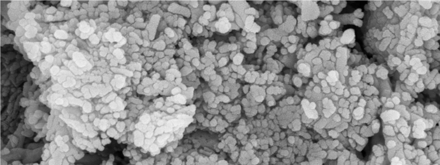
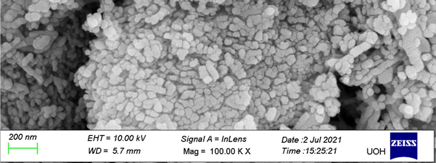
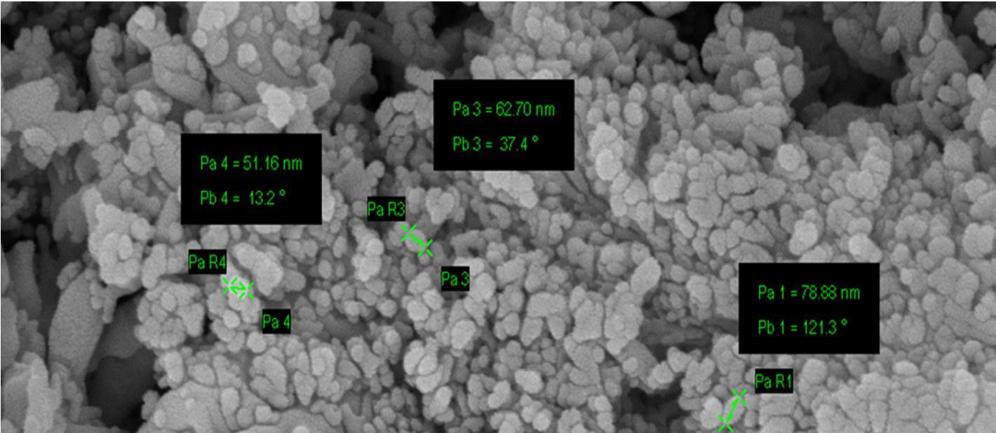
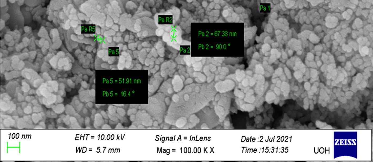
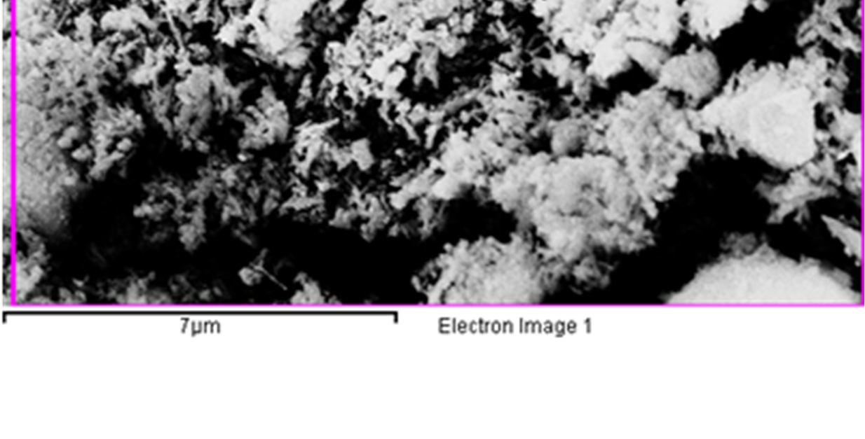
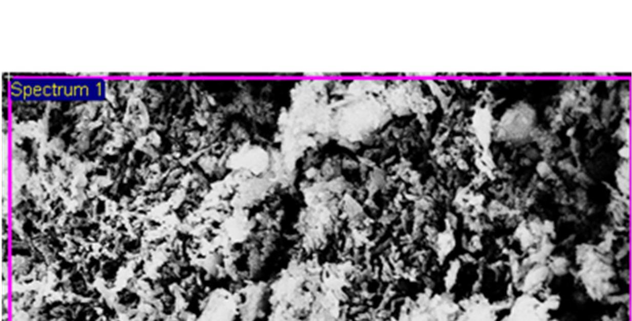
The Field-Emission Scanning Electron Microscope (FESEM) image shows that the synthesized nano-crystals are in the size range of 51nm 80nm.
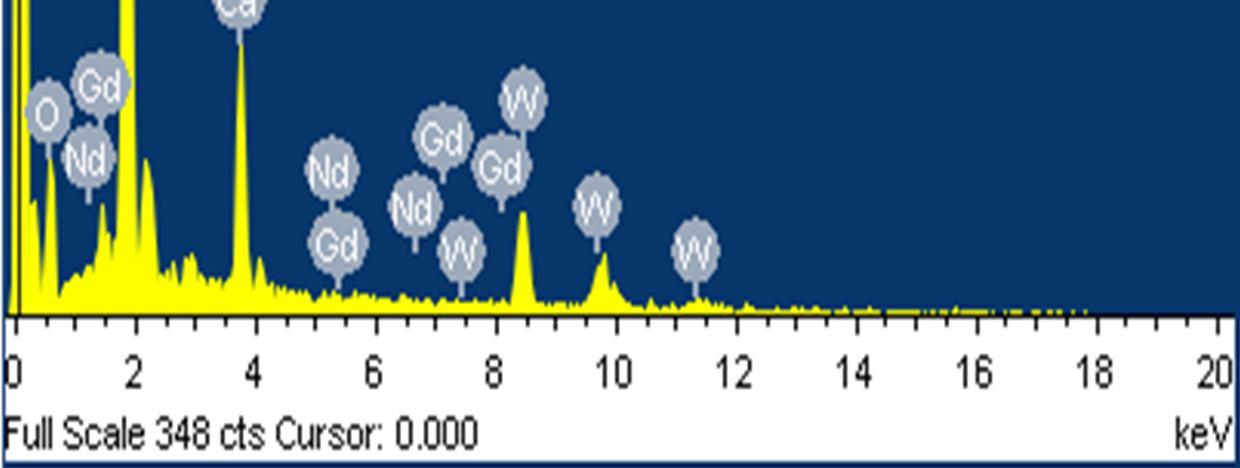
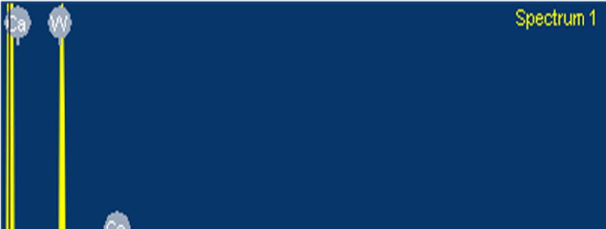
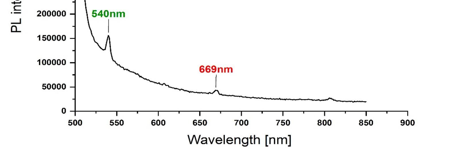
The presence of Gd and Nd rare earth ions in the GD Nd co doped CaWO4 nano crystals was confirmed by the energy dispersive analysis of the X-rays (EDAX). The photoluminescence properties of the Gd-Nd co-doped CaWO4 nano-crystals were compared with the undoped CaWO4 nano crystals and are reported. The synthesized nano crystals exhibit emissions at (i) 369nm, 392nm, 435nm and 477nm for an excitation wavelength of 280 nm and (ii) 540nm and 669nm for an excitation wavelength of 490nm.
The emission wavelengths clearly indicate the blue light and green light generation, which is significant for developing luminescent materials of tungstate systems.
Keywords: Nano crystals, X ray diffraction, Field Emission Scanning Electron Microscopy and Photoluminescence.
The physical parameters such as shape, size and crystallographic structure shows a great impact on the luminescence properties of nanophosphors[1 5] .The morphological changes could lead to a change in the able population of luminescent activators undergoing excitation or emission and are manifested in their quantum yields, luminescence intensities, and lifetimes.Tungstates of Scheelite structure , with the typical AWO4 formula , have potential applications as scintillators, in catalysis, in optoelectronics, lasers, drug delivery, etc.[6,7].
In their purest state self activating phosphors, can absorb at a certain wavelength range and then emit at a particular wavelength. For lanthanide based luminescence, these phosphors are very cost effective and convenient materials. Calcium tungstate (CaWO4) is a self activating phosphor which absorbs in the mid ultraviolet (mid UV) region at around 250 nm and emits blue light (420 nm). The blue emission of CaWO4 is due to its tetrahedral WO4 2 group. Its physical and chemical stability, intrinsic blue emission and wide band gap semiconducting nature make it a potential host for luminescence [8 10]. Calcium tungstate (CaWO4) produce multicolour emissions on being doped with certain lanthanide ions due to the energy transfer from the host to the activator lanthanide ions. By tuning the size, concentration of lanthanide ions or mixing of the dopants there can be a controlled energy transfer from the host to the activator [11 14].
Over the past few decades, several papers were reported on synthesis of single rare earth doped CaWO4 nano crystals by hydrothermal process [15 17]. In recent years, codoping of rare earth ions in various hosts using different methods are being carried out to enhance the luminescence properties and improve their applications in biological imaging, white light generation and in optoelectronic devices. The rare earth co doped system exhibits higher emission intensity than those of the single doped systems [18 20]. As a part of the synthesis of series of co doped nano crystals, in this paper for the first time we report the synthesis of Gd and Nd rare earth ions co doped CaWO4 nano crystals by modified hydrothermal synthesis method using sodium oleate and oleic acid as the capping agents. The synthesized nano crystals were characterized by X ray diffraction (XRD) with energy dispersive analysis of the X rays (EDAX) and Field emission scanning electron microscopy (FESEM), and photo luminescence excitation studies

Journal for Research in Applied Science & Engineering Technology (IJRASET)
ISSN: 2321 9653; IC Value: 45.98; SJ Impact Factor: 7.538 Volume 10 Issue IX Sep 2022 Available at www.ijraset.com
The Gd Nd rare earth co doped CaWO4 nano crystals were synthesized by the following slightly modified hydrothermal process. The chemicals used in this process are of analytical grade and were purchased from Sigma Aldrich. At first a solution was prepared by adding 0.91 g of sodium oleate (C18H33NaO2) to the mixture of 0.25 g ammonium meta tungstate hydrate (NH4)6H2W12O40 and 15 ml double distilled water by stirring for 20min.Then, another alcoholic solution was prepared by adding 2 ml of oleic acid (C18H34O2) with 8 ml of ethyl alcohol under stirring for half an hour. Now the alcoholic solution so obtained is added to the first prepared solution by stirring for 20 min. Then, the obtained mixture is slowly introduced to a solution formed by 1 mol% RE(NO3)3 (RE = Gd3+ and Nd3+), 8 ml of double distilled water and 1 gm of Ca (NO3)2 4H2O and stirred for half an hour. The final obtained solution was transferred into a 50 ml Teflon jar and placed in an autoclave. The autoclave was sealed and maintained at 180 °C for 12 h. The resultant product was centrifuged at room temperature for 15min at 4500rpm. The obtained sample was placed in a muffle furnace at 6000 C for 5min, resulting in a white foamy powder.
To estimate the size of the nano crystals synthesized and to confirm the incorporation of rare earth ions into the host lattice (CaWO4) of nano crystals different techniques were used. The phase of the formed nano crystals was identified by X ray diffraction (XRD)analysis using an automated X ray powder diffractometer at an operating voltage of 40kV and current of 30 mA with scanning rate of 4.0 deg min 1 , within a range from 10° to 80° The size of the nano crystals formed was determined by using FESEM(Carl Zeiss Model no : Ultra 55).The EDAX (Energy dispersive X ray analysis )was carried out using Oxford: INCAX act model, device and the incorporation of rare earth ions was clearly identified. Further, the photoluminescent properties of the Gd Nd co doped CaWO4 nano crystals were determined at different excitation wavelengths using HORIBA FL3 21 TCSPC model spectrofluorometer.
In this present modified hydrothermal synthesis process oleic acid and sodium oleate act as the capping reagents. The role of oleic acid is to separate out the particles from the supersaturated solution and restrict the growth of the CaWO4 nano crystals and sodium oleate is used to avoid further nucleation, and acts as a coordinating agent to mediate the nucleation process[17].
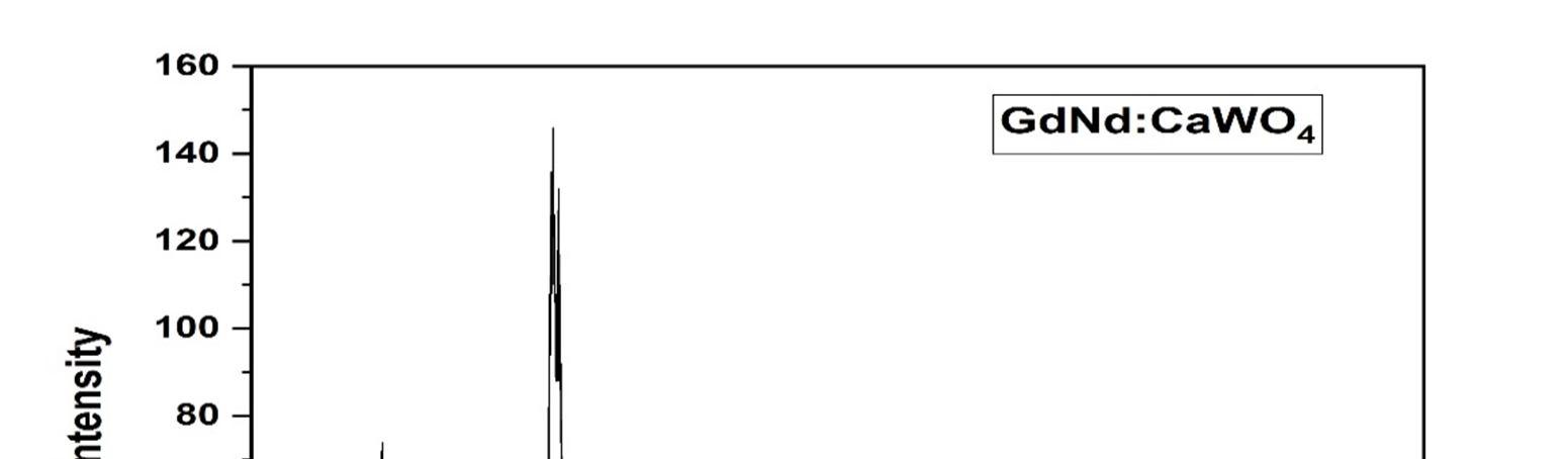
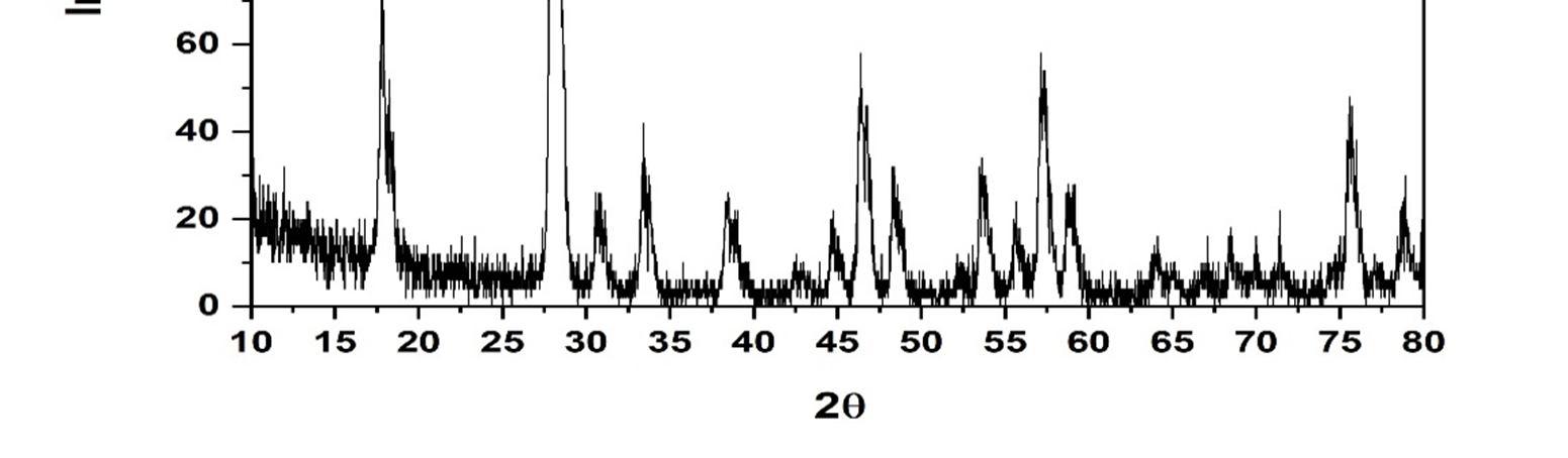
The XRD provides information related to the phase of the material. The XRD pattern obtained reveals the crystallinity and phase purity of the Gd and Nd co doped CaWO4 nano crystals, with sharp diffraction peaks as shown in figure 1; All the phases were indexed to scheelite structure.
Figure 1: XRD pattern of the sample, confirming the formation of pure CaWO4 phase.
The
The photoluminescent
of
Suneetha
Gd Nd
with the undoped CaWO
in the range
were
1450nm using modified
2321 9653;
Value:
Sep
at www.ijraset.com
International Journal for
in
Science & Engineering Technology (IJRASET)
ISSN: 2321 9653; IC Value: 45.98; SJ Impact Factor: 7.538
Volume 10 Issue IX Sep 2022 Available at www.ijraset.com
In summary, the Gd Nd co doped CaWO4 nano crystals were synthesized successfully by the hydrothermal synthesis route using oleic acid and sodium oleate act as the capping reagents, for the first time and further a series of lanthanides were chosen for future scope of work. The average size of the nano crystals synthesized using this method is within 51nm 80 nm range as observed by FESEM. The presence of Gd3+ and Nd3+ rare earth ions in Gd Nd: CaWO4 nano crystals is confirmed by EDAX. PL studies were used to further confirm the presence of Gd and Nd ions and shows the emission wavelengths within the range of 369 nm to 670nm at excitation wavelengths 280nm and 490nm respectively. The emission wavelengths clearly indicate the blue light and green light generation. This work shows that, by doping a small amount of Gd3+ and Nd3+ into CaWO4 host materials, can improve the emission intensity [22] and the multiple luminescence emission can be realized which is significant for developing luminescent materials of tungstate systems. This may be due to the energy transfer from Gd3+ to Nd3+ ions or vice versa and needs to be confirmed by further spectroscopic techniques The results clearly indicate that the nano crystals under study can be used for the white light generation applications with suitable modifications.
The authors would like to thank the Department of Physics, Osmania University, School of Physics, University of Hyderabad, Department of Physics, NIT Warangal for extending the experimental facilities.
[1] Brenier, G. Jia and C. Tu, J. Phys.: Condens. Matter, 2004; 16:9103 9108.
[2] Y. Hu, W. Zhuang, H. Ye, D. Wang, S. Zhang and X. Huang,J. Alloys Compd., 2005; 390: 226 229.
[3] J.Ninkovic´, G. Angloher, C. Bucci, C. Cozzini, T. Frank,D. Hauff, H. Kraus, B. Majorovits, V. Mikhailik, F. Petricca,F. Pro¨bst, Y. Ramachers, W. Rau, W. Seidel and S. Uchaikin,Nucl. Instrum. Methods Phys. Res., Sect. A, 2005; 537: 339 343.
[4] Z. Shan, Y. Wang, H. Ding and F. Huang, J. Mol. Catal. A: Chem., 2009; 302:54 58.
[5] X. Zhai,M.Yu, Z. Cheng, Z. Hou, P.Ma,D.Yang, X. Kang,Y.Dai,D. Wang and J. Lin, Dalton Trans., 2011; 40:12818 12825.
[6] T. J. Boyle, P. Yang, K. Hattar, B. A. Hernandez Sanchez, M. L. Neville and S. Hoppe, Chem. Mater., 2014;26: 965 975.
[7] M. J. Treadaway and R. C. Powell, J. Chem. Phys., 1974; 61: 4003 4011.
[8] Y. Xu, Y. Wang, L. Shi, L. Xing and X. Tan, Opt. LaserTechnol., 2013;54:50 52.
[9] G. S. Ningombam and R. S. Nongmaithem, Int. Nano Lett.,2017;7:133 140.
[10] G. S. Ningombam, N. R. Singh and R. S. Ningthoujam, Colloids Surf., A, 2017; 518, 249 262.
[11] C. Burda, X. Chen, R. Narayanan, and M. A. El Sayed, Chem.Rev., 2005;105:1025 1102.
[12] H. G. Yang, C. H. Sun, S. Z. Qiao, J. Zou, G. Liu, S. C. Smith, H. M. Cheng, and G. Q. Lu, Nature, 2008, 453, 638.
[13] V. M. Longo, L. Gracia, D. G. Stroppa, L. S. Cavalcante,M. Orlandi, A. J. Ramirez, E. R. Leite, J. Andre´s, A. Beltra´n, J. A.Varela and E. Longo, J. Phys. Chem. C, 2011; 115: 20113 20119.
[14] G. S. Ningombam, T. S. David and N. R. Singh, ACS Omega,2019;4: 13762 13771.
[15] Lingna Suna,b, Minhua Caoc, Yonghui Wangc, Genban Suna,b, Changwen Hua Journal of Crystal Growth 289 (2006) : 231 235.
[16] P Suneetha, Ch Rajesh and M V Ramana Mater. Res. Express 4 (2017): 045013
[17] Wang W, Yang P, Gai S, Niu N, He F and Lin J 2010 J. Nanopart. Res. 12 2295
[18] Qi Fan, Xiaoxia Cui, Haitao Guo, Yantao Xu,Guangwei Zhang and Bo Peng, Journal of Biomaterials Applications 0(0): 1 27.
[19] N. F. Andrade Neto, J. M. P. Silva, R. L. Tranquilin, E. Longo,J. F. M. Domenegueti,M. R. D. Bomio1 F. V. Motta1 Journal of Materials Science: Materials in Electronics J. Mater Sci: Mater Electron 31, 13261 13272 (2020).
[20] R. L. Tranquilin,L. X. Lovisa,C. R. R. Almeida,C. A. Paskocimas, M. S. Li, M. C. Oliveira, L. Gracia, J. Andres, E. Longo,F. V. Motta, and M. R. D. Bomio: J. Phys. Chem. C 2019; 123: 18536−18550
[21] P Suneetha, Ch Rajesh and M V Ramana, Mater. Res. Express 4 (2017): 085020
[22] Guiqiang Chen , Fengli Wang , Wenchao Ji , Yanxia Liu , Xiao Zhang , Superlattices and Microstructures 90 (2016): 30 37.
[23] N. Thejokalyani, S.J. Dhoble, Renew. Sustain. Energy Rev. 32 (2014): 448 467.