
10 VII July 2022 https://doi.org/10.22214/ijraset.2022.46031

International Journal for Research in Applied Science & Engineering Technology (IJRASET) ISSN: 2321 9653; IC Value: 45.98; SJ Impact Factor: 7.538 Volume 10 Issue VII July 2022 Available at www.ijraset.com 4639©IJRASET: All Rights are Reserved | SJ Impact Factor 7.538 | ISRA Journal Impact Factor 7.894 |
I would also like to thank my beloved coordinator Dr. Chetan K. R, Professor, Dept. ofComputer Science and Engineering. I am highlyindebted to them for their coordination aswell as for providing necessary information regarding the project.
On the very outset of this report on “Pneumonia detection in X ray chest images basedon CNN and data augmentation”, I would like to extend our sincere & heartfelt obligationtowards all the persons who have helped me in this endeavor. I am grateful to my institutionJ N N College of Engineering and the Department of Computer Science and Engineering for imparting us the knowledge with which I can do my best.
Pranathi Patel1, Hiriyanna GS2 1, 2JNNCE, India
Abstract: Pneumonia is a life threatening infectious disease affecting one or both lungs in humanscommonly caused by bacteria called Streptococcus pneumonia. One in three deaths in India is caused due to pneumonia as reported by World Health Organization (WHO). Chest X Rays which are used to diagnose pneumonia need expert radiotherapists for evaluation. Thus, developing an automatic system for detecting pneumonia would be beneficial for treating the disease without any delay particularly in remote areas. Due to the successof deep learning algorithms in analyzing medical images, Convolutional Neural Networks(CNNs) have gained much attention for disease classification. In addition, features learnedby pre trained CNN models on large scale datasets are much useful in image classificationtasks. In this work, appraise the functionality of pre trained CNN models utilized as feature extractors followed by different classifiers for the classification of abnormal and normal chest X-Rays. We analytically determine the optimal CNN model for the purpose.Statistical results obtained demonstrates that pretrained CNN models employed along withsupervised classifier algorithms can be very beneficial in analyzing chest X ray images, specifically to detect Pneumonia.
ACKNOWLEDGEMENT
I would like to thank my beloved guide Mr. Hiriyanna G. S, Asst. Professor, Dept. of Computer Science and Engineering. I am highly indebted to them for their guidance and constant supervision as well as for providing necessary information regarding the project & also for their support in completing the project.
Pneumonia Detection in X-Ray Chest Images Based on CNN and Data Augmentation
I would also like to express my gratitude to Dr. Poornima K. M, Professor and Head ofthe Department, Computer Science and Engineering and Dr. K Nagendra Prasad, Principal, J N N College of Engineering, Shivamogga for all their support and encouragement. I am thankfultoandfortunateenoughtogetconstantencouragement, support andguidancefromall Teaching staffs of the Department of Computer Science and Engineering which helpedme in successfully completing my project. Also, I would like to extend mysincere regardsto all the non teaching staff of Department of Computer Science and Engineering for theirtimely support. Thanking you all, Pranathi Patel HE4JN20SCS04 I. INTRODUCTION
Pneumonia causes inflammation of lungs especially air sacs which may filled with fluidor pus causing cough and difficulty in breathing. More than 150 million people especially children below 5 years get infected with pneumonia yearly world wide. The mortality ratedue to pneumonia is specifically higher in children below years in developing nations. Thismandates the detection of pneumonia on time so that proper treatment can be provided to the person. The most practiced method world wide to detect pneumonia is using chest X ray images. To detect pneumonia, careful examination of chest Xray images is required which in turn necessitate experienced and knowledgeable radiologist/ experts. This makesthe process of pneumonia detection a challenging task. Moreover, it is a time consuming task and a little error can have fatal consequences.


The above describes the simplest type of deep neural network in the simplest terms. However, deep learning algorithms are incredibly complex, and there are di erent types of neural networks to address specific problems or datasets. For example, Convolutional neural networks (CNNs), used primarily in computer vision and image classification applications, can detect features and patterns within an image, enabling tasks, like object detection or recognition. In 2015, a CNN bested a human in an object recognition challenge for the first time. Recurrent neural network (RNNs) are typically used in natural language and speech recognition applications as it leverages sequential or times series data.
Deep learning maybe used in manyfields like, a) Robotics: Many of the recent developments in robotics have been driven by advances in AI and deep learning. For example, AI enables robots to sense and respond to their environment. This capability increases the range of functions they can perform, from navigating their way around warehouse floors to sorting and handling objects that are uneven, fragile, or jumbled together. Something as simple as picking up a strawberry is an easy task for humans, but it has been remarkably difficult for robots to perform. As AI progresses, that progress will enhance the capabilities of robots. Developments in AI mean we can expect the robots of the future to increasingly be used as human assistants. They will not only be used to understand and answer questions, as some are used today. They will also be able to act on voice commands and gestures, even anticipate a worker’s next move. Today, collaborative robots already work alongsidehumans, with humans and robots each performing separate tasksthatarebest suitedtotheirstrengths.
1) Deep Learning
Deep learning (also known as deep structured learning) is part of a broader family of machine learning methods based on artificial neuralnetworks with representation learning.Learning can be supervised, semi supervised or unsupervised. Deep learning architectures such as deep neural networks, deep belief networks, deep reinforcement learning, recurrent neural networks and convolutional neural networks havebeen applied to elds including computer vision, speech recognition, natural language processing, machine translation, bioinformatics, drug design, medical image analysis, climate science, material inspection and board game programs, where they have producedresults comparable to and in some cases surpassing human expert performance. Artificial neural networks (ANNs) were inspired by information processing and distributed communication nodes in biological systems. ANNs have various differences from biological brains. Specifically, artificial neural networks tend to be static and symbolic, while the biological brain of most living organisms is dynamic (plastic) and analogue.
The adjective "deep" in deep learning refers to the use of multiple layers in the network. Early work showed that a linear perceptron cannot be a universal classifier, but that a network with a nonpolynomial activation function with one hidden layer of unbounded width can. Deep learning is a modern variation which is concerned with an unbounded number of layers of bounded size, which permits practical application and optimized implementation, while retaining theoretical universality under mild conditions. In deep learning the layers are also permitted to be heterogeneous and to deviate widely from biologically informed connectionist models, for the sake of efficiency, trainability and understandability, whence the "structured" part. Deep learning neural networks, or artificial neural networks, attempts to mimic the human brain through a combination of data inputs, weights, and bias. Theseelements worktogether to accuratelyrecognize, classify, and describe objects within the data. Deep neural networks consist of multiple layers of interconnected nodes, each buildingupon the previous layer to re ne and optimize the prediction or categorization. This progression of computations through the network is called forward propagation. The inputand output layers of a deep neural network are called visible layers. The input layer is where the deep learning model ingests the data for processing, and the output layer is where the nal prediction or classification is made.
International Journal for Research in Applied Science & Engineering Technology (IJRASET) ISSN: 2321 9653; IC Value: 45.98; SJ Impact Factor: 7.538 Volume 10 Issue VII July 2022 Available at www.ijraset.com 4640©IJRASET: All Rights are Reserved | SJ Impact Factor 7.538 | ISRA Journal Impact Factor 7.894 | A. Pneumonia Detection Due to the tedious nature of X ray image analysis task, many computer algorithms and computer aided diagnostic tools have been proposed by researchers in order to analyze X ray images; however, they were not proved very significant in assisting experts in makingdecisions. Recently, handcrafted techniques and deep learning techniques are being appliedsuccessfully by researchers in the area of medical imaging to analyze and classify medicalimages for detection of various diseases like skin cancer, breast cancer, tuberculosis, braintumor etc. This motivated us to propose a deep convolution neural network (CNN) that helps to extract features from chest X rayimage. The extracted features are further used.
Another process called backpropagation uses algorithms, like gradient descent, to calculate errors in predictions and then adjusts the weights and biases of the function by moving backwards through the layers in an e ort to train the model. Together, forward propagation and backpropagation allow a neural network to make predictions and correct for any errors accordingly. Over time, the algorithm becomes graduallymore accurate.
c) Medical Imaging and Healthcare: Deep learning has been particularly effective in medical imaging, due to the availability of high quality data and the ability of convolutional neural networks to classify images. For example, deep learning can be as effective as a dermatologist in classifying skin cancers, if not more so. Several vendors have alreadyreceived FDA approval for deep learning algorithms for diagnostic purposes,including image analysis for oncology and retina diseases. Deep learning is also making significant inroads into improving healthcare quality by predicting medical events from electronic health record data.
Secondly, the activation layer plays the role of adding nonlinear factors, which activatethe whole feature map through an activation function, such as ReLU, Sigmoid, and Tanh. When Convolutional Neural Network classifies the processed output vector of the originalimage. These vectors usually have a very high dimension, which could not separate by a line. Thus, the nonlinearity quality is thenecessity. Also, the function of the pooling layer, also called the downsampling layer, is samplingfrom the feature map and reducing the computation burden, which picks out the representative attribute of the feature map, such as the maximum value or the average value. “Image based convolutional networks typically use a pooling layer which summarizes the activations of many adjacent filters with a single response. Such pooling layers may summarize the activations of groups of units with a function such as their maximum, mean, or L2 norm. These pooling layers help the network be robust to small translations of the input.”
Convolutional neural networks are composed of multiple layers of artificial neurons. Artificial neurons, a rough imitation of their biological counterparts, are mathematical functionsthat calculatetheweighted sum ofmultiple inputs and outputsan activation value. When you input an image in a ConvNet, each layer generates several activation functions that are passed on to thenext layer.
International Journal for Research in Applied Science & Engineering Technology (IJRASET) ISSN: 2321 9653; IC Value: 45.98; SJ Impact Factor: 7.538 Volume 10 Issue VII July 2022 Available at www.ijraset.com 4641©IJRASET: All Rights are Reserved | SJ Impact Factor 7.538 | ISRA Journal Impact Factor 7.894 |
b) Agriculture: AI has the potential to revolutionize farming. Today, deep learning enables farmers to deploy equipment that can see and differentiate between crop plants andweeds. This capability allows weeding machines to selectively spray herbicides on weeds and leave other plants untouched. Farming machines that use deep learning enabled computer vision can even optimize individual plants in a field by selectively spraying herbicides, fertilizers, fungicides, insecticides, and biologicals. In addition to reducing herbicide use and improving farm output, deep learning can be further extended to other farming operations such as applying fertilizer, performing irrigation, andharvesting.
A Convolutional Neural Network, also known as CNN or ConvNet, is a class of neuralnetworks that specializes in processing data that has a grid like topology, such as an image.A digital image is a binary representation of visual data. It contains a series of pixels arranged in a grid like fashion that contains pixel values to denote how bright and what color each pixel should be.
2) CNN CNN’s were first developed and used around the 1980s. The most that a CNN could doat that time was recognize handwritten digits. It was mostly used in the postal sectors to read zip codes, pin codes, etc. The important thing to remember about any deep learning model is that it requires a large amount of data to train and also requires a lot of computingresources. This was a major drawback for CNNs at that period and hence CNNs were only limited to the postal sectors and it failed to enter the world of machine learning. In deep learning, a convolutional neural network (CNN/ConvNet) is a class of deep neural networks, most commonly applied to analyze visual imagery. Now when we think of a neural network wethink about matrixmultiplications but that isnotthecasewith ConvNet. It uses a special technique called Convolution. Now in mathematics convolution is a mathematical operation on two functions that produces a third function that expresses howthe shape of one is modified by the other.
ConvolutionalNeuralNetwork(CNN)consistsof4parts, convolutionallayer, activationlayer, polling layer, and fully connected layer.
Finally, a fully connected layer is computing the output by weighting the attribute vectors generated by hidden layers via the vector product and generating the final classification result.
Firstly, the convolutional layer is using a filter(convolutional kernel) to scan the originalimage, compute the product between the original pixel and the weight on the filter. Then, generating the feature map by average the sum of the product.
The human brain processes a huge amount of information the second we see an image.Each neuron works in its own receptive field and is connected to other neurons in a way that they cover the entire visual field. Just as each neuron responds to stimuli only in the restrictedregion ofthe visual field calledthereceptive field in thebiological vision system,each neuron in a CNN processes data only in its receptive field as well. The layers are arranged in such a way so that they detect simpler patterns first (lines, curves, etc.) and morecomplex patterns (faces, objects, etc.) further along. Byusinga CNN, onecan enablesight to computers.
Contributions: The primary aim of this study was to examine and test the performance of Machine Learning techniques and Convolutional Neural Network (CNN) for the classi cation of Pneumonia, which is based on Chest X ray Images to achieve high accuracy. Another aim is to provide radiologists and medical experts with a tool that is a lower cost.This tool could help them read theX rayimagesandtocreateabasis for amodel toanalyzemore complex data like CT images.
Advantages: In this work, we have oered a model for detecting and classifyingPneumonia from Chest X ray Images using Machine Learning methods based on Convolutional Neural Network (CNN).
Limitations: The two networks also show the contrast gap between sensitivity and spesi city which caused by unbalance dataset. Using more complex network structure and augmenting the unbalance dataset may also possible in the future so that we can get the best architecture for pneumonia CAD system.
Contributions: Each of trained architecture also shows the contrast difference betweensensitivity and specificity. The difference may caused by the unbalanced data between pneumonia and non pneumonia classes which can cause the architecture tend to missed classify the pneumonia class. The false classification of pneumonia class is not desired for CAD since it can cause the patients tend to have normal lungs or other abnormalities.
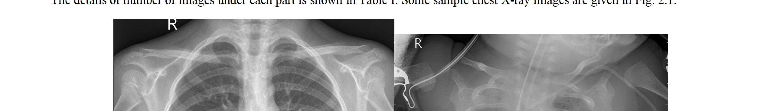
Methodology: The system is composed of several steps, including the data collection and pre processing description, building the classification models and extracting the required features. These steps can be divided into four phases: Data Set, data pre processing, building and validating classification models, and feature extraction. The chest X ray image dataset. It was the Chest X ray Images, including normal chest X Ray and Pneumonia chest X Ray. Here, we have taken 5,856 sample images of the dataset to use later on for recognition and classification of Pneumonia. 1,583 of which are normal XRaylung images, and 4,273 of which are X Ray lung images with Pneumonia. These data set was used for training in Deep Learning method and testing data using Pandas for predicting Pneumonia. The X ray images were divided into train, test, and validation groups. 80% of data was used for training, 10% used for testing, and 10% used for validations. The data set was randomized in advance to avoid potential bias. Trained the dataset using Keras on the top of Tensor Flow. There were various Machine Learning models used for classifying Pneumonia X Ray images from normal ones. The module aims to provide a binary classification of presence absence of Pneumonia within a chest x ray and to select The Machine Learning algorithms are applied according to the featuresselected and extracted manually, while the Deep neural network learns to extract the mostrepresentative or complex features and patterns on its own according to the input. Movingfrom Machine Learning to Deep Learning. CNN was one of the best models for image recognition and classification. Therefore, the following sections were focusing on describing the CNN model that was used for this data set. The models that were implemented for comparison were Random Forest, Xgboost, K nearest neighbor, DecisionTree, Gradient Boost, Adaboost, and Convolutional Neural Network (CNN).
Kaggle RSNA Pneumonia Detection Challenge dataset from the Radiological Society of North America. The dataset consists of 26.684 data, which are in DICOM format, dividedinto 21.684 training data, 2.000 validation data, 3.000 testing data. The dataset is providedas a set ofpatient Ids, bounding boxes, andtarget values. In addition, Thedatasetis groupedinto two groups, pneumonia and non pneumonia. However, the non pneumonia groups contain another two groups which are normal and non pneumonia abnormal lungs. The residual mapping can be used when the input and output are in the same dimension. Batchnormalization is used right after each convolution and activation. We use Adam optimizerwith 32 mini batches. Cosine annealing is used for learning rate to optimize the training process. For the loss function, we combine IoU and binary cross entropy.
B. Literature Survey 1) Pneumonia Detection with Deep Convolutional Architecture
Methodology: Deep residual network which was introduced by Microsoft researchers in ILSVRC 2015 and mask RCNN by Facebook AI researchers. Those networks are usedto develop CAD for pneumonia detection with thorax x ray images. We also represent, compare and evaluate the performance of two architectures with simple evaluation, yet respectable.
International Journal for Research in Applied Science & Engineering Technology (IJRASET) ISSN: 2321 9653; IC Value: 45.98; SJ Impact Factor: 7.538 Volume 10 Issue VII July 2022 Available at www.ijraset.com 4642©IJRASET: All Rights are Reserved | SJ Impact Factor 7.538 | ISRA Journal Impact Factor 7.894 |
Mask Regional CNN (mask RCNN) is the further development of Faster Regional CNN algorithm. This algorithm usually used in object localization and recognition inan image by combining object detection and semantic segmentation. The objective of object detection is to localize each object on the image using a bounding box. Meanwhile,semantic segmentation goal is to classify each pixel intoaxedset ofcategoriesusingobjectdelineation.
Advantages: In the pneumonia detection, residual network shows better performance than mask RCNN. It may caused by the poor performance of mask RCNN’s RPN algorithm to find which of the proposed RoIs have an object since the pneumonia features are difficult to find comparing with widely used object detection dataset such as COCO.
2) Investigation of the performance of Machine LearningClassifiers for Pneumonia Detection in Chest X ray Images
International Journal for Research in Applied Science & Engineering Technology (IJRASET) ISSN: 2321 9653; IC Value: 45.98; SJ Impact Factor: 7.538 Volume 10 Issue VII July 2022 Available at www.ijraset.com 4643©IJRASET: All Rights are Reserved | SJ Impact Factor 7.538 | ISRA Journal Impact Factor 7.894 | In particular, comparing all seven models based on a test accuracy score, F score, and ROC curve, it appears that CNN outperforms all other models by a small margin with a test accuracy score of 98.46%. Random forest performs surprisingly well with a test accuracy score of 97.61%. It should note that the CNN modelwas run with only 100 epochs due to its long running and waiting time. The result of our model indicates that our model outperforms other methods, which use no extra training data on chest radiography and shows that image training may be sufficient for general medical image recognition tasks. Moreover, the cooperation between a Machine Learning based medical system and the detection of Pneumonia will improve the outcomes and bring benefits to clinicians and their patients.
Contributions: The work aims to detect COVID 19 and Pneumonia patients using deeplearning techniques as Normal, Infected, Pneumonia patients. Transfer learningtechniquescan be used for COVID 19 detection.
Advantages: CNN is the model used to detect Normal and Infected patients like COVID 19 Infected lungs, Pneumonia Infected lungs. The accuracy of detection of COVID 19 is 95 % which a plus about this model and the current requirement of this pandemic with limited cost and computations. This model is more appropriate for medicalpractitioners, researchers etc.
Methodology: The complex nature of COVID 19 and pneumonia radiological features canbe described based on the formation of GGO and consolidations, which are inconsistent in sizes and locations within the lung regions. Therefore, the probability of a positive true label CT 2D axial slice with absence of pneumonia findings can be high when 3D CTdata are decomposed in a 2D system. This scenario is more commonly happened among the early course infected lung, in which the pneumonia in ltration is barely manifested and hardly visible. Hence, such a data space condition is defined as ambiguously labeled samples (AL CT) in this study. It is hypothesized that theutilityofAL CT slices for modeltraining can complicate the learning process and consequently lead to poor performances as the AL CT slices share similar spatial representation with the normal lung CT slices.
The suitableConvolutional Neural Network Model is selected for the identified dataset. The model detects COVID 19 patients and Pneumonia patients on the real world dataset of lung X Ray images. Images are pre processed and trained for various classifications like Normal,COVID 19 and Pneumonia. After pre processing, the detection of the disease is doneby selecting the appropriate features from the images in each of the datasets. The result indicates that accuracy of detection of COVID vs Normal and COVID vs Pneumonia. Among those two, COVID vs Normal is with better accuracy than COVID vs Pneumonia.This method detects not only COVID or Pneumonia, but also the subtypes of Pneumonia as bacterial or Viral Pneumonia with 80% and 91.46% respectively. The detection of COVID, Bacterial Pneumonia and Viral Pneumon+ia using the proposed model helps in rapid diagnosis and to distinguish COVID from Pneumonia and its types which facilitates to use appropriate and fast solutions.
Limitations: With ensembleofdifferent models to improve the accuracyofdetection ofdifferent classes but with the cost of time and computation.
3) An Adaptive Data Processing Framework for Cost EffectiveCOVID 19 and Pneumonia Detection
Limitations: Furthermore, this study can be applied in the diagnosis of some diseases such as COVID 19. As future work, we hope to build a larger database so we can apply more Deep Learning techniques to train and test the system to predict better results.
Contributions: Cost effective data processing framework for COVID 19 and pneumonia detection to effectively distinguish COVID 19 and other pneumonia infected lungs from healthy lungs. Community acquired pneumonia (CAP) samples and clinical confirmed COVID19 samples are considered as a single class label in this study due to the invariant radiographic features of GGO that are shared between COVID 19 and CAP CT samples. At the present time, more discriminative multi class classification task requires further scientific investigation due to the complicated radiography characteristics of COVID 19 and the continuous emergence of new COVID 19 variants. Advantages: It is worth mentioning that the data processing framework can be extended easily to different medical ML/DL applications, especially in attempts of utilizing 3D datain a 2D based solution. That is, it can be referenced as data pre processing procedures for relatable applications to improve data diversity and quality for more efficient neural network development in a cost effective setting. Limitations: Training a 3DCNN model is much complex and exhausting compared to a 2DCNN model. Moreover, 3D training samples with good feature descriptions are exceptionally scarce.
4) Analysis of COVID 19 and Pneumonia Detection in Chest X RayImages using Deep Learning Methodology: The on going COVID 19 outbreak made healthcare systems across the globeto be in the edge of the battle. Recent stats indicate that more than 140+ million confirmedcases are diagnosed globally as of April 2021. The cases are increasing day by day.The early and auto diagnosis helps people to be precautious. The proposed work aimsto detect COVID 19 patients and Pneumonia patients from X Rays which is one of the medical imaging modes to analyze the health of patient’s lung inflammation.
Limitations: Implemented in healthcare system to supplement the diagnosis of X ray pneumonia images of pediatric patients.
6) Convolutional Neural Network for AutomaticPneumonia Detection in Chest Radiography
Methodology: The training dataset was augmented to develop the dataset to avoid over fitting. The augmentation strategy consists of resizing (cropping), ip, and rotation. The training dataset is divided into training datasets and validation randomly. The validation dataset is used to provide a model evaluation that does not favor the training dataset. The resulting model will be used for the testing process (prediction). Before the prediction process is carried out, the data testing is pre processed. Pre processing of the testing dataset consists of the centered crop, ip, and rotation. The dataset used in this paper uses 5,856 X ray radiographic images of Kaggle in JPEG format with dimensions of 10241024pixels and grouped into two classes: NORMAL and PNEUMONIA, which is stored into a different folder. The dataset is divided into training and testing data with a composition of 90% and 10% randomly. Composition of training and testing dataset, 10% of training dataset are randomly selected as validation data to provide the skills of the model sought by comparing and selecting suitable models. The convolutional layer is the core building of CNN. Image input passes through a convolutional layer which consists of a set of filters with certain parameters. The filter consists of a height and weight that is smaller than the input size. Filters are built in 3x3 size to ensure a detailed feature reading. Each filter will calculate the ReLu activation value and be pooled to summarize the convolution resultinformation. The output from the convolution process is used as input in the classificationprocess by first flattening.
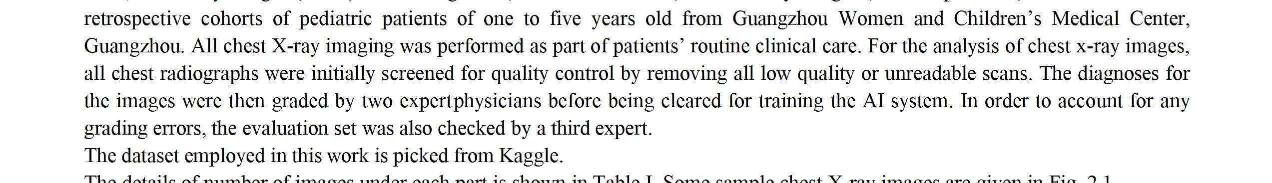
Methodology: The dataset of 5863 analyzed X ray images comes from Kaggle.com web service [Attribution 4.0 International (CC BY 4.0)], which unites the community ofartificial intelligence enthusiasts as well as holds machine learning competitions. These images are of pediatric patients between one and years old from Guangzhou Women and Children’s Medical Centre, Guangzhou. Images of different resolutions, with the majorityof approximately 1500 x 1000 pixels, are given in JPEG format. All of the images are in grayscale at 8 bpp. According to Kaggle’s information all of the radiographs have been initially screened for quality by removing unusable scans and classified to one of the categories (pneumonia/normal) bytwo experienced radiologists and only then cleared for training neural networks. The downloaded images were sorted into two categories as the contest announced on Kaggle.com required differentiation of healthyand infected cases.
International Journal for Research in Applied Science & Engineering Technology (IJRASET) ISSN: 2321 9653; IC Value: 45.98; SJ Impact Factor: 7.538 Volume 10 Issue VII July 2022 Available at www.ijraset.com 4644©IJRASET: All Rights are Reserved | SJ Impact Factor 7.538 | ISRA Journal Impact Factor 7.894 |
Contributions: Different deep convolution neural network architectures with an augmentation strategy to classify the pneumonia detection from the chest X ray images. We use three convolution layers and three classification layers (fullyconnected)
7) A Pneumonia Detection Method Based on Improved Convolutional Neural Network Methodology: For traditional machine learning and image processing, it is di cult to extract features, the quality of feature extraction will directly affect the classification accuracy, another problem is that the large difference between the original datasets and thetarget datasets in the transfer learning, lead to unsatisfactory results. In addition, the original convolution model network is too shallow, recognition rate is not high; Therefore, this paper presents an improved convolutional neural network method for pneumonia detection based on deep learning model. First, the image size in the original datasets are xed and the appropriate batch size are used as the input of the network, Then theconvolution layers and pooling layers are added on the basis of lenet 5 model, Secondly, a feature integration layer is added to construct an optimal model for accurate classification.The proposed method not only avoids the complex feature extraction process but also has fewer parameters than other classical convolutional neural networks. By using two public datasets, the second which provided by the RSNA joint Kaggle medical image pneumoniarecognition competition, after a series of experiments of single dataset and fusion of two datasets, the final accuracy rate reached 98.83% and 98.44% respectively, the test accuracyalso reached 97.26% and 91.41% respectively. Compared with existing transfer learning, GoogleNet Inception V3+Data Augmentation(GIV+DA), GIV3+RF models.
results with the augmentation strategy and without the augmentation strategy shows that the proposed CNN’s architecture can train small datasets.
Advantages: CNN’s architecture with an augmentation strategy to detect the pneumonia disease using a chest X ray image. The experiment result shows that the augmentation strategy on the proposed CNN’s architecture achieves better results than without Limitations:augmentation.Prediction
5) Pneumonia detection in X ray chest images based onconvolutional neural networks and data augmentation methods
Contributions: The purpose of this study is to apply CNN into medical imaging environment to ascertain how accurate and sensitive these methods prove to be in recognizing different types of pneumonia (viral or bacterial). Advantages: Standard CNN is able to provide quite accurate classification (85%) of X ray pneumonia images solving true three class problem (distinguishing between viral, bacterial and control cases).
Contributions: It isabletoassist doctors taking appropriate medicine toshorten thecuretime of pneumonia and improve the cure rate of Advantages:pneumonia.This paper first introduces the models and results proposed by some previous researchers, and brie y introduces some basic knowledge of convolutional neuralnetworks Then, on the basis of relevant research, a convolutional neural network based onimproved Lenet was proposed to realize the detection of pneumonia images, the model in this paper is based on the original classical lenet 5 model by adding convolutional layer and pooling layer as well as feature integration layer, the obtained features were further highly abstracted, and finally excellent results were obtained on two public datasets, not only on the training set but also on the test set, which shows the proposed model has goodrobustness. In the following research work, further studies will be conducted on the detection of pneumonia types in our team, which will be detected according to types. at the same time, using convolutional neural network segment the lung area and locate the lesion area. Using CapsNet reconstruct the blurred images of lesion area (CapsNet has been proven that reconstructed images have very useful functions such as smoothing the noise) then, using appropriate neural networks for detection.
8) Feature Extraction and Classification of Chest X Ray ImagesUsing CNN to Detect Pneumonia
Limitations: still seems to be a little inadequate, the recognition rate is not high, proneto over fitting series of problems;
Limitations: we plan to use different optimizers and other data augmentation techniques in an attempt to further improve the classification accuracy of the proposed CNN architecture with data augmentation. We also plan to use early stopping and batch
Methodology: Here, we present two CNN architectures one with a dropout layer and another without a dropout layer. Both CNN consist of convolution layer, max pooling and a classification layer. A series of convolution and max pooling layers act as a feature extractor that is divided into two parts. The first part consists of two Convolution layers with 32 32 units each along with a max pooling layer of size 3 3 and a Relu activator. While the other also has two Convolution layers but with 64 and 128 units respectively along witha max pooling layer of size 2 2 and a Relu activator. Relu is a popular activation function which is generally used in neural networks especially in CNNs. Relu layer introduces nonlinearity to the model. Features extracted from the feature extractor part of the CNN aregiven as input to the dense layer which classifies the image. Before feeding the extracted features to the dense layer, a flatten layer a used. As the dense layer takes 1 Dimensional input, hence, flatten layer flattens the feature data and gives a 1 Dimensional output whichis fed to the dense layer. While training a CNN it might be possible that output through a certain layer is more dependent on a few selected neural units. To reduce this dependencyand prevent over fitting, the concept of dropout is introduced. During training, in each epoch, a neuron is momentarily dropped with a dropout probability p. Due to this, all the inputs and outputs to this neuron become disabled in the current epoch which results in some loss of data enhancing regularization in the model so that at times it would predict with much higher accuracy. The dropped out neurons are resampled with probability p at everytrainingstep, soadropped outneuron atonestepmaybecomeactivein thenextstep. A dropout probability of 0.5, corresponds to 50% of the neurons being dropped out. In the proposed CNN architecture with dropout layer, we apply dropout at two places. First, it is applied at the feature extractor part i.e. after convolution and max pool layers. As, the convolutional layers have not too many parameters, hence over fitting is not an issuein this case. Hence, here we take a low drop probability of 0.2. Second, it is used at dense layer. Dropout in the lower layers helps because it provides noisy inputs for the higher fully connected layers which prevents them from over fitting. At dense layers a drop probability of usually 0.5 is used as it has been observed that dense probability of 0.5 gives best regularization in most of the cases. We also use a drop probability of 0.5 here. The model summary of CNN with and without dropout. The architecture of CNN with dropout. The dataset employed in this work is picked from Kaggle. It consists of 5863 images of chest X ray. The dataset is split into three portions namely training dataset,validation dataset and testing dataset. Initially, the images in the dataset are of varying sizes. However, all the input images given to CNN should be of same size. To solve this problem, all the images in the dataset areresized to 64 64. To avoid over fitting and toenhancethegeneralization capabilitiesoftheproposedCNNarchitectures, variousin placedata augmentation techniques such as rescaling, flipping etc. are used in this paper. Use ofthis type of data augmentation ensures that our CNN see new variations of data at each andevery epoch during Contributions:training. CNN architecture from scratch instead of using transfer learning. In this paper, we propose various CNN architectures and these are trained on chest X ray image dataset
International Journal for Research in Applied Science & Engineering Technology (IJRASET) ISSN: 2321 9653; IC Value: 45.98; SJ Impact Factor: 7.538 Volume 10 Issue VII July 2022 Available at www.ijraset.com 4645©IJRASET: All Rights are Reserved | SJ Impact Factor 7.538 | ISRA Journal Impact Factor 7.894 |
Advantages: we propose two CNN architectures that are designed from scratch to detect pneumonia from images of chest X ray. To avoid over fitting, data augmentation techniques are used. The result of experiments performed to assess the performance of the proposed architectures and the effect of data augmentation on the performance of the proposed CNN’s show that CNN with dropout trained on augmented data outperforms the other models.
Advantages: Unlike showing the application and performance of one model, this work not only generates the results but also visualizes the activation map of the original picture tocompare the differences in which the models capture the features. This work demonstratesthe mechanism of Convolutional Neural Network via a concrete way.
Advantages: Presence of expert radiologists is the topmost necessity to properly diagnose any kind of thoracic disease. This paper primarily aims to improve the medical adeptness in areas where the availability of radiotherapists is still limited. Our study facilitate the early diagnosis of Pneumonia to prevent adverse consequences (including death) in such remote areas. So far, not much work has been contributed to specifically to detect Pneumonia from the mentioned dataset. The development of algorithms in this domain can be highly beneficial for providing better health care services. In this regard, we have proposed a model architecture for detecting Pneumonia from frontal chest X ray images with the utilization of Densenet as feature extractors and SVM as classifier. We observed the performance of various pretrained CNN models along with distinct classifiersand then on the basis of statistical results selected DenseNet 169.
9) Pneumonia Detection Using CNN based Feature Extraction Methodology: Pneumonia is a life threatening infectious disease affecting one or both lungs in humans commonly caused by bacteria called Streptococcus pneumoniae. One in three deaths in India is caused due to pneumonia as reported by World Health Organization (WHO). Chest X Rays which are used to diagnose pneumonia need expert radiotherapists for evaluation. Thus, developing an automatic system for detecting pneumonia would be beneficial for treating the disease without any delay particularly in remote areas. Dueto the success of deep learning algorithms in analyzing medical images, Convolutional Neural Networks (CNNs) have gained much attention for disease classification. In addition, features learned by pre trained CNN models on large scale datasets are much useful in image classi cation tasks. In this work, we appraise the functionality of pre trained CNN models utilized as feature extractors followed by different classifiers for the classificationof abnormal and normal chest X Rays. We analytically determine the optimal CNN modelfor the purpose. Statistical results obtained demonstrates that pretrained CNN models employed along with supervised classifier algorithms can be very beneficial in analyzing chest X ray images, specifically to detect Contributions:Pneumonia.The
normalization instead of dropout layer to see their effect in avoiding over fitting.
10) Comparative Experiment of Convolutional Neural Network(CNN) Models Based on Pneumonia X ray Images Detection
Limitations: In this experiment, it also has some Deficiency, first is over fitting problems. Accuracy of VGG16, the accuracy is dropping down rapidly because of over fitting; it is better to add regularization in the loss function to prevent over fitting problems. Second, there are also some disruptive factors in the datasets. The model cannotcompletely capture the location of the infected area. Sometimes the model captures the shoulder or outside the body, and the shoulder has no relationship with pneumonia. It is better to preprocess the datasets to reduce the disruptive factors.
Limitations: Although the results were overwhelming, there were still some limitations in our model which we believe are vital to keep in consideration. The first biggest limitation is that there is no history of the associated patient considered in our evaluation model. Secondly, only frontal chest X rays were used but it has been shown that lateral view chest X rays are also helpful in diagnosis. Thirdly, since the model exercises a lot of convolutional layers, the model need very high computational power otherwise it’ll eatup a lot of time in computations.
classification used with high rich extracted features exhibit improved performance in classifying images
Methodology: This paper aims toreveal therelationship between theConvolutional NeuralNetwork (CNN) model’s behavior and the depth of the model. Due to the worldwide coronavirus pandemic, the training dataset is the chest x ray images of the lungs, which are infected by pneumonia. The contrastive study incorporates three models: a classic model, which is the imitation of LeNet5, VGG16, and Residual Network 50. This research is based on pneumonia detection, and it can give people a deeper understanding of CNN’smechanism rather than only focusing on the result of di erent models. The explainable analysis visualizes the loss value and accuracy curves, CAM & Grad CAM images, and activation maps. It leads to three conclusions: rst, as the depth of the model increases, the average loss values of a given epoch will decrease; secondly, the accuracy will increase with the increasing depth; third, theextractedattributes becomemoreabstractin thedeeperhidden layers.
International Journal for Research in Applied Science & Engineering Technology (IJRASET) ISSN: 2321 9653; IC Value: 45.98; SJ Impact Factor: 7.538 Volume 10 Issue VII July 2022 Available at www.ijraset.com 4646©IJRASET: All Rights are Reserved | SJ Impact Factor 7.538 | ISRA Journal Impact Factor 7.894 |
Contributions: CNN is the most effective means, nowadays, to deal with various pictorial tasks. This work shows the basic idea and functions of different models. We hopeother people can gain enlightenment and do the improvement based on this work.
A. CNN Architecture
II. METHODOLOGY
C. Problems Statement
The idea is to identify Pneumonia Infected patients using chest X Ray images with the helpof CNN. The identification can be classified as Identification of Pneumonia affected lungs. Identification ofNormalandnot affected lungs.
D. Objectives
1) Organization of The Report: The organization of the paper is as follows, Chapter 1 contains introduction to pneumonia detection and CNN with deep learning, Objectives and Problem Statement. Chapter 1 also contains a literature survey of relevant research papers of the topic pneumonia detection using CNN. Chapter 2 provides the Methodolgy and fetaures of dataset considered. Chapter 3 provides the followed System Architecture. Chapter 4 provides the Result snapshots and analysis of the results. Lastly, Chapter 5 gives the Conclusion and future scope of the proposed report.
Motivation is to build a CNN architecture from scratch instead of using transfer learning. Various CNN architectures and these are trained on chest X ray image dataset. Later the CNN model is trained and will be able to detect whether pneumonia is present or pneumonia is not present by the input Chest X ray.
1) Convolutional Layer: It is the building block of the CNNs. Convolution operation is done in mathematics to merge two functions. In the CNN models, the input image is first converted into matrix form. Convolution filter is applied to the input matrix which slides over it, performing element wise multiplication and storing the sum. This creates a featuremap. 3 × 3 filter is generally employed to create 2D feature maps when images are black and white. Convolutions are performed in 3D when the input image is represented as a 3Dmatrix where the RGB color represents the third dimension. Several feature detectors are operated with the input matrix to generate a layer of feature maps which thus forms the convolutional layer.
2) Activation Functions: All four models presented in this paper use two different activation functions, namely ReLU activation function and softmax activation function. The ReLU activation function stands for rectified linear function . It is a nonlinear function that outputs zero when the input is negative and outputs one when the input is positive. The ReLU function is given by the following formula: This type of activation function is broadly used in CNNs as it deals with the problem of vanishing gradients and is useful for increasing the nonlinearity of layers. ReLU activation function has many variants such as Noisy ReLUs, Leaky ReLUs and ParametricReLUs. Advantages of ReLU over other activation functions are computational simplicity and representational sparsity. Softmax activation function is used in all four models presented in this paper. This broadly used activation function is employed in the last denselayer of all the four models . This activation function normalizes inputs into a probability distribution. Categorical cross entropy cost function is mostly used with this type of activation function
The model of convolutional neural network consists of convolution layers, pooling layers and full connection layers. The convolution layer and pooling layer are superimposed alternately. After passing through the full connection layers, another softmax layer is connected to map the probability of each category to the output of the network CNN models are feed forward networks with convolutional layers, pooling layers, flattening layers and fully connected layers employing suitable activation functions.
4) Flattening Layer and Fully Connected Layers: After the input image passes through the convolutional layer and the pooling layer, it is fed into the flattening layer. This layer flattens out the input image into a column, further reducing its computational complexity. This is then fed into the fully connected layer/dense layer. The fully connected layerhas multiple layers, and every node in the first layer is connected to every node in the second layer. Each layer in the fully connected layer extracts features, and on this basis, the network makes a prediction. This process is known as forward propagation. After forward propagation, a cost function is calculated. It is a measure of performance of a neural networkmodel. Thecostfunctionusedinallfour models is categorical cross entropy. Afterthe cost function is calculated, back propagation takes place. This process is repeated untilthe network achieves optimum performance. Adam optimization algorithm has been used in all four models.
International Journal for Research in Applied Science & Engineering Technology (IJRASET) ISSN: 2321 9653; IC Value: 45.98; SJ Impact Factor: 7.538 Volume 10 Issue VII July 2022 Available at www.ijraset.com 4647©IJRASET: All Rights are Reserved | SJ Impact Factor 7.538 | ISRA Journal Impact Factor 7.894 |
3) Pooling Layer: Convolutional layers are followed by pooling layers. The type of pooling layer used in all four models is max pooling layers. The max pooling layer having a dimension of 2 × 2 selects the maximum pixel intensity values from the window of the image currently covered by the kernel. Max pooling is used to down sample images, hence reducing the dimensionality and complexity of the image . Two other types of pooling layers can also be used which are general pooling and overlapping pooling. The models presented in this paper use max pooling technique as it helps recognize salient features in the image.


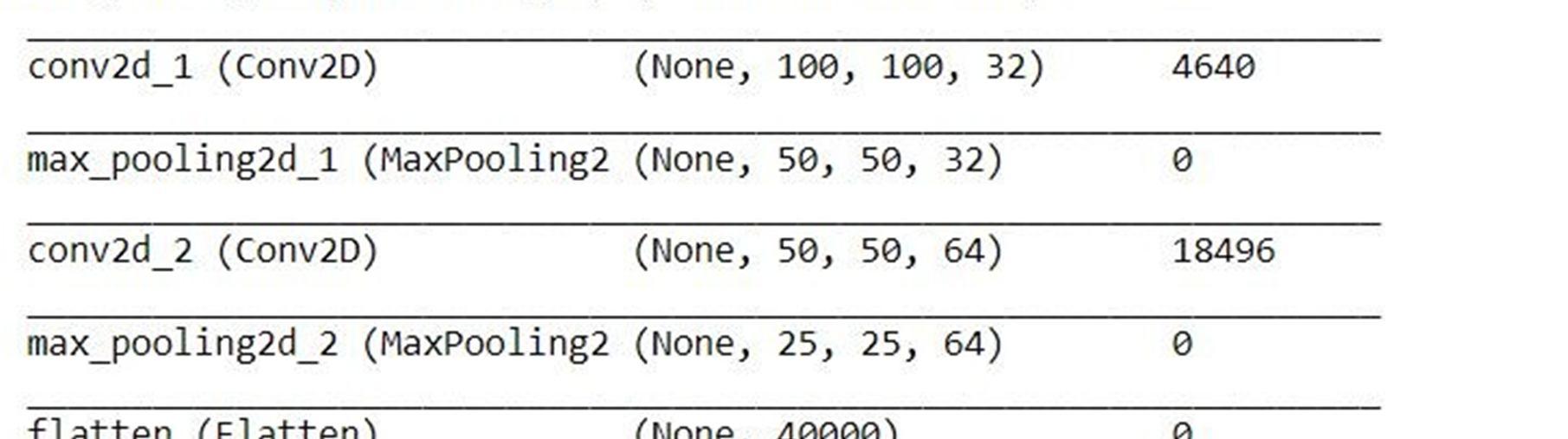
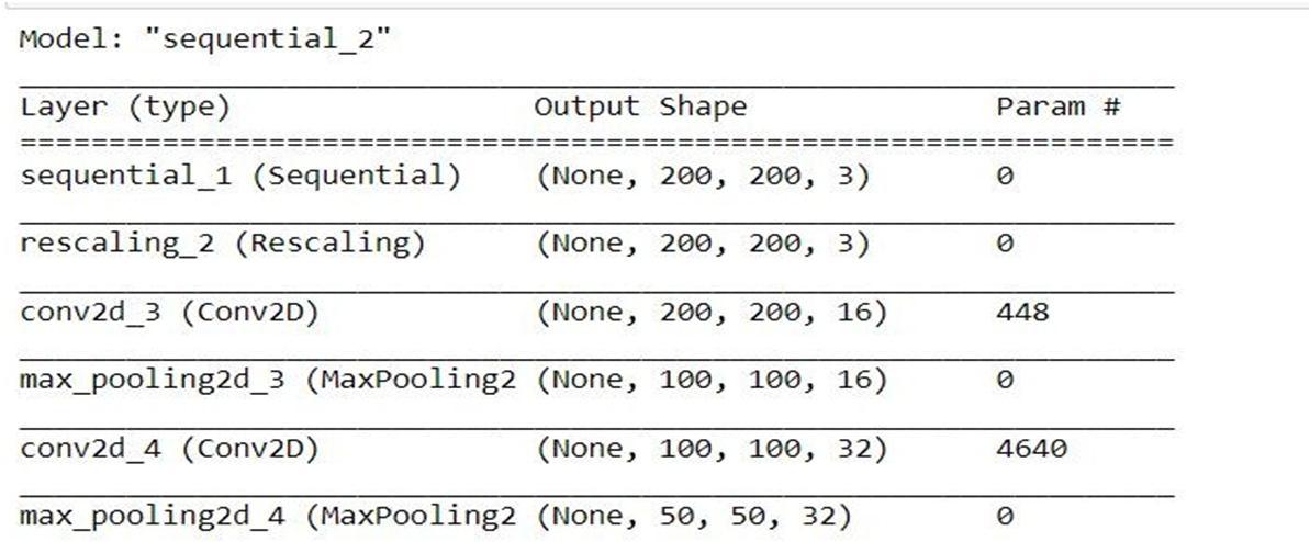
The size of each image is (200, 200, 3), Keras then appends an extra dimension for processing multiple batches, i.e., to train multiple images in every step of a single epoch. Since batch size can vary, its size is represented by None. Hence, the input shape becomes (None, 200, 200, 3). a (200, 200) image with a (4, 4) filter, with strides and dilation rate of 1, and ’valid’ padding, results in an output Since you have 200 such filters, the output shapebecomes (200, 200, 16).
C. Model Summary
Convolving
.
The first convolution has an output with shape (None, 100, 100, 16) Flatten layer, which takes the images and transform them into a single vector, output shape is (None, 40000), where None is still the batch size untouched, the 40000 are all elements you had in the input tensor, now in a single vector, one vector per sample in the batch. Then Denselayers, the first with 128 units, the second with 2 units.
The final output shape of your model is (None, 82). It outputs 82 values per sample in the batch.
Each layer has a number of parameters (which are generally the weights). Theparameters that are trainable will be updated with backpropagation.
Figure Model SummaryWithoutaugmentation
2.2:
The parameters that are not trainable will remain static or will be updated with a different method (only a few layers such as BatchNormalization has parameters that are updated with different methods) Your model has a total of 5143970 weights, all trainable.
ThedefaultMaxPoolingkernel hasashape of(16, 16)andstrides of(16, 16). Applyingthat to a (100, 100) image results in an image This pattern can be extended toall Conv2D and MaxPooling layers.
The Flatten layer takes all pixels along all channels and creates a 1D vector (not considering batch size)= 40000 values maxpooling layer that takes the output of the convolution as input. The output of the pooling has shape (None, 50, 50, 32), so it divided the size of your image by two, leaving therest as it was first layer is a convolution, which takes an unknown input shape (it’s known by you, you defined it somewhere with input_shape.
International Journal for Research in Applied Science & Engineering Technology (IJRASET) ISSN: 2321 9653; IC Value: 45.98; SJ Impact Factor: 7.538 Volume 10 Issue VII July 2022 Available at www.ijraset.com 4649©IJRASET: All Rights are Reserved | SJ Impact Factor 7.538 | ISRA Journal Impact Factor 7.894 |


A. System Architecture
The pooling operation is processed on every slice of the representation individually. Thereare several pooling functions such as the average of the rectangular neighborhood, L2 norm of the rectangular neighborhood, and a weighted average based on the distance fromthe central pixel. However, the most popular process is max pooling, which reports the maximum output from the Neuronsneighborhood.inFull
During the forward pass, the kernel slides across the height and width of the image producing the image representation of that receptive region. This produces a two dimensional representation of the image known as an activation map that gives the response of the kernel at each spatial position of the image. The sliding size of the kernel is called a stride. Trivial neural network layers use matrix multiplication by a matrix of parameters describing the interaction between the input and output unit. This means that every output unit interacts with every input unit. However, convolution neural networks have sparse interaction. This is achieved by making kernel smaller than the input e.g., an image can have millions or thousands of pixels, but while processing it using kernel we can detect meaningful information that is of tens or hundreds of pixels. This means that we need to store fewer parameters that not only reduces the memory requirement of the model but also improves the statistical efficiency of the model.
Connected layer have full connectivity with all neurons in the preceding and succeeding layer as seen in regular FCNN. This is why it can be computed as usual bya matrix multiplication followed by a bias effect. The FC layer helps to map therepresentation between the input and the output.
Figure 3.1: System Architecture
The convolution layer carries the main portion of the network’s computational load.This layer performs a dot product between two matrices, where one matrix is the set of learnableparameters otherwise known as a kernel, and the other matrix is the restricted portion of the receptive field. The kernel is spatially smaller than an image but is more in depth. Thismeans that, if the image is composed of three (RGB) channels, the kernel height and widthwill be spatially small, but the depth extends up to all three channels.
International Journal for Research in Applied Science & Engineering Technology (IJRASET) ISSN: 2321 9653; IC Value: 45.98; SJ Impact Factor: 7.538 Volume 10 Issue VII July 2022 Available at www.ijraset.com 4650©IJRASET: All Rights are Reserved | SJ Impact Factor 7.538 | ISRA Journal Impact Factor 7.894 | Figure 2.3: Model SummaryWith augmentation III. DESIGN AND IMPLEMENTATION
The pooling layer replaces the output of the network at certain locations by derivinga summary statistic of the nearby outputs. This helps in reducing the spatial size of the representation, which decreases the required amount of computation and weights.


International Journal for Research in Applied Science & Engineering Technology (IJRASET) ISSN: 2321 9653; IC Value: 45.98; SJ Impact Factor: 7.538 Volume 10 Issue VII July 2022 Available at www.ijraset.com 4651©IJRASET: All Rights are Reserved | SJ Impact Factor 7.538 | ISRA Journal Impact Factor 7.894 | B. Implementation Here, we present two CNN architectures one with a dropout layer and another without a dropout layer. Both CNN consist of convolution layer, max pooling and a classification layer. A series of convolution and max pooling layers act as a feature extractor that is divided into two parts. The first part consists of two Convolution layers with 32 32 unitseach along with a max pooling layer of size 3 × 3 and a Relu activator. While the other also has two Convolution layers but with 64 and 128 units respectively along with a max pooling layer of size 2 × 2 and a Relu activator. Relu is a popular activation function whichis generally used in neural networks especially in CNNs. Relu layer introduces nonlinearityto the model. Features extracted from the feature extractor part of the CNN are given as input to the dense layer which classifies the image. Before feeding the extracted features to the dense layer, a flatten layer a used. As the dense layer takes 1 Dimensional input, hence, flatten layer flattens the feature data and gives a 1 Dimensional output which is fedto the dense layer. While training a CNN it might be possible that output through a certainlayer is more dependent on a few selected neural units. To reduce this dependency and prevent overfitting, the concept of dropout is introduced. During training, in each epoch,a neuron is momentarily dropped with a dropout probability p. Due to this, all the inputs and outputs to this neuron become disabled in the current epoch which results in someloss of data enhancing regularization in the model so that at times it would predict with much higher accuracy. The dropped out neurons are resampled with probability p at everytraining step, so a dropped out neuron at one step may become active in the next step.A dropout probability of 0.5, corresponds to 50% of the neurons being dropped out. In the proposed CNN architecture with dropout layer, we apply dropout at two places. First, it is applied at the feature extractor part i.e. after convolution and max pool layers. As, the convolutional layers have not too many parameters, hence overfitting is not an issuein this case. Hence, here we take a low drop probability of 0.2. Second, it is used at dense layer. Dropout in the lower layers helps because it provides noisy inputs for the higher fully connected layers which prevents them from overfitting. At dense layers a dropprobability of usually 0.5 is used as it has been observed that dense probabilityof 0.5 givesbest regularization in most of the cases Figure 3.2: Model of CNN With Data Agumentation


consequences of
To
methods on
Figure 4.1: Model 1 Without Augmentation, for 50 epochs Above figure contains the training accuracy and validation accuracy and loss values against 50 epoch values are being seen through the graph for the model without augumentation.
International Journal for Research in Applied Science & Engineering Technology (IJRASET) ISSN: 2321 9653; IC Value: 45.98; SJ Impact Factor: 7.538 Volume 10 Issue VII July 2022 Available at www.ijraset.com 4652©IJRASET: All Rights are Reserved | SJ Impact Factor 7.538 | ISRA Journal Impact Factor 7.894 |
are given .TYPES
is
IV. RESULTS AND
Figure 3.3: Model of CNN Without Data Agumentation ANALYSIS assess the data augmentation the performance of proposedCNN architectures, we trained the two CNN’s with the original dataset as well as the augmented dataset. The detail of CNN’s with the type of dataset used for training them OF MODEL Model Dataset and CNN architectureModel 1 Without Augmentation, for 50 epochs Model 2 With Augmentation, for 50 epochs Model 3 Without Augmentation, for 20 epochsModel 4 With Augmentation, for 20 epochs The 2 models are trained for 50 epochs and other 2 models are trained for 20 epochs. The batch size 82.

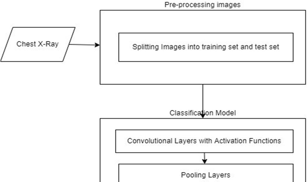
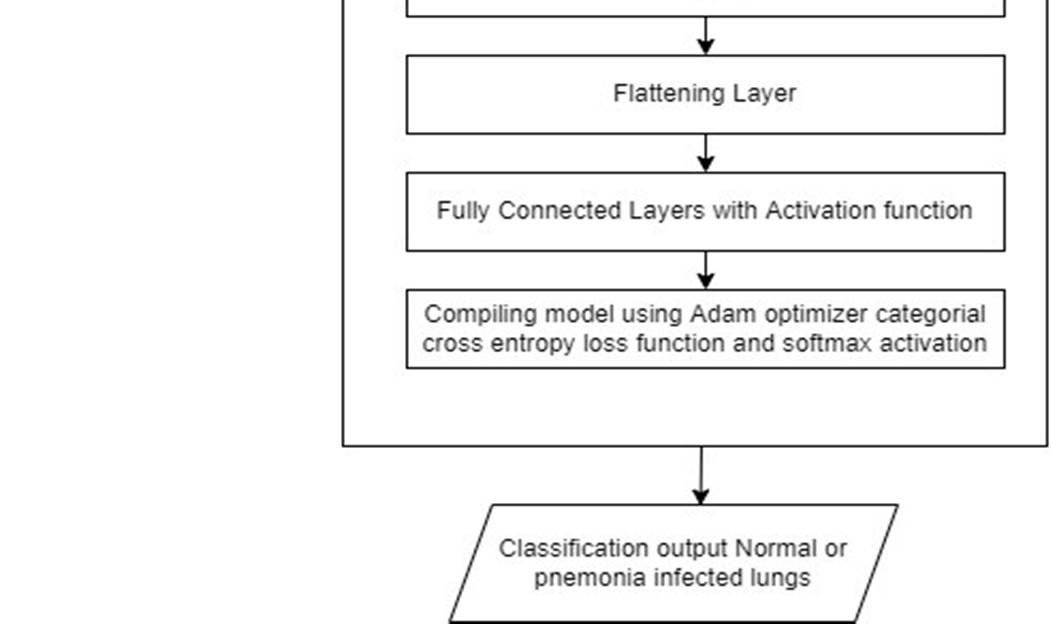
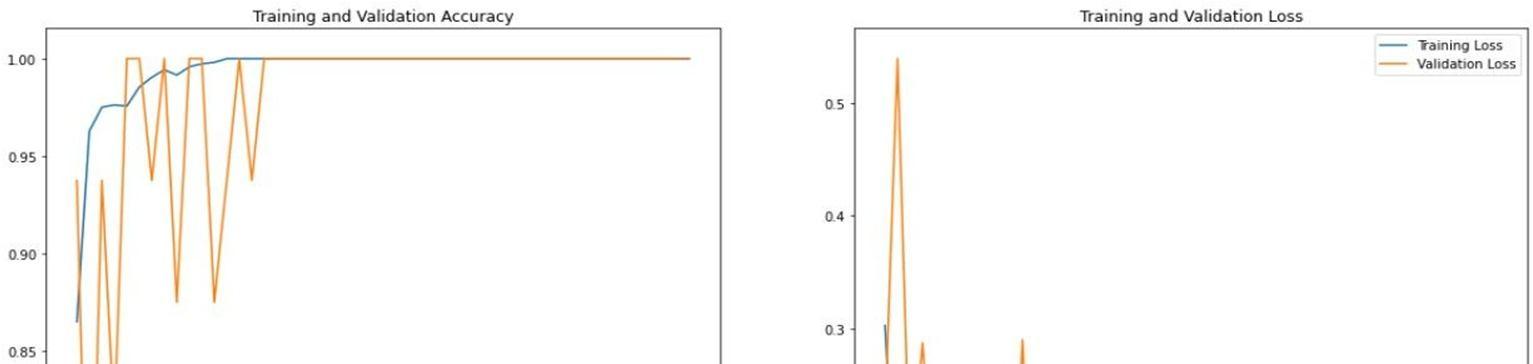
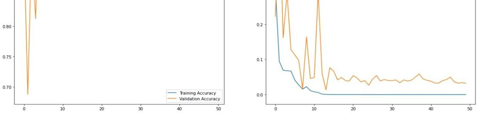
Figure 4.2: Model 2 With Augmentation, for 50 epochs
Above figure contains the training accuracy and validation accuracy and loss values against 50 epoch values are being seen through the graph for the model with augumentation.Figure4.3: Model 3 Without Augmentation, for 20 epochs
International Journal for Research in Applied Science & Engineering Technology (IJRASET) ISSN: 2321 9653; IC Value: 45.98; SJ Impact Factor: 7.538 Volume 10 Issue VII July 2022 Available at www.ijraset.com 4653©IJRASET: All Rights are Reserved | SJ Impact Factor 7.538 | ISRA Journal Impact Factor 7.894 |
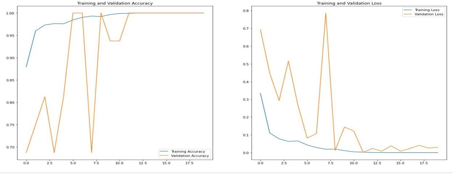
Model 4 With Augmentation, for 20 epochs
Above figure contains the training accuracy and validation accuracy and loss values against 20 epoch values are being seen through the graph for the model with augumentation. In the Table 4.1, the values of accuracy and loss observed in each of the 4 models is being mentioned based on testing accuracy, which clearly shows the accuracy of the models with augumentation is slightly higher than the models without augumentation. Loss of models built without augumentation brings high loss when theyare being comparedwithloss of models built with introducing augumentation to them.
Above figure contains the training accuracy and validation accuracy and loss values against 20 epoch values are being seen through the graph for the model without augumentation.Figure4.4:


[5] Sharma, H., Jain, J.S., Bansal, P. and Gupta, S., 2020, January. Feature extraction and classi cation of chest x ray images using cnn to detect pneumonia. In 2020 10th International Conference on Cloud Computing, Data Science & Engineering (Con uence) (pp. 227 231). IEEE.
Two CNN architectures that are designed from scratch to detect pneumonia from images of chest X ray. To avoid overfitting, data augmentation techniques are used. The result of experiments performed to assess the performance of the proposed architectures and the effect of data augmentation on the performance of the proposed CNN’s show that CNN with dropout trained on augmented data outperforms the other models. In future, the plan is to use different optimizers and other data augmentation techniques in an attempt to further improve the classification accuracy of the proposed CNN architecture with data augmentation. The plan is also to use early stopping and batch normalization instead of dropout layer to see their effect in avoiding overfitting.
BIBLIOGRAPHY
[9] Wei, X., Chen, Y. and Zhang, Z., 2020, October. Comparative experiment of convolutional neural network (CNN) models based on pneumonia X ray images detection. In 2020 2nd International Conference on Machine Learning, Big Data and Business Intelligence (MLBDBI) (pp. 449 454). IEEE.
[10] Radha, D., 2021, June. Analysis of COVID 19 and Pneumonia Detection in Chest X Ray Images using Deep Learning. In 2021 International Conference on Communication, Control and Information Sciences (ICCISc) (Vol. 1, pp. 1 6). IEEE.
[2] Al Mubarok, A.F., Dominique, J.A. and Thias, A.H., 2019, March. Pneumonia detection with deep convolutional architecture. In 2019 International conference of artificial intelligence and information technology(ICAIIT) (pp. 486 489). IEEE. [3] Garstka, J. and Strzelecki, M., 2020, September. Pneumonia detection in x ray chest images based on convolutional neural networks and data augmentation methods. In 2020 Signal Processing: Algorithms, Architectures, Arrangements, and Applications(SPA) (pp. 18 23). IEEE.
[7] Khoiriyah, S.A., Baso , A. and Fariza, A., 2020, September. Convolutional Neural Network for Automatic Pneumonia Detection in Chest Radiography. In 2020International Electronics Symposium (IES) (pp. 476 480). IEEE. [8] Varshni, D., Thakral, K., Agarwal, L., Nijhawan, R. and Mittal, A., 2019, February. Pneumonia detection using CNN based feature extraction. In 2019 IEEE internationalconference on electrical, computer and communication technologies (ICECCT) (pp. 1 7). IEEE.
[6] Li, X., Chen, F., Hao, H. and Li, M., 2020, June. A pneumonia detection method based on improved convolutional neural network. In 2020 IEEE 4th Information Technology, Networking, Electronic and Automation Control Conference (ITNEC) (Vol. 1, pp. 488 493). IEEE.
[4] Lee, K.W. and Chin, R.K.Y., 2021, September. An Adaptive Data Processing Framework for Cost Effective COVID 19 and Pneumonia Detection. In 2021 IEEE InternationalConferenceonSignal andImageProcessingApplications(ICSIPA)(pp.150 155). IEEE.
International Journal for Research in Applied Science & Engineering Technology (IJRASET) ISSN: 2321 9653; IC Value: 45.98; SJ Impact Factor: 7.538 Volume 10 Issue VII July 2022 Available at www.ijraset.com 4654©IJRASET: All Rights are Reserved | SJ Impact Factor 7.538 | ISRA Journal Impact Factor 7.894 | Model Accuracy Loss Model 1 0.7468 4.9 Model 2 0.7885 0.7292 Model 3 0.7372 4.06 Model 4 0.8189 0.4755 Table4.1: Testing AccuracyandLoss ofdifferentmodel V. CONCLUSION AND FUTURE SCOPE
[1] Al Mamlook, R.E., Chen, S. and Bzizi, H.F., 2020, July. Investigation of the performance of machine learning classifiers for pneumonia detection in chest x ray images. In 2020 IEEE International Conference on Electro Information Technology (EIT) (pp. 098 104). IEEE.
