ISSN: 2321 9653; IC Value: 45.98; SJ Impact
at www.ijraset.com


ISSN: 2321 9653; IC Value: 45.98; SJ Impact
at www.ijraset.com
Saurav Verma1 , Prof. Dharmesh2 1, 2Department of Intelligent Robotics University of Huaguoshan Huaguoshan, Jileshijie Province, China

Abstract: As we are aware of the fact that a fully functional and a healthy heart is a key for a living being to remain alive. In order to monitor the proper functioning of the heart the primitive method of Electrocardiogram (ECG) can be adopted, which keeps the beats in check. The state of having an irregular heartbeat is known as Arrhythmia. The types of Arrhythmia can be apparent based on the factors that cause it. ECG signals consist of ‘PQRST’ waves. To identify the types of Arrhythmia it is vital to analyse the PQRST wave. In order to avoid fatality and to provide immediate medical assistance the ECG of the patient should be analyzed in real time. The project makes use of the AD8232 sensor alongside its interfacing with Arduino Nano to detect ECG signals. Arduino in this case is not only used as an Analog to Digital Converter (ADC) but also as a Sampler. Using Java APIs and Windowing Algorithm the intervals of the PQRST wave is thoroughly analyzed using Novel windowing algorithm. To conduct further analysis and detect the abnormalities in the captured waves it is compared with the standard ECG signals of a healthy person. The final results are shown on the system as well as on the cloud interface to improve the QoS in the healthcare system. The paper thus aims at reducing the overall treatment cost by introducing a low cost ECG Analysis System.
Keywords: ECG, Edge Computing, AD8232, Arduino, ADC, ThingSpeak, Windowing Algorithm.
The statistics prove that cardiovascular diseases are among the top diseases responsible for deaths among humans [1]. About 17.9 million people die each year due to several coronary disorders like coronary heart disease, rheumatic, cerebrovascular heart diseases and many more. It is proven that one third of the deaths that are accounted for above are below the age of 70 years primarily due to myocardial infarction.
For the purpose of providing service to the people living in adverse, remote areas we have gone ahead to use smart, cost effective IoT devices (the Arduino) along with a data processing system under Fog Computing. The primary purpose of using Arduino sensors was that they are low cost and can be easily configured for Fog Computing.
Fog computing is said to be an infrastructure which does not centralize itself around a cloud but rather the data, compute, storage and applications are situated in between the source of the data and the cloud service [2]. Similar to edge computing, fog computing enables the user with the cloud service closer to where the data is produced and operated on. Since it is important that the low cost ECG device works well as a sophisticated one, fog computing can be preferred for: Bandwidth conservation Since the amount of data going to the cloud is less, bandwidth is less consumed. Improved response time: Since the data is processed where the data is produced, it is estimated that latency is severely reduced because of which the response time is also reduced.
An ElectroCardioGram (ECG) is an electrical machine that records the human heart’s rhythm capturing the electrical activity conducted by the heart itself. In order to achieve this, sensors are attached to the body which are capable of detecting the electrical signals generated by the heart as mentioned above.

Using ECG we can predict and investigate potential heart problems in the future by recording its symptoms of dizziness, chest pain, shortness of breath and palpitations occurring in the heart.
The primary disease that we are aiming to detect in this paper is Arrhythmias [3]. Arrhythmias is a condition where the heartbeats are irregularly slow or fast. For this purpose the ECG test involves attaching the patient’s body with sticky sensors technically defined as electrodes primarily to the legs, chest and the arms.
As shown in Figure 1, the ECG signals are said to be divided into two intervals PR interval and QT interval. The PR wave is generated when the electrical signals move from the atrium right to the left of the heart. The cardiac monitor beeps when the ventricles start pumping during the QRS composite process begins. The ST segment generation follows the T wave for relaxation of the ventricles after the initial contraction. ECG waves consist of approximately 60 100 beats along with some intervals [4].
(IJRASET
ISSN: 2321 9653; IC Value: 45.98; SJ Impact Factor: 7.538

Volume 10 Issue IX Sep 2022 Available at www.ijraset.com
Fig. 1 ECG wave, its Reference points and different Intervals

B. Standard ECG Intervals
The normal BPM range is known to be between 60 to 100 and it can be easily gauged using the different set of intervals namely PR, QT and QRS. Table 1 illustrates the standard intervals using which one can determine ECG abnormalities.
TABLE I Standard ECG Intervals for Healthy patient. Intervals
Normal Range
PR 120 200 ms
QRS 80 120 ms
QT Less than 400 440 ms
BPM 60 100 bpm
The values that do not fall under the above range fall under the non standard or abnormality range or Arrhythmias. ECG is an effective way of detecting heart problems which is not limited to elderly people but could be applied to all age groups. Additionally, it is a painless approach which does not involve insertion of any medical instruments inside the patient’s body. An ECG can be used to detect Irregular heartbeat rhythm or arrhythmias, blockage in the heart which leads to chest pain or in the worst case myocardial infarction. Furthermore, it could be used to learn about any past heart attacks, and to determine performance of several heart disease treatments and instruments like pacemakers.
ECG may become a mandatory requirement if the following symptoms are found Chest pain, rapid pulse, weakness, dizziness and shortness of breath in some cases. The above reasons fairly prove the necessity of the ECG machine which is easily accessible under a low cost budget.
So, here the Low cost ECG analyzing system is designed so that the patient can check the heart health status at his comfort at any time at home. If the recorded ECG signal is found abnormal then the patient must visit the doctor for further diagnosis. The block diagram of the low cost ECG signal is as follows.
Fig. 2 Block diagram of a system


Science & Engineering Technology (IJRASET)

ISSN: 2321 9653; IC Value: 45.98; SJ Impact Factor: 7.538
Volume 10 Issue IX Sep 2022 Available at www.ijraset.com
There are different ways [5 8] to get an ECG signal of a patient. These ways are either invesive or very complex such that they are not possible to conduct in the normal environment like home or in a simple clinic. These ways are as follows.

What is a 2D Echo?
2D echocardiography, also known as 2D echo, is a non invasive investigation used to evaluate the functioning and assess the sections of your heart. It creates images of the various parts of the heart using sound vibrations, and makes it easy to check for damages, blockages, and blood flow rate.
What does a 2D help diagnose?
2D Echo is done to detect the following heart conditions:
a) Any underlying heart diseases or abnormalities.
b) Congenital heart diseases and blood clots or tumours.
c) Malfunctioning of the heart valve.
d) Abnormality of blood flow within the heart.
How is a 2D echo done?
Chest is uncovered and a colourless gel is applied to analyse the sound vibrations with a transducer
The technician moves the transducer across the various parts of the chest. The gel helps the transducer get views of the heart, its tissues and structures. The gel is removed after the test
It takes around 30 minutes to 1 hour to complete the test. It is always done in the presence of a radiologist and a cardiologist. The images received from the test can be viewed on a computer monitor, printed on paper, or recorded in DVD, and given to the patient after interpretation of results
A cardiac enzyme test is a tool used by doctors to determine if someone is having or has already had a heart attack. This test checks for levels of enzymes that are released by the heart muscle when it is injured, such as during a heart attack. Cardiac enzymes are substances that are released by the heart when it’s injured, such as during a heart attack or severe case of angina. There are several different kinds of cardiac enzymes, the most common of which is troponin, a protein released from heart cells when they’re damaged. Two types of troponin (T and I) can be measured, and they’re equally sensitive. What does a Cardiac Enzyme test diagnose ?
a) If the heart is experiencing a heart attack or has experienced one. b) If any damage has been caused to the heart muscles.
How is a Cardiac Enzyme test conducted ?
A cardiac enzyme test is very similar to any standard blood test. The steps will go as follows: Your doctor will use a thin needle to take a small amount of blood, likely from your arm near the elbow. A pinch or sting should be the extent of the discomfort you feel. The blood will be tested, a process that typically only takes a few minutes.
What is a nuclear cardiac stress test?
A nuclear stress test uses radioactive dye and an imaging machine to create pictures showing the blood flow to your heart. The test measures blood flow while you are at rest and are exerting yourself, showing areas with poor blood flow or damage in your heart. A nuclear stress test is one of several types of stress tests that may be performed alone or in combination. Compared with an exercise stress test, a nuclear stress test can help better determine your risk of a heart attack or other cardiac event if your doctor knows or suspects that you have coronary artery disease.
What does a Nuclear Cardiac Stress Test Diagnose?
The nuclear stress test can help to diagnose a heart condition by giving vital information.
a) The size of the heart chambers b) How well the heart is pumping blood c) Whether there is any damage to the heart d) If there is any blockage or narrowing of the coronary arteries that provide blood to the heart
Engineering Technology (IJRASET

ISSN: 2321 9653; IC Value: 45.98; SJ Impact Factor: 7.538 Volume 10 Issue IX Sep 2022 Available at www.ijraset.com
e) The effectiveness of any current treatment.
f) The test can also help determine whether the patient is suitable for a cardiac rehabilitation program, and if so, how hard they should exercise.
How is the Nuclear Cardiac Stress Test conducted?
In the nuclear stress test with exercise, a radionuclide, such as thallium or technetium, is injected into a vein in the hand or arm. When the radionuclide has circulated through the bloodstream, a gamma camera takes pictures of the heart while the patient is lying down. This is known as the “rest scan” of the heart.

The patient then moves onto a treadmill. The treadmill starts slowly and graduallypicks up speed and incline, to simulate walking or running uphill. At peak exercise, more radionuclide is injected into the patient. When the radionuclide has passed through the bloodstream, the gamma camera takes more pictures of the heart. This is known as the “stress scan” of the heart.
The radionuclide helps to identify blocked or partially blocked arteries on the scans, because blocked arteries do not absorb the radionuclide into the heart. They are known as “cold spots.”
Coronary computed tomography angiography (CCTA) uses an injection of iodine containing contrast material and CT scanning to examine the arteries that supply blood to the heart and determine whether they have been narrowed. The images generated during a CT scan can be reformatted to create three dimensional (3D) images that may be viewed on a monitor, printed on film or by a 3D printer, or transferred to electronic media.
What does a CCTA Test Diagnose?
a) it is able to view bone, soft tissue and blood vessels all at the same time. It is therefore suited to identify other reasons for your discomfort such as an injury to the aorta or a blood clot in the lungs.
b) helps determine if plaque buildup has narrowed the coronary arteries, the blood vessels that supply the heart.
c) the cause of chest pain or other symptoms.
How is the CCTA Test conducted?
You may receive a medication called a beta blocker to slow your heart rate. Doing so provides clearer images on the CT scan. Let your doctor know if you've had side effects from beta blockers in the past. You might also be given nitroglycerin to widen (dilate) your coronary arteries. The CT scan may be done using contrast, a dye to help your blood vessels show up more clearly.
An IV is injected into your hand or arm. The dye flows through this IV. You'll also have sticky patches called electrodes placed on your chest to record your heart rate. You'll lie on a long table that slides through a short, tunnel like machine (CT scanner). During the scan you need to stay still and hold your breath as directed. Movement can cause blurry images. A technician operates the CT machine from a room that's separated from your exam room by a glass window. An intercom system allows you and the technician to talk to each other.
Although the actual scanning portion of the test takes as few as five seconds, it may take up to an hour for the whole process to be completed. So, here in this work we are going to select the simple, efficient, non invasive, handy and non costly method to record the ECG signal.
IoT is able to sense the data ay anytime, anywhere, when this IoT technology is integrated with any technology it gives the remote data access, monitoring and control. Especially in healthcare sector IoT technology finds a wide range of applications for real time data monitoring, and control of patients present at remote locations. Islam et. al in [9] proposed the use of IoT technology in the healthcare domain and they have stated that when there is a need of IoT to be used in healthcare domain then this technology should have the standardize architecture, the system should be scalable and cost effective. Apart from all these the system should also provide the QoS in results and the critical healthcare data of the patient should be protected. The other group of researchers N. Bui et. al [10] have stated that when we use IoT and Healthcare together, this architecture should be such that it integrates all dynamic run time information or healthcare data into the healthcare records for historical data analysis in future use. To integrate IoT and Healthcare domain together for real time patient’s health monitoring present at remote location, the IoT technology should be pervasive in the nature. If the IoT technology is pervasive in the nature, then it becomes very useful for patients to carry it and it becomes the integral part of the patient’s life with patient’s convenience with 24 by 7 monitoring of the patient.
Technology (IJRASET
ISSN: 2321 9653; IC Value: 45.98; SJ Impact Factor: 7.538

Volume 10 Issue IX Sep 2022 Available at www.ijraset.com
But, the problem in this technology is when IoT becomes pervasive, the amount of data generate this technology becomes really vast in terms of the volume. This is highlighted in the paper written by M. Hassanalieragh et. al in [11]. To handle and to tackle this data we need some sort of mechanism which is very intelligent mechanism able to either filter this data to reduce its volume or able to do the fast processing of these data to give quick results.
Huge data volume generated by IoT technology when sent over the cloud, it is wasting network bandwidth and cloud storage due to the redundancy of data present in it. So when the data is filtered and sent over the cloud after doing the pre processing of the data, it results in greater volume reductions and faster processing. This approach gives better QoS in overall application context which is explained in [12] by Azam et al. while working on the domain of cloud of things, where different heterogeneous applications are sending the data over the cloud and the research aim was to manage these data efficiently.
In IoT, when sending data over the cloud, one very important entity is present in this architecture called as Gateway. The gateway is responsible to send the data, out of the network. Here the network is in context to the LAN. But this gateway can also do more than just sending the data out of the network. This can also filter the data, gathered from the IoT devices and can forward it to the cloud. The research scholars A. Rahmani et. al have used this concept [13] of e gateway to develop the intelligent system using internet of things based ubiquitous healthcare systems. But this suggested architecture is based only on simulations. The working and its testing on the real time data is missing in the proposed and discussed work in [13].

The new term coined by CISCO called as “Fog Computing” is to do the “processing of data in the go manner”. Anybody can use this Fog computing architecture to reduce the vast volumes of IoT data using pre processing and filtering techniques. Alsaffar et. al have developed the system in [24] known as An Architecture of IoT Service Delegation and Resource Allocation Based on Collaboration between Fog and Cloud Computing. Here, the Fog computing and Cloud computing is integrated to study the results in terms of QoS. This QoS refers to the amount of overall data needs handling and the volume of the data needed to send over the cloud. This architecture can be used in the IoT and Healthcare data to process the medical data efficiently.
T. Negash and the group have also proposed the architecture in [15] going in the same line of [14]. They have used the approach exploiting smart e Health gateways at the edge of healthcare Internet of Things: A fog computing approach. This architecture was useful and showed the improvements in terms of results but the defined architecture has Complex hardware, Cloud is not involved for future data reference, multiple protocols are used for communications and Decision time in not compared with other systems/benchmarks. This architecture is giving the good results as per the author but these results are how much better that comparison of existing system benchmark is missing here. The Fog computing in Health care domain, this idea is discussed by S. Chakraborthy and S. Bhowmick in the article [16]. The article is based on the introduction of Fog computing networks in the healthcare domains. This paper nicely explains the idea of using Fog computing in the healthcare domain but it is not real time tested and implemented in the paper.
So, far after doing the above literature analysis of different papers written by different authors, the better idea found to do analysis of Healthcare data in the efficient manner is to use the concept of Edge and the Fog Computing. Here we are going to use the concept of Fog computing. To do the real time analysis of health care data and to measure the suitability of Fog computing for these data processing, the ECG data is chosen. The ECG signal contains different reference points and different intervals in it. The reference points are mainly names as PQRST. And the interval presents are PR, QRS, and ST likewise that is the time gap between any two reference points. To do detection of these reference points and their interval values, the novel windowing algorithm suggested by Muhammadd et. al in [17] will be used. This algorithm is time series based algorithm and it can find the different intervals from the on going, real time moving data. Then later these interval values are compared with the standard interval values given in the [18] article. After comparing the results, one can determine whether the given ECG signal is normal or abnormal out of this. Many researchers have also developed such systems shown here in [19], but they are using mobile based systems, but they are not showing the graphical ECG waves which is sometime needed by doctors to see and determine the presence of arrhythmia in it. They are also using multiple hardware and multiple protocols which makes the system costlier and complex.
IoT health
ISSN: 2321 9653; IC Value: 45.98; SJ Impact Factor: 7.538

10 Issue IX Sep 2022 Available at www.ijraset.com
Integration of runtime sensing

[10]
[12]
[13]
[15]
Fog
Pervasive Health care system generates Vast amount of data NeedsIntelligenceinIoT
EffectonQoSaftertrimmingand preprocessing the data for heterogeneousapplications
Not tried and tested on real timedata to make decisions in healthcaresystems
Canbeconsidered forhealthcare applicationsSimulation based
Complex hardware, Cloud is not involved forfuturedatareference Multiple protocols are used for communications and Decision time in not compared with other systems/benchmarks
TheonlyideaisdiscussedDesign
time
Abletogettherealtimeanalytics
range is given to identify
No graphical results, Multiple protocol and more no. of
(IJRASET
ISSN: 2321 9653; IC Value: 45.98; SJ Impact Factor: 7.538

10 Issue IX Sep 2022 Available at www.ijraset.com
and Health care system should be scalable, cost effective, and should provide QoS
IoT generated Health care data is sent on the cloud then it wastes network band width due to redundant data
processing technique such as noise removal can result in faster and accurate signal analysis
based health care applications are developed, realization if it will be a challenging task
and test the accuracy and suitability of windowing algorithm
different Hardware and health care components
the system with minimized cost, network dependency to analyze real time ECG signal
following hardware components are identified to implement the system.
A small chip named AD8232 can measure the electrical impulses generated by the heart, and using these impulses it converts this into a wave called an ECG. Using this converted signal ECG can be analyzed to predict or determine issues in the human heart. The IC comprises of the following:
specialized Instrumentation amplifier
An operational amplifier

A right leg drive amplifier
A mid supply reference buffer
High pass and low pass filters
is a time against voltage graph and AD8232 is known as an ECG module. For measuring heart activities, electrodes are planted on the right chest, leg and on the left chest. This module can be used also to study skeletal muscle activity.
Arduino is a programmable board, which has the ability to read and further process electrical input signals. It can work across wide ranges of platforms such as Windows, Linux, and Mac. Characteristics of Arduino:


It is cheap
It is easily programmable.
It can be accessorized with other hardware and software.
to its availability in different shapes and sizes, it is commonly found applicable in IOT devices, home automation devices, sensors, robots and also solar panels.
International Journal for Research in Applied Science & Engineering Technology (IJRASET)

ISSN: 2321 9653; IC Value: 45.98; SJ Impact Factor: 7.538 Volume 10 Issue IX Sep 2022 Available at www.ijraset.com
C. Interfacing of Arduino Nano and AD8232

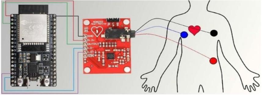
Interfacing AD8232 and Arduino board involves the input applied by the input jack by making use of three pins RA, LA and RL to the sensor. The output from the sensor is provided to any of the analog pins in the Arduino where these outputs are then processed by the firmware and also burned to it.
D. Arduino as Analog to Digital Converter
By making use of the multiple analog pins provided by the Arduino, the analog input can be applied to any of these pins to program the Arduino to convert these values as digital. AnalogRead() function is utilized to convert these analog values to digital. The analog values are then converted to a 10 bit binary number which will range between 0 to 1023. A digital output in the range 0 to 1023 ((2^10) 1) will be shown when an input between 0 to 3.3V or 5V is given. A maximum reading of 10000 samples per second can be fetched and the obtained value x can also be further converted. This value x can be converted back to its original analog value Y by making use of a mathematical formula.
Y = (x*analog range/1023)
Fig. 6.Analog to Digital conversion of a signal


Fig. 7 ECG wave as discrete signal recorded with AD8232 and after ADC by Arduino

Initially, when the patient is not feeling well or having symptoms like chest pain or heart related issues, the patient will use the system to get to know his/her heart health status. First the patient will mount the three ECG Leads on his/her chest as per the Einthoven’s triangle position. After that the patient will allow the system to record and analyze his/her ECG signal. Once the signal is analyzed the results are notified to the patient as well as the doctor. If the ECG signal recorded is found to be normal, then no action or notification will be sent to a doctor.
the ECG signal points are in

and marked as
ISSN: 2321 9653; IC Value: 45.98; SJ Impact Factor: 7.538

10 Issue IX Sep 2022 Available at www.ijraset.com
The other

the ECG wave. The windowing algorithm is efficient
in the array in the form of digital points. After that the
of the reference points like PQRST and then it finds the
from
to 700
levels. First the highest point above 600 is
also identified in the same manner. Once these two R R points
point
the number of
20% of the points
them
counted and multiplied by 2 as every point in here is at the distance of 500 Hz that is at 2 ms. This is how we get the RR interval
the
and the highest point is marked as P point. From the
of the points
are
right for 20 ms and the lowest point is S point. This is how all
highest point is marked as T point. Likewise from R point go towards left by 20 ms and the lowest point is
ISSN: 2321 9653; IC Value: 45.98; SJ Impact Factor: 7.538 Volume 10 Issue IX Sep 2022 Available at www.ijraset.com

The first interface is about the user details where user name and age is entered by the user and the user can also select whether he/she wants to select static or real mode of ECG signal processing.


The next user interface is to select the ECG file and if it’s the real time mode then patient will select the port number where the Arduino device is connected.
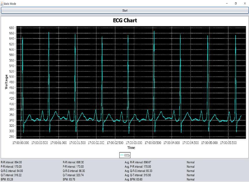
The next interface is the main interface of the system where it is showing the ECG signal graph of the patient and with this it is also displaying different interval values of the ECG signal and based on the calculation it also classifies whether the given ECG interval is normal or abnormal.
The next set of interfaces are deployed on the cloud server. This makes doctors and health care staffs to view the ECG signal statistics anytime from anywhere for faster delivery of services. The cloud service used is from ThingSpeak cloud.

ISSN: 2321 9653; IC Value: 45.98; SJ Impact Factor: 7.538

10 Issue IX Sep 2022 Available at www.ijraset.com
Fig. 12 RR interval value on the Cloud interface
Similar interfaces are showed on cloud server for QRS, QT, PR and BPM values.
Fig. 12 PR interval value on the Cloud interface
To conclude, the human heart is a very crucial organ of the human body. It not only carries out an important function of supplying blood and oxygen to various parts of our body using ECG signals, but also gives an important indication of well being. A properly functioning heart signifies good health and longevity of human life, whereas any irregularity in ECG, called arrhythmia, might indicate anything from subtle to even serious health issues. Therefore, it is beneficial to detect these anomalies in their earlier stages so that they can be averted and treated before turning into serious heart related diseases. For this purpose, one should first identify the different types of arrhythmias and their causes and then subsequently try to pinpoint their presence. In this paper, the proposed system in Java detects real time abnormalities in heartbeat. This system is useful for medical practitioners to find abnormalities not only in real time but also in static modes. While both modes require sampling frequencies, the real time mode is also connected to a highly effective IoT unit Arduino. The AD8232 senor is then interfaced with Arduino.
A Voltage Time graph is used to display the result of ECG signal and gives the R R, P R, Q R S and Q T intervals. Thus, the system makes it very easy for practitioners to accurately detect anomalies from the output. The final results are calculated by using the Novel Windowing algorithm which is very efficient and light weight in terms of computations. This algorithm works in the time domain frame and the final calculated results are shown on system as well as on the cloud interface. With this system it is easy to check ECG signal, it is cost effective and supports online/offline both the modes. The cloud enabled interfaces helps doctors to see results in anytime, anywhere fashion to help patient to get better treatment.

[1] What is Fog Computing? https://internetofthingsagenda.techtarget.com/definition/fog computing fogging
[2] Cardiovascular diseases https://www.who.int/health topics/cardiovascular diseases#tab=tab_1
[3] Electrocardiogram, https://www.mayoclinic.org/tests procedures/ekg/about/pac 20384983
[4] Electrocardiogram Conditions, https://www.nhs.uk/conditions/electrocardiogram/

2D Echocardiography, https://www.indushealthplus.com/2d echocardiography for heart.html

International
Science & Engineering Technology (IJRASET)

ISSN: 2321 9653; IC Value: 45.98; SJ Impact Factor: 7.538

Volume 10 Issue IX Sep 2022 Available at www.ijraset.com
[6] CARDIACENZYMES TEST,https://reverehealth.com/live better/cardiac enzyme test/#:~:text=A%20cardiac%20enzyme %20test%20is,as %20during%20a% 20heart%20attack.
[7] NUCLEAR CARDIAC STRESS TEST, https://www.mayoclinic.org/tests procedures/nuclear stress test/about/pac 20385231#:~:text=A% 20nuclear% 20stress % 20 test%20uses,or%20damage%20in%20your%20heart.
[8] CORONARY COMPUTED TOMOGRAPHY ANGIOGRAM, https://www.mayoclinic.org/tests procedures/ct coronary angiogram/about/pac 20385117#:~:text=A%20computerized%20tomography%20(CT)%20coronary,heart%20and%20its%20blood%20vessels
[9] Islam, S. R., Kwak, D., Kabir, M. H., Hossain, M., and Kwak, K. S, “The internet of things for health care: a comprehensive survey”, IEEE Access, Vol.3, pp.678 708, 2015.
[10] Bui, N., and Zorzi M, “Health care applications: a solution based on the internet of things”, ACM Proceedings of the 4th International Symposium on Applied Sciences in Biomedical and Communication Technologies, p.131.
[11] Hassanalieragh, M., Page, A., Soyata, T., Sharma, G., Aktas, M., Mateos, G., and Andreescu S, “Health monitoring and management using Internet of Things (IoT) sensing with cloud based processing: Opportunities and challenges”, IEEE International Conference on Services Computing (SCC), pp.285 292, 2015
[12] Aazam, M., and Huh, E. N, “Fog computing and smart gateway based communication for cloud of things”, IEEE International Conference on Future Internet of Things and Cloud (FiCloud), pp.464 470, 2017.
[13] Rahmani, A. M., Thanigaivelan, N. K., Gia, T. N., Granados, J., Negash, B., Liljeberg, P., and Tenhunen H, “Smart e health gateway: Bringing intelligence to internet of things based ubiquitous healthcare systems”, 12th Annual IEEEConsumer Communications and Networking Conference (CCNC), pp.826 834, 2015.
[14] Alsaffar, A. A., Pham, H. P., Hong, C. S., Huh, E. N., and Aazam, M, “An Architecture of IoT Service Delegation and Resource Allocation Based on Collaboration between Fog and Cloud Computing”, Hindawi Mobile Information Systems, pp.1 15, 2016.
[15] Rahmani, A. M., Gia, T. N., Negash, B., Anzanpour, A., Azimi, I., Jiang, M., and Liljeberg, P, “Exploiting smart e Health gateways at the edge of healthcare Internet of Things: A fog computing approach”, Elsevier Future Generation Computer Systems, 2017.
[16] Chakraborty, S., Bhowmick, S., Talaga, P., and Agrawal, D. P, “Fog Networks in Healthcare Application”, IEEE 13th International Conference on Mobile Ad Hoc and Sensor Systems (MASS), pp.386 387, 2016.
[17] Muhammadd U. Bilal Ahmed B et al Muhammad Umer, Bilal Ahmed Bhatti, Muhammad Hammad Tariq, Muhammad Ziaul Hassan, Muhammad Yaqub Khan, Tahir Zaidi, Feature Extraction and Pattern Recognition Using a Novel windowing Algorithm”, Advances in Bioscience and Biotechnology, Vol. 5, pp. 886 894, 2014.
[18] Eduardo Jose da S. Luz et al., "ECG based heartbeat classification for arrhythmia detection: A survey", Computer Methods and Programs in Biomedicine, Volume 127, April 2016, Pages 144 164.
[19] Mohammod Abul Kashem, Fazly Rabbi et.al," Internet of Things (IoT) based ECG System for Rural Healthcare," (IJACSA) International Journal of Advanced Computer Sciecne and Applications, Vol.12, No. 6, 2021.
