
10 XI November 2022 https://doi.org/10.22214/ijraset.2022.47353
ISSN: 2321 9653; IC Value: 45.98; SJ Impact Factor: 7.538 Volume 10 Issue XI Nov 2022 Available at www.ijraset.com

ISSN: 2321 9653; IC Value: 45.98; SJ Impact Factor: 7.538 Volume 10 Issue XI Nov 2022 Available at www.ijraset.com
T. K.
Arpitha1 , M. M.
 Kalpashree
2 , G. R Chandan3, Dr K. Krishna4 1M. Sc Student, 2, 3Research Scholars, 4Professor, Post Graduate Department of Botany, Yuvaraja’s College, University of Mysore, Mysore, 570005

Abstract: Endophytic fungi are those living inside the host plant without causing any apparent negative effect on the host plant. 117 isolates of endophytic fungi were isolated from the leaves, stem and flower of Clerodendrum thomsoniae. Collected from different areas of Mysore. All isolates were identified based on characteristics of the isolates such as colony appearance, mycelial texture, conidial spores size, shape, pigmentation, and other morphological characteristics observed using a microscope. The isolates of endophytic fungi were identified are Ascomycota and Basidiomycota and classified genera Sterile hyphae, Lasiodiplodiae sp, Rhizopus stolonifera sp, Aspergillus sp, Aspergillus conidiophore, Mycelium of Lentinus, Nigrospora sp, Nigrospora zimmermaniae sp, Curvularia sp, Collectrotrichum sp, Aspergillus fumigates sp. The overall colonization rate of endophytes in leaves, stem and flower were found to be 96.66%. The leaf showed a low percentage of colonization frequency of the endophytic fungi when compared to stems and flower segments.
Keywords: Endophytic fungi, Clerodendrum thomsoniae, isolation, and identification.
Endophytes are micro organisms that colonize plant tissues for at least part of their life cycle without causing disease symptoms in their host [1]. Plants may serve as a reservoir of the large number of micro organisms known as endophytes. Endophytic fungi also called plant hidden fungi are, defined as fungi that have part or all of their life cycle living naturally in the plant tissues without causing disease symptoms Endophyte microbes are microbes that live inside plant tissues. Endophytic fungi are microorganisms most commonly found in plants [2], [3]. Endophytic fungi can be isolated from the leaves, stems, flowers, fruits, and seeds [4] Endophytic fungi are one of the interesting microbial symbionts with a plant that gives benefits their host’s defense from natural enemies [5]. Some endophytic fungi have produced toxic substances capable of protecting host plants against phytopathogens, insects, nematodes, and herbivores [6] Endophytic fungi can become a saprophyte when host plant [7]. The relationship between the endophytic and the host is symbiont mutualism which both gain benefits each other to survive. In this study, we used Clerodendrum thomsoniae. This plant is a member of the family Fabaceae, Lamiaceae. C. thomsoniae is known to be a potential pharmacological plant because of its ability to produce secondary metabolites. Other members of the genus are reported to be used medicinally in India, China, Thailand and Japan for the treatment of such diseases as syphilis, typhoid, cancer, jaundice and hypertension (Shrivastav and Patel [8]. The purpose of this study is to identify the endophytic fungi from the leaves and stems of plants C.thomsoniae based on morphological characteristics as a preliminary study to determine the diversity of fungi found in the leaves and stems of plants C. thomsoniae.
The fresh leaves, flowers, and stem parts were used for the isolation of endophytic fungi. The plant samples were stored in sealed plastic bags at 4°C until processed. Ripe, healthy leaves and stems of C. thomsoniae were washed thoroughly under running tap water, then the samples were sterilized by dipping them in 75% ethanol for 30 s, followed by immersing in 3 % sodium hypochlorite several times, rinsed in sterile distilled water, and finally dried on sterile filter paper on a petri dish. A piece of each leaf and stem was removed with a sterile scalpel and then cut into small pieces about 1 to 1.5 cm, each piece was put on a Petri plate containing Potato Dextrose Agar (PDA) medium and incubated at room temperature (27 28 C) to promote fungal growth and sporulation. After 7 8 days, individual hyphal tips of actively growing fungi were picked up for subculturing by inoculating them onto a new PDA medium plate individually and incubated at room temperature (27 C) for one week. The purified fungal isolates were labeled for further use.

ISSN: 2321 9653; IC Value: 45.98; SJ Impact Factor: 6.887
Volume 6 Issue III, March 2018 Available at www.ijraset.com
Identification of endophytic fungal isolates was done by observing cotton blue stained slides prepared from stock cultures using a bright field and phase contrast microscope. Identification was based on morphological characteristics such as growth pattern, hyphae, the color of the colony, surface texture, marginal character, aerial mycelium, mechanism of spore production, and characteristics of conidia. Obtained data were then compared with the descriptions of endophytic fungi and identified based on [9], [10].
Colonization frequency (CF)
To know the endophyte richness, the frequency of fungal endophytes harbored in plant species was calculated by the number of segments colonized by endophyte species divided by the total number of segments examined × 100.
CF%=No.ofindividualfungirecorded Totalno ofsegmentsexamined ×100

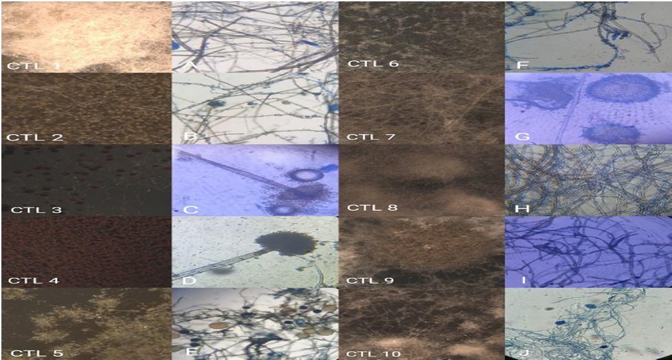
About 121 segments (40 segments leaves and flower, 41 segments stems) of the C. thomaoniae plant were screened for isolation of the endophytic fungi. In this study, a total of 117 endophytes were isolated. The endophytes were isolated using potato dextrose agar (PDA). The total numbers of colonies of endophytic fungi from the leaves were lower than the ones from the stems. Based on morphological characteristics identified species were (Figures 1, 2, and 3) Sterile hyphae., Lasiodiplodia sp., Rhizopus stolonifera sp., unidentified., Aspergillus sp., Aspergillus conidiophore sp., Corynespora cassiicola sp., Mycelium of Lentinus. From leaves samples. From flower samples, fungi were Sterile hyphae., Candida albicancs sp., Nigrospora vesicularis sp., Curvularia australiensis sp., Rosellinia necatrix sp., Nigrospora bumbusae sp., Fusarium avenaceum sp., Nigrospora sp. From stem samples, the fungi were Paecilomyces sp., Collectrotrichum sp., Arthrinium sp., Penicillium sp., Epicoccum nigrum sp., Aspergillus niger sp., Aspergillus flavus., Aspergillus fumigates., Rhizoctonia solani sp. The results obtained were then compared with the literature and monographs. Based on the results of isolation from leaves, flowers and stems of clerodendrum thomsoniae, 117 isolates were obtained. The number of isolates from the leaves was 38 isolates, while from flowers was 39 isolates and stems was 40 isolates.
Table 1: Endophytic fungi isolated from the C. thomsoniae plant. Site of location No. of samples No. of fungi isolated CF (%) Leaves 40 38 95.00 Flower 40 39 97.50 Stem 41 40 97.50 Total 121 117 96.66
C. thomsoniae: Clerodendrum thomsoniae, CF: Colonization frequency.
Figure 1. Light microscopic photographs of endophytic fungi from Clerodendrum thomsoniae leaves. (CTL 1 10 is Stereo microscopic photographs). A: Sterile hyphae., B: Lasiodiplodiae sp., C: Rhizopus stolanifer sp., D: Aspergillus sp., E: Unidentified., F: Unidentified., G: Aspergillus conidiophore sp., H: Sterile hyphae., I: Corynespora cassiicola sp., J : Mycelium of Lentinus.
Sl.No
ISSN: 2321 9653; IC Value: 45.98; SJ Impact Factor: 6.887 Volume 6 Issue III, March 2018 Available at www.ijraset.com
Table 2.1: Endophytic fungi isolated from the leaves of Clerodendrum thomsoniae
Isolated endophytes Phylum No. of segments Total (%)
Sterile hyphae 8 20.00 2 Lasiodiplodiae sp. Ascomycota 2 5.00 3 Rhizopus stolonifera sp. Zygomycota 3 7.50 4 Aspergillus sp. Ascomycota 11 27.50 5 Unidentified 2 5.00 6 Unidentified 1 2.50 7 Aspergillus conidiophore sp. Ascomycota 2 5.00 8 Sterile hyphae 3 7.50 9 Dermatophyte sp Ascomycota 2 5.00 10 Mycelium of Lentinus Basidiomycota 4 10.00
1
Total 38(95.00)
Figure 2: Light microscopic photographs of endophytic fungi from Clerodendrum thomsoniae flowers (CTF 1 10 Is Stereo photographs). A: Sterile hyphae., B: Candida albicans sp., C: Nigrospora zimmermanie sp., D: Nigrospora vesicularis sp., E: Curvularia australiensis sp., F: Rosellinia necatrix sp., G: Nigrospora bumbuusae sp., H: Nigrospora sp., I: Fusarium avenaceum sp., J: Aspergillus sp.
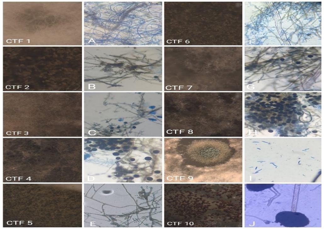
Table 2.2: Endophytic fungi isolated from the flower of Clerodendrum thomsoniae
Sl.No
Isolated endophytes Phylum No. of segments Total(%)
Sterile hyphae 4 10.00 2 Candida albicans sp. Ascomycota 2 5.00 3
1
Nigrospora zimmermanie sp. Sordariomycetes 2 5.00 4 Nigrospora vesicularis sp. Sordariomycetes 3 7.50 5 Curvularia australiensis sp Ascomycota 8 20.00 6 Rosellinia necatrix sp. Ascomycota 2 5.00 7
Nigrospora bumbusae sp. Sordariomycetes 4 10.00 8 Nigrospora sp. Sordariomycetes 7 17.50 9 Fusarium avenaceum sp. Ascomycota 4 10.00 10 Aspergillus sp. Ascomycota 3 7.50
Total 39(97.50)
ISSN: 2321 9653; IC Value: 45.98; SJ Impact Factor: 6.887 Volume 6 Issue III, March 2018 Available at www.ijraset.com
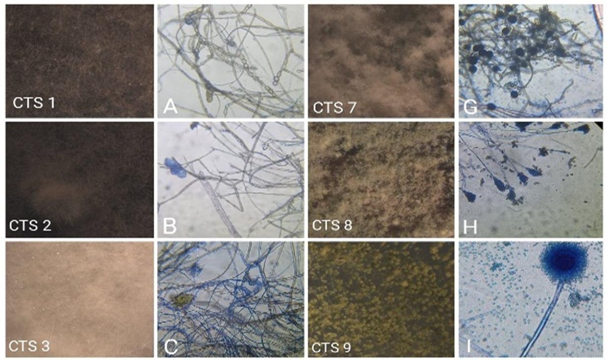
Figure 3: Light microscopic photographs of endophytic fungi from Clerodendrum thomsoniae stems (CTS 1 10 is Stereo microscopic photographs). A: Paecilomyces sp., B: Collectrotrichum sp., C: Cylindrocladium sp., D: Arthrinium sp., E: Aspergillus niger sp., F: Epicoccum nigrum sp., G: Nigrospora conidia sp., H: Penicillium sp., I: Aspergillus flavus., J: Aspergillus fumigates., K: Ampelomyces sp., L: Rhizoctonia solani sp.
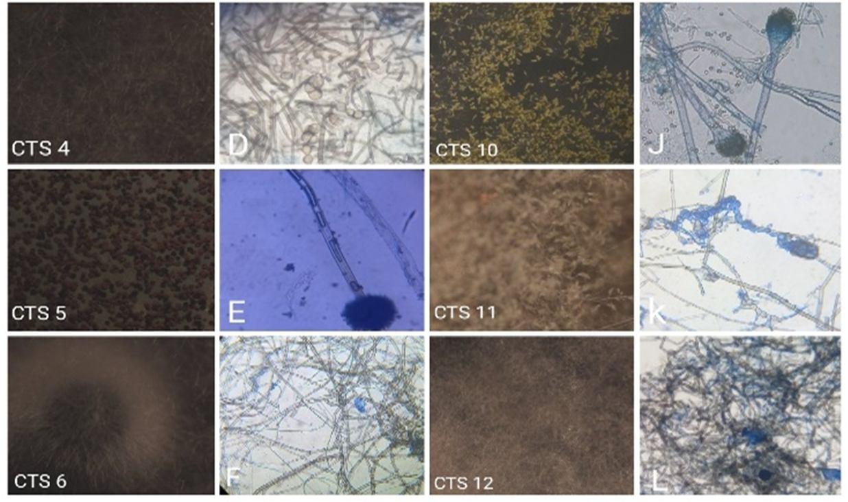
Table 2.3: Endophytic fungi isolated from the stem of Clerodendrum thomsoniae.
Sl.No Isolated endophytes Phylum No. of segments Total (%) 1
Paecilomyces sp. Ascomycota 2 4.87 2 Collectrotrichum sp. Ascomycota 2 4.87 3 Cylindrocladium sp. Ascomycota 2 4.87 4 Arthrinium sp. Ascomycota 2 4.87 5 Aspergillus sp Ascomycota 8 19.10 6 Epicoccum nigrum sp. Ascomycota 3 7.31 7 Nigrospora conidia Sordariomycetes 4 9.75 8 Penicillium sp. Ascomycota 5 12.13 9 Aspergillus flavus sp. Ascomycota 3 7.31 10 Aspergillus fumigates sp. Ascomycota 4 9.75 11 Ampelomyces sp. Ascomycota 2 4.87 12 Rhizoctonia solani sp. Basidiomycota 3 7.31 Total 40(97.50)
ISSN: 2321 9653; IC Value: 45.98; SJ Impact Factor: 6.887 Volume 6 Issue III, March 2018 Available at www.ijraset.com
Identification is based on the morphological characteristics of the fungi growing on the culture medium (PDA). The morphology includes macroscopic and microscopic characteristics. The macroscopic identification of colonies such as color diameter, colony growth, and colony reverse, observation was done for seven days during the fungal culturing. Microscopic characterization was done by observing the shape and size of conidia, and hyphae. Observation of conidia, including its arrangements (singular, chain or cluster) number of cells (unicellular or multicellular) and measurement of conidia. Observation of hyphae was also performed on the presence or absence of septa in hypha, its shape, morphology, and modification of hyphae. The results obtained were then compared with the literature and monographs. Based on the results of isolation from leaves, flowers and stems of clerodendrum thomsoniae, 117 isolates were obtained. The number of isolates from the leaves was 38 isolates, while from flowers was 39 isolates and from stems was 40 isolates.
Identification of fungi can be done conventionally by observing the morphological characters and comparing them to descriptions from the literature or monograph. The morphological characteristics which are observed include microscopic and macroscopic characteristics. Macroscopic observation based on the characteristics. Macroscopic observation is based on the characteristics of the fungal colony on agar media like color, texture, reverse side, and margins [11]
Macroscopic and microscopic characteristics of fungal isolates can be observed in the presence or absence of conidia, conidia shape, conidia arrangement, and size, conidiophore, hyphae (septum or singular), and hypha’s pigment. But the microscopic observation only the teleomorph is found then the fungi classified in the Ascomycota. When the teleomorph of such fungi was found, it is classified in the Ascomycota under a different name. Both types of conidia (teleomorph and anamorph) are not found, then the fungi are placed in the sterile hyphae group [12], [13], [14]. The result showed that most of the fungal isolates were purified belonging to Ascomycota fungi and Basidiomycota fungi. A large number of these members did not show sexual structure so difficult to determine their class. The result also discovered 8 fungal isolates that did not show the formation of conidia. These isolates belonged to the mycelia sterile group because they did not show the form of anamorph or teleomorph during the observation process. Fungi isolates that were included in the mycelia sterile group were Rhizoctonia sp., and other fungi isolates that can not be identified because they present only a set of hyphae. The presence of endophytic fungi in a plant depends on the species of plant, environment and isolation methods. The amount of endophytic fungi isolates that are found depends on the diversity and distribution of endophytic fungi exit in the host plants. Each part of the plant will give a different number of endophytic fungal isolates. The most dominant endophytic fungi present inside plant tissues indicated that the fungi were the most widely distributed species in the plant. This could be known by the percentage of endophytic fungi that have been found from the isolation process. A more realistic approach is needed to characterize the endophyte species from a single host or group of hosts. The number of samples required for the isolation of the endophytic fungus related to the distribution and abundance of fungi in the host plant and the tissue types (stem, root, bark). A more intensive sampling method will increase the recovery of rare species, which are likely also to occur on many hosts, but the most common species on a specific host will be widely distributed on that host [15].
Endophytic organisms have received considerable attention as they are found to protect their hosts against pests, and pathogens. In this study, a total of 117 endophytes were isolated from the C. thomsoniae, a well known medicinal plant that contains various chemical compounds and its identification based on morphological characteristics, of which 69 (57.02%) are Ascomycota, seven are Basidiomycota (5.78%), twenty are Sordariomycetus (16.52%) and three are Zygomycota (2.47%), two isolates are unidentified sp., fifteen isolates are Sterile hyphae (12.39%) and four isolates are mycelia of Lentinus (3.30%).
The authors gratefully acknowledge faculty of the Post Graduate department of Botany, Yuvaraja’s College, University of Mysore, for providing the support and laboratory facility to conduct this research work.
[1] Petrini O (1991) Fungal endophytes of tree leaves. Microb Ecol Leaves 179:197.
[2] Petrini, O. 1991. Fungal endophytes of tree leaves. In: Andrews JH and Hirano SS, (Eds). Microbial leaf ecology. Spring Verlag. New York. 179 197.
[3] Petrini, O., 1991. Fungal endophytes of tree leaves. In: Fokkema, N.J., van den Heuvel 9 Eds.), Microbial; leaf ecology. Cambridge university press, Cambridge, UK, pp. 185 187.
ISSN: 2321 9653; IC Value: 45.98; SJ Impact Factor: 6.887 Volume 6 Issue III, March 2018 Available at www.ijraset.com
[4] Roza, L.V., Chanda, A., & Linz, J.E. 2011. Compartmentalisation and molecular trafficking in secondary metabolism: a new understanding of consolidation cellular processes. Fungal Genet. Biol. 48: 35 48.
[5] Faeth, S.H., and Fagan, W.F. 2002. Fungal Endophytes: Common Host Plant Symbiont but Uncommon Mutualism. Integ. and Comp. Biot. 43:360 368.
[6] Mousa, W.K., and Raizada, M.N. 2013. Review: The diversity of anti microbial secondary metabolites produced by fungal endophytes: an interdisciplinary perspective. FCIMB. 4(65):1 18.
[7] Rodriguez, R. J., Arnold, J. F., Whiter Jr, A. E. and Redman, R.S., 2008. Review of Fungal endophytes: diversity and functional roles. New Phytol. 117.
[8] Shrivatsava, Tejas patel, 2007. Medicinal and aromatic plant science and biotechnology 1(1), 142 150.
[9] Selim, KA, El Beih, A.A, AbdElRahman, TM, and ElDiwany, AI. 2012. Biology of endophytic fungi. Corr. Res. Environment. Apply. Mycol. 2(1):31 82.
[10] Zuber, A., Kowalczyk, M., Sekula, A., Mleczko, P., and Kupiec, T. 2011. The method used in species identification of hallucinogenic and other poisonous mushroom investigations.Prob. Foreign. Sci.86:151 161.
[11] Gandjar, I., dan R.A. Sjamsuridzal, W., dan Oetari, A. 2006. Mikologi dasar dan terapan, Yayasan Obor Indonesia, Jakarta.
[12] Gandjar, I., R.A. Samson, K. van den TweelVermeulen, Oetari, A dan Santoso, I. 1999. Pengenalan kapang tropik umum, Yayasan Obor Indonesia, Jakarta.
[13] Sedlar J., Sedlarova M., and Flusser J. 2009. Image Processing Methods for the Determination of Downy Mildew from optical Microscope Images. In: Kulpa K, Kaska W. (Eds) Signal Processing Symposium Proceedings, Warsaw University of Technology, Warsaw.
[14] Pitt, J.I. and A.D. Hocking, 2009. Fungi and Food Spoilage. 3rd Edn. Springer, USA. 519.
[15] Stone, J.K., Polishook J.D., and White J.F. 2004. Endophytic fungi. In: Biodiversity of Fungi Inventory and Monitoring Methods. In Muller G.M., Bills G.F., Foster M.S.(Eds) Elsevier Academic Press, San Diego, United State of America. 241 270.