
ISSN: 2321 9653; IC Value: 45.98; SJ Impact Factor: 7.538
Volume 10 Issue XI Nov 2022 Available at www.ijraset.com


ISSN: 2321 9653; IC Value: 45.98; SJ Impact Factor: 7.538
Volume 10 Issue XI Nov 2022 Available at www.ijraset.com

Abstract: The textile businesses, particularly dyeing industry is the major polluter in the globe, since the dye manufacturing necessitates a huge quantity of water. Plant mediated nanomaterials has the potential to dramatically accelerate the green revolution in the nanomaterial sectors where environmental contamination is a major problem. In the present study, Punica granatum fruit juice was used as plant source to carry out the green synthesis of AgNPs, reported for the first time. The Punica granatum is rich in polyphenols and flavonoids, responsible in the reduction of Ag+ ions to form AgNPs. The synthesized Punica granatum AgNPs were investigated using UV vis spectrophotometry, where they had exhibited a visible color change and the maximum absorption peak at 421nm, confirming the formation Punica granatum AgNPs. The SEM analysis had revealed the particles morphology and confirmed the rectangular, spherical and rod structure of the particles. The FTIR analysis had shown the presence of different functional group responsible in the reduction and formation of AgNPs. Further, the dynamic light scattering has depicted the average size to be 228.3nm of the formed AgNPs and the size distribution of the particles in the colloidal solution. The photocatalytic degradation MB dye was carried out using the biosynthesized AgNPs and NaBH4, resulting in 99.4% degradation activity, with an increased concentration of the colloidal Punica granatum AgNPs. The quantitative detection of heavy metal salt solution was performed, with the use of Punica granatum AgNPs as a biosensor, showed its sensitivity towards mercury. The antibacterial activity performed against the following bacteria Escherichia coli, Pseudomonas aeruginosa, Bacillus subtilis, and Proteus sp., which had resulted in the formation of a clear zone of inhibition. Punica granatum AgNPs had also showed maximum of 80.94% of DPPH scavenging activity.

Keywords: Punica granatum, AgNPs, Photocatalytic activity, Biosensor
Contamination of water is presently one of the world's significant issues, owing to improper dumping of untreated water from industry into the environment, increased use of chemical fertilisers in agricultural areas, and other structures (Donkadokula et.al., 2020).
Textile businesses, particularly dye operating textile sectors, are at a fork in the road due to strict restrictions and consent limitations imposed by environmental concerns in several industrialized and emerging nations for letting out the wastewater including colourant, hazardous chemical into the water channels (Hassan et.al., 2018). Dyeing is an intermediate step in the textile industry that involves the application of dyes to fibres as well as the inclusion of other chemicals to improve dye fibre binding (Fouda et.al., 2021). In view of chemical and biological oxygen demand, colour, suspended particles, salinity, and a wide range of pH, the textile and dye processing sector effluents coming in contact with the waterways has detrimental effects on the environment (Khan et.al., 2020). The dyes are organic compounds having water solubility, particularly those that are reactive, direct, basic, or acidic, creating it strong to detach them employing the conventional processes, due to the existence of chromophoric classes in their molecular pattern. Dyes have the capability to impart colour to a given substrate (Lellis et.al., 2019). At various phases of the process taking place in textile sector, such as softening, sizing, de sizing, etc, textile companies use a huge number of highly harmful chemicals. On the other hand, utilised dyes does not bond strongly to fabric and are dumped into aquatic habitats as effluent accompanying wastewater (Al Tohamy et.al., 2022). The deposition of these dyes on the water surface prevents sunlight from penetrating the water, resulting in eutrophication and a rise in chemical oxygen demand, harming the aquatic ecology. (Mehta et.al., 2021). The vivid hue of textile dye house effluent inhibits light penetration through water, inhibiting photosynthetic activities to generate the chemical element, air in water by submerged plants, and so, reducing the survivability of water living creatures and plants (Hassan et.al., 2018).
ISSN: 2321 9653; IC Value: 45.98; SJ Impact Factor: 7.538 Volume 10 Issue XI Nov 2022 Available at www.ijraset.com
Nanoparticles have novel properties not seen in bulk materials which have been employed in the reductive destruction of hazardous and toxic colours. In reductive processes, nanoparticles operate as efficient catalysts (Nandhini et.al., 2019). In terms of hazardous nature of nanoparticles, bioinspired agents are being used to synthesize them which is currently recognised as an environmentally acceptable method (Bibi et.al., 2019). In contrary to other conventional methods, photocatalytic degradation received considerable attention of the scientific community, because of its high efficiency and practicality, it is ideal for such applications. These benefits, however, cannot be fully realised without a photocatalyst with a greater surface area, durability, photocatalytic activity, and biocompatibility. Only nanomaterials based photocatalysts were discovered to have such capabilities, and they are now being considered for efficient degradation of textile colours (Bhuiyan et.al., 2020).
Metallic nanoparticles are known to have a variety of physiochemical properties that enable researchers to use them in a variety of applications (Gola et.al., 2021). Because of their particular features linked to a surface area to volume ratio, noble metal nanoparticles have strong catalytic properties towards the synthesis of various organics. In an air environment, meanwhile, they are instable and can quickly agglomerate to reduce their surface energy, resulting in a considerable drop in their catalytic efficacy (Jebakumar et.al., 2020).
Chemical and physical approaches are put back by green fabrication methods, in which microorganisms and plant extracts are used as a simple and practical alternative. Plant resources could be more favourable for nanoparticle synthesis because they don't require complicated procedures like intracellular synthesis and several purification steps, or the management of microbial cell cultures (Shanmugavadivu et.al., 2014).
Green fabrication, a bottom up approach, is identical to chemical reduction, except that for the fabrication of metal or metal oxide NPs, pricey reducing agents are substituted by extracts of a bioactive components such as leaves of trees or fruits. These biological entities contain capping and stabilising chemicals, which act as growth inhibitors and prevent aggregation/agglomeration (Din et.al., 2019).
Punica granatum’s therapeutical properties mainly comes from the presence of large number of essential phytochemicals. The peel, leaves, juice, and oil of pomegranates contain flavones, alkaloids, luteolin, tannins, and many other chemicals. These essential phytochemicals have been shown to have antioxidant, antibacterial, anticancer effects in numerous studies (Khan et.al., 2021).
Through use of antioxidant plant extracts in the production of AgNPs has reduced their toxicity significantly. The plant extract serves as the reducing agent and the capping agent in the green fabrication of AgNPs. Silver had been investigated for a longer time in relation to many applications, such as medicine and treatment of water. Silver nanoparticles, on the other hand, have given silver a new lease on life.
The efficacy of silver nanoparticles in various applications is determined by the parameters that influence their synthesis. Metal precursors, solvents, and reducing agents are among them (Marimuthu et.al., 2020). In terms of nanomedicine, AgNPs are amongst the most potent antimicrobial materials. AgNPs can make contact with the cell wall of microorganisms, resulting in reactive oxygen species and cell death (Garbio et.al., 2020). In terms of dye degradation by AgNPs, the transport of electrons from NaBH4 to the dye molecule via bio synthesized AgNPs is a likely mechanism of dye degradation produced by AgNPS in the presence of NaBH4. First, the dye molecule and the Bohorhydride ion adsorbed onto the surface of AgNPs. In addition, the BH4 ion works as a nucleophilic agent, donating an electron to bio synthesized AgNPs. Now, the dye molecules adsorbed onto the surface of AgNPs, on the other hand, will operate as an electrophilic agent, capturing the electron available on the AgNPs surface. The dye molecule is broken down into a colourless component by this overall process, which starts a dye degradation reaction (Kaushik et.al., 2022).
Pomegranate fruits were purchased from a local market in Vasanth Nagar, and authenticated in the Department of Biotechnology, Mount Carmel College, Bangalore, which were used for the synthesis of silver nanoparticles.


For the extract preparation, the pomegranate fruits were separated from the peel, which were then grinded using an electrical mixer. The obtained juice was filtered using a stainless strainer and was further filtered with a what man filter paper and transferred to a clean glass beaker.
ISSN: 2321 9653; IC Value: 45.98; SJ Impact Factor: 7.538

Volume 10 Issue XI Nov 2022 Available at www.ijraset.com
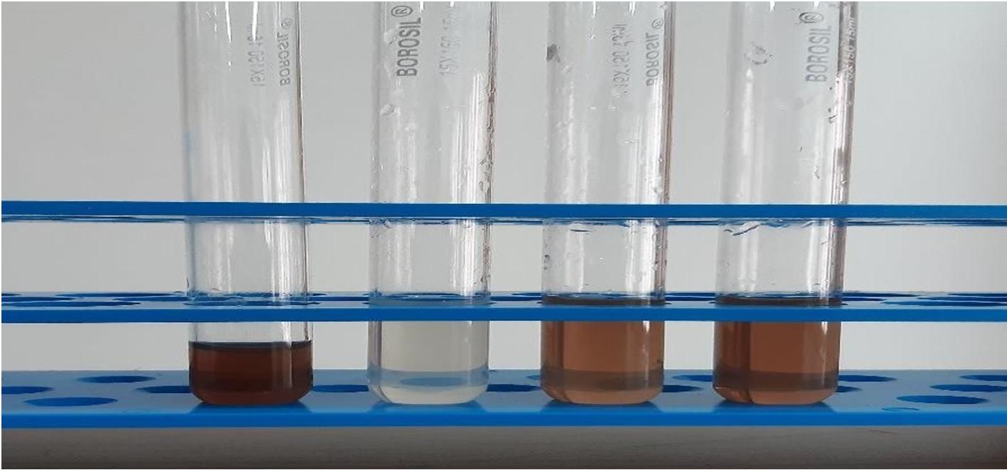
Silver nanoparticles were synthesised using silver nitrate as a source. 1mM of silver nitrate was added to 300 mL of ultra pure water. To the prepared AgNO3 solution, 3mL of extracted pomegranate juice was added and the resultant mixture was kept in magnetic stirrer with continuous stirring for 1 hour 15 minutes, below the boiling point at 600 660 rpm, until the whitish shade of the solution changed to honey brown colour suggesting the development of silver nanoparticles.

1) UV Vis Spectrophotometer: The Ag+ ions reduction to AgNPs were confirmed visually by observing the color change and was later confirmed by measuring the absorbance spectrum of the colloidal suspension in the range of 250 700nm in UV vis spectrophotometry. The nanoparticles suspension was taken in a quartz cuvette and the ultra pure water was taken as a reference The maximum absorption peak was recorded.
2) Scanning Electron Microscope: The SEM analysis was done to learn the external morphology, the size and crystalline structure of the formed AgNPs through direct visualization under the scanning electron microscope.
3) Dynamic Light Scattering Study: DLS was carried out to determine the average size and the dispersion of the formed AgNPs in the colloidal nanoparticle suspension.
4) Fourier Transform Infrared Spectrophotometric Analysis: FTIR analysis was carried out to analyze the functional group that are responsible in the formation PG AgNPs in the range 4000cm 1 to 400cm 1. It also determines the biomolecules present in Punica granatum, is accountable for the reduction and stabilization of the formed PG AgNPs.
5) Photocatalytic activity of PG AgNPs: The photocatalytic activity of AgNPs were carried out against Methylene blue (1mg/100mL) aqueous solution. The Methylene blue stock solution was prepared by dissolved 1mg of the dye in 100ml of distilled water. To the prepared stock of MB, different concentration of colloidal solution of AgNPs and NaBH4 solution (0.1M) were added in different concentration (5mL/100mL, 7mL/100mL, 10mL/100mL). The MB dye containing different concentration of NaBH4 (5mL, 7Ml and 10mL) was taken as a control. The blank was set up with distilled water. Dye degradation activity was determined using UV Vis spectrophotometry at regular intervals at the range from 300 800nm. The dye degradation percentage was estimated through the equation, C0 Ct
Degradation efficiency (%) = × 100 C0
In which, C0 is the concentration of the dye at initial/0 time and Ct is the concentration at time t of the methylene blue dye.
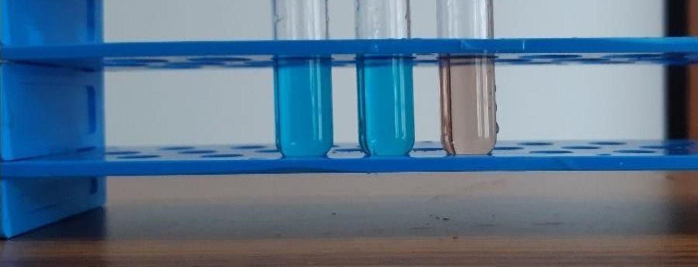
6) Biosensor Development: To 2mL of freshly prepared pomegranate AgNPs, 2mL of the milli molar solution of heavy metal salts such as lead nitrate, mercury chloride and Nickel sulfate were added and the following colour change was noticed.
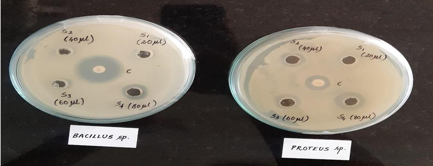
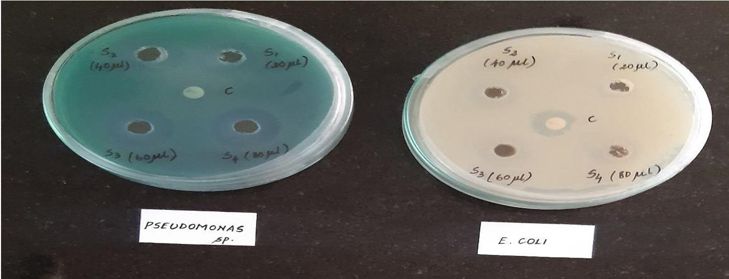
7) Antibacterial activity of PG AgNPs: Agar well diffusion method was used to asses the antibacterial activity of biosynthesized AgNPs. The nutrient agar plate was spread with pathogenic bacterial species such as E. coli, P. aeruginosa, Bacillus subtilis, and Proteus sp. Four wells were bored in the nutrient agar plate and different concentration of the PG AgNPs (20µL, 40µL, 60µL, 80µL) was added to the respective wells. Chloramphenicol disc served as a positive control and the plates was incubated at 37°C for 24 hrs, to observe zone of inhibition.
8) Antioxidant activity of PG AgNPs: The antioxidant activity for the free radical scavenging capability of PG AgNPs were investigated using DPPH (1, 1 dipheny 2 picrylhydrazyl). Different concentration of the PG AgNPs (0.2, 0.4, 0.6, 0.8, 1, and 1.2 mL) were taken in the test tubes and made up to 3 mL with methanol. 1mL DPPH was added to each of the test tubes. 3mL of methanol along with 1mL of DPPH were served as a control, while the cuvette containing only methanol served as blank. Ascorbic acid standard was taken as a positive control (Renuka et.al., 2018). The absorbance was measured after 1hr of incubation in the dark, using of UV Vis spectrophotometry at 517nm. The percentage of DPPH radical scavenging were determined with the formula,
(Absorbance of control Absorbance of sample)
% DPPH radical scavenging = × 100 (Absorbance of control)
ISSN: 2321 9653; IC Value: 45.98; SJ Impact Factor: 7.538 Volume 10 Issue XI Nov 2022 Available at www.ijraset.com

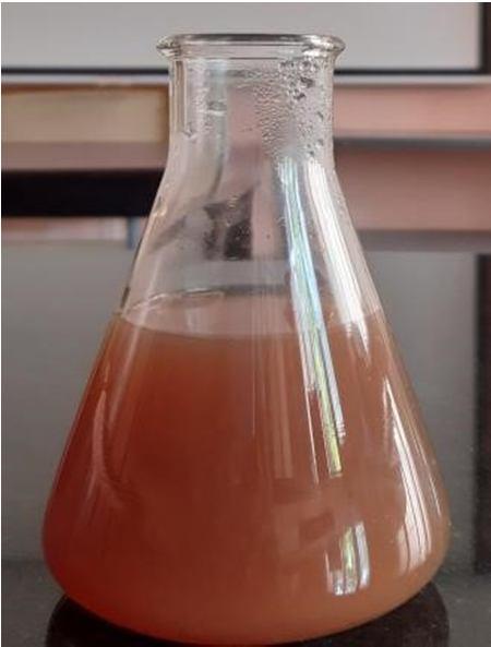


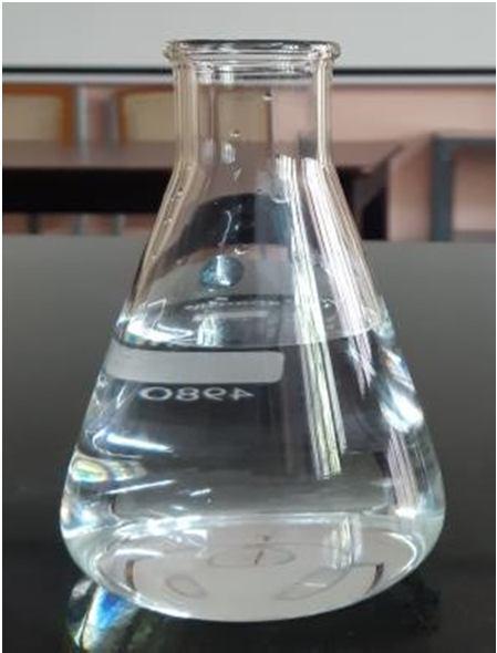
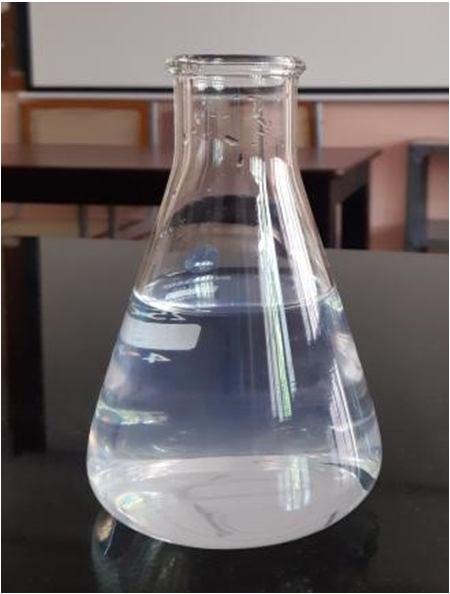
Silver ions are reduced to generate silver nanoparticles are indicated by the visible switch in the color of the solution, from colorless to white to honey brown, is displayed in the Figure 1(a), 1(b) and 1(c). Silver nanoparticles are formed, when the plant metabolites like polyphenols acts as a reducing agent, that reduces the silver ions. Pomegranate fruits are rich in polyphenol compounds, including flavonoids which are the major phytochemicals that are involved in the reduction process and provides high stability to the synthesized AgNPs.

Figure 2, shows a peak at 421nm, conforming the formation of AgNPs. The peaks appearances reveal the features of silver nanoparticle surface plasmon resonance.
2: UV Visible spectra of synthesized PG AgNPs
ISSN: 2321 9653; IC Value: 45.98; SJ Impact Factor: 7.538 Volume 10 Issue XI Nov 2022 Available at www.ijraset.com

The depth of the absorbance bands rose as the extraction temperature increased, indicating a rise in silver nanoparticle concentration. This is consistent with the findings that phenolic content in pomegranate extract increased with extraction temperature, implying that phenolic chemicals are the primary reducing agents. Because of electrons in metallic nanoparticles oscillate mutually in resonance with light waves, metallic nanoparticles containing free electrons, produces a surface plasmon resonance absorption band.


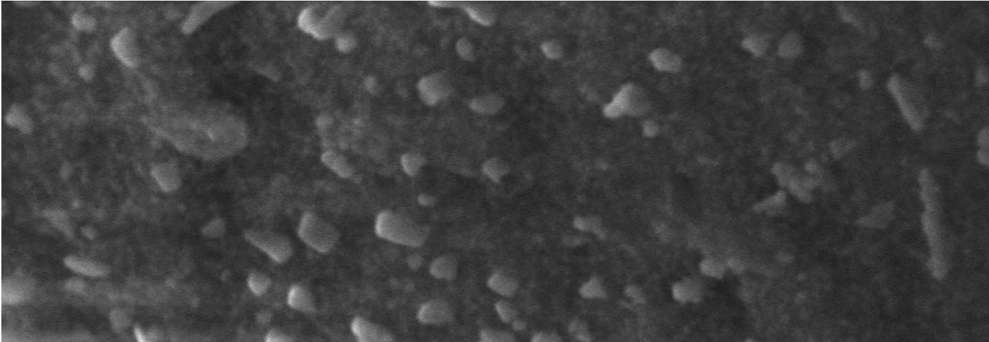
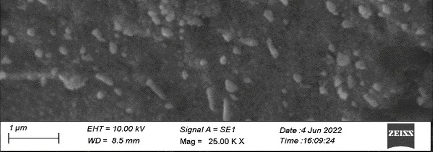
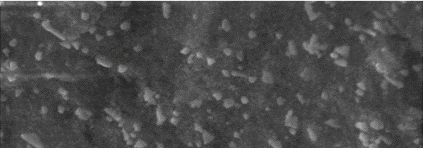
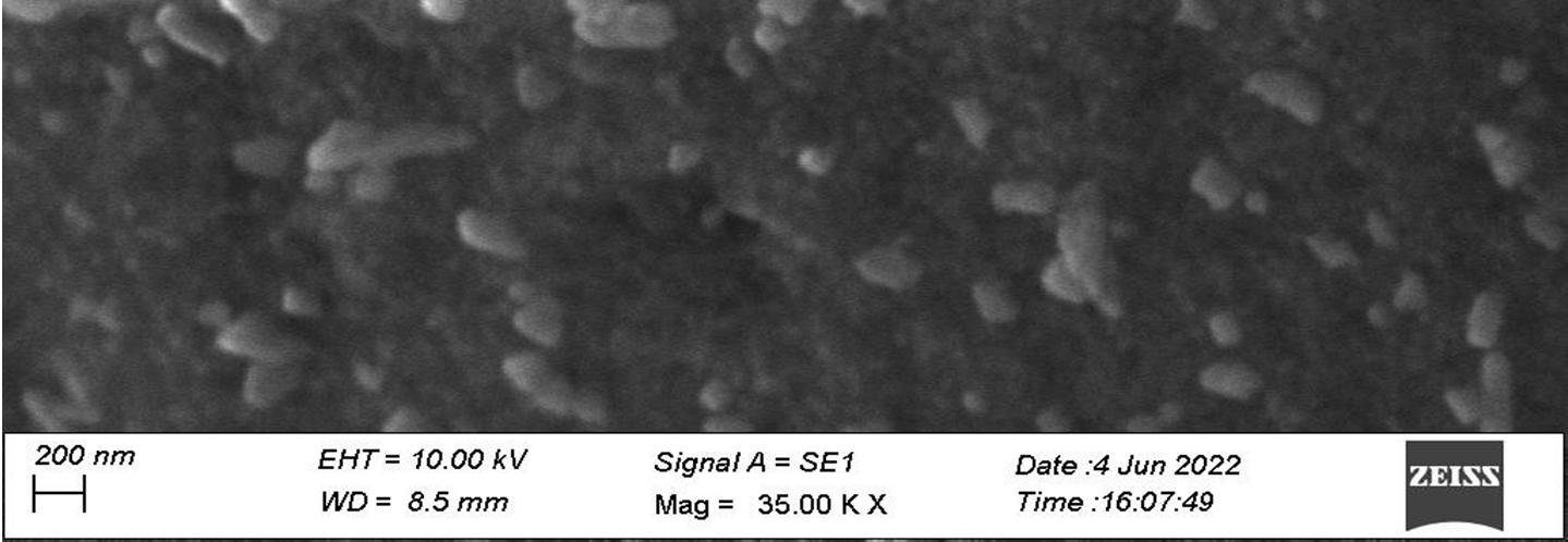
Figure 3, shows the shape and the size of the resultant synthesized AgNPs, containing dispersive nanoparticle of an average size of 85.71nm, 3(a) reveals the presence of 1D nanorods and 3(b) reveals, the formed nanoparticles were rectangles and spherical in structure.

a b
Figure 3(a) and 3(b): SEM images of synthesized PG AgNPs
ISSN: 2321 9653; IC Value: 45.98; SJ Impact Factor: 7.538 Volume 10 Issue XI Nov 2022 Available at www.ijraset.com

The size and the size distribution of the bio synthesized AgNPs in the colloidal suspension were measured using dynamic light scattering. Figure 4, depicts the range of synthesized AgNPs distributed from 10nm to 1000nm, found to have an overall average size of 228.3nm.


Figure 5, shows the different stretches of bonds at different peaks corresponding to various functional groups accountable for the reduction and formation of PG AgNPs. The obtained peaks at different cm 1 corresponds to different functional groups. The broad peak obtained at 3457.90 cm 1 corresponds to the O H stretching alcohol, the peak obtained at 2923.48 cm 1 and 2852.62 cm 1 corresponds to C H stretching alkane, the peak at 2092.62 cm 1 corresponds to N=C=S stretching isothiocyanate, the narrow peak obtained at 1639.68 cm 1 corresponds to C=C stretching alkene the following peak at 1384.26 cm 1 corresponds to C H bending alkane (gem dimethyl) and the further peak at 651.91 cm 1 corresponds to C Br stretching halo compounds. cm 1
NameDescription
MCC FTIR PS
Figure 5: FTIR image of the synthesized PG AgNPs.
1081.06cm
ISSN: 2321 9653; IC Value: 45.98; SJ Impact Factor: 7.538 Volume 10 Issue XI Nov 2022 Available at www.ijraset.com
Thus, the FTIR analysis reveals the formation of the functional groups such as alcohol, alkane, isothiocyanate, a bending alkane and a halo compounds respectively, involved in the formation of PG AgNPs. The shifts in the stretch frequency between functional groups, resulting in lower wavenumbers to disappearance of the resonance may be because of their binding with silver nanoparticle surface.


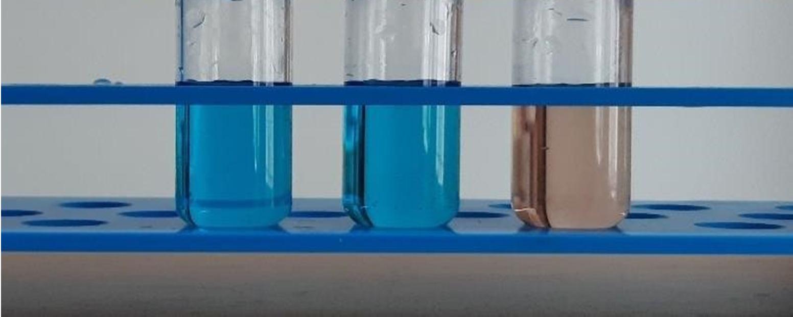
The efficiency of the biosynthesized AgNPs as a nano catalyst in degrading the methylene blue dye solution was carried out by subjecting the dye to UV irradiation. NaBH4 acts as a reducing agent in the degradation activity, but it was observed that the degradation rate of the MB dye increased after the addition of synthesized AgNPs. The decrease in the absorption peak and decolorization of the MB dye, indicates the degradation activity, was shown in the Figure 6(a), 6(b), 6(c), 7(a), 7(b), 7(c) and 8(a), 8(b) and 8(c).

The maximum degradation activity of 99.4% was observed with the maximum concentration that was10ml of PG AgNPs and NaBH4, 93.7% and 90.6% of degradation with 7ml and 5 ml of PGAgNPs and NaBH4 respectively. Based on the obtained result, increase in the concentration of nanoparticles, increases the dye degradation activity with less exposure time to UV. The dye molecule and the borohydride ion were both adsorbed onto the surface of AgNPs. Moreover, the BH4 ion serves as a nucleophilic agent, contributing an electron to AgNPs. So, the dye molecules adsorbed onto the surface of AgNPs, on the other hand, will operate as an electrophilic agent, capturing the electron available on the AgNPs surface. The resulting dye molecule is broken down into a colourless component by this overall process, which starts a dye degradation reaction.
2.5
2
1.5
1
0.5
0
0.5
Figure 6(a): MB dye degradation in the presence of NaBH4 (10ml concentration)
ISSN: 2321 9653; IC Value: 45.98; SJ Impact Factor: 7.538 Volume 10 Issue XI Nov 2022 Available at www.ijraset.com


Figure 6(b): MB dye degradation in the presence of PG AgNPS+ NaBH4 (10ml concentration).

ISSN: 2321 9653; IC Value: 45.98; SJ Impact Factor: 7.538 Volume 10 Issue XI Nov 2022 Available at www.ijraset.com

1.8
1.6
1.4
1.2
1
0.8
0.6
0.4
0.2
0
0.2
2 0 100 200 300 400 500 600 700 800 900 Wavelength (nm)


Figure 7(a): MB dye degradation in the presence of NaBH4 (7ml concentration)
1.8
1.6
1.4
1.2
1
0.8
0.6
0.4
0.2
C (0 time) MB+NaBH4 (3 mins) 0.2
2 0 100 200 300 400 500 600 700 800 900 Wavelength (nm)
0
C (0 time) MB+NaBH4+PG AgNPs (3 mins)
Figure 7(b): MB dye degradation in the presence of PG AgNPS+ NaBH4 (7ml concentration)

ISSN: 2321 9653; IC Value: 45.98; SJ Impact Factor: 7.538 Volume 10 Issue XI Nov 2022 Available at www.ijraset.com


Figure 7(c): Decolorization of MB dye at 7ml concentration

ISSN: 2321 9653; IC Value: 45.98; SJ Impact Factor: 7.538 Volume 10 Issue XI Nov 2022 Available at www.ijraset.com


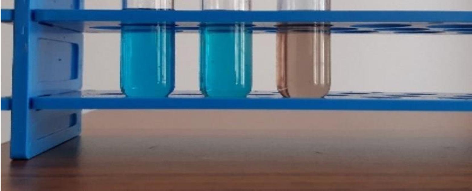
The freshly prepared Ag nanoparticles are mixed with the milli molar solution of heavy metal salts such as lead nitrate, mercury chloride and Nickle sulfate for their quantitative detection. Figure 15, shows instant vanishing of color in the test tube containing mercury. Since the synthesized nanoparticles are sensitive to mercury, there is change in color of the solution, this had led to the development of biosensor
ISSN: 2321 9653; IC Value: 45.98; SJ Impact Factor: 7.538

Volume 10 Issue XI Nov 2022 Available at www.ijraset.com

The bio synthesized AgNPs were investigated for antibacterial activity upon three gram negative bacteria, E. coli, Proteus sp., Pseudomonas sp., respectively and Bacillus sp., a gram positive bacteria. With varied concentration of bio synthesized pomegranate AgNPs, the corresponding zone of clearance was observed are displayed in the Figure 10. The zone of clearance was measured using a measuring scale. The antibacterial activity was increased for all the bacterial species upon increasing the concentration of bio synthesized AgNPs. The highest zone of inhibition of the following bacterial species E. coli, Proteus sp., P aeruginosa, at 80µL was found to be more (±10.75mm, ±10.05mm, ±11mm) than the control (±4.1mm, ±6.05mm, ±4.95mm). Further, the gram negative bacteria’s (E. coli, Proteus sp., P aeruginosa,) exhibited better antibacterial activity compared to gram positive bacteria (B. subtilis). In this case, silver nanoparticles can consistently emit silver ions, which might be a bacteria killing mechanism. On the account of electrostatic pull and affinity for sulfur proteins, silver ions can cling to the cell wall and cytoplasmic membrane. The associated ions can cause the bacterial envelope to be ruptured by increasing the permeability of the cytoplasmic membrane (Yin et.al., 2020).
ISSN: 2321 9653; IC Value: 45.98; SJ Impact Factor: 7.538 Volume 10 Issue XI Nov 2022 Available at www.ijraset.com
TThe antioxidant activity of bio synthesized AgNPs has revealed that the DPPH radical scavenging activity was increased with increase in the concentration of the sample. Thishappenswhen the free DPPH radical combines with hydrogen donors, it gets decreased. The colour of theradical changes from purple to yellow throughout this reduction due to a drop in abs orbance at 517nm. The bio synthesized PG AgNPs had shown the maximum DPPH scavenging activity at the concentration of 1.2 mL or 1200µl.
90
80
70
60
50
40
30
20
10
0
100 0.2 0.4 0.6 0.8 1 1.2
Concentration (mL) % DPPH radical scavenging activity of ascorbic acid % DPPH radical scavenging activity of PG AgNPs
DPPH free radical scavenging activity of ascorbic acid and biosynthesized AgNPs


The fruit extract of Punica granatum had been used to successfully synthesize AgNPs in an environmentally benign manner, is reported for the first time. The biosynthesized AgNPs were characterized by UV Visible spectrophotometer, SEM, DLS and FTIR. The UV Visible spectrophotometry, showed the maximum absorption peak, which is distinct for AgNPs, the SEM analysis revealed the rectangular, spherical and rod shaped structure of the AgNPs, the DLS analysis depicted the overall average size of the particle and the size distribution in the colloidal AgNPs solution and the FTIR result showed the potential of the plant extract, responsible in the conversion and formation of AgNPs. PG AgNPs are found to be a favorable photocatalyst in the degradation of methylene blue with the help of NaBH4. The PG AgNPs also paves way for the quantitative detection of mercury through the development of biosensor. The AgNPs displayed a significant zone of inhibition against the gram negative as well as gram positive bacterial species. The antioxidant activity of AgNPs, resulted in the maximum DPPH scavenging activity. When it comes to photocatalytic, antibacterial and antioxidant activity, the obtained results show that as concentration of PG AgNPs increases, activity increases as well. To conclude, the use of plant source for the biosynthesize of nanoparticles gives all applications a new dimension.
[1] Abdulrahman A. Almehizia, Mohamed A. Al Omar, Ahmed M. Naglah, Mashooq A. Bhat, Nasser S. Al Shakliah, (2022), Facile synthesis and characterization of ZnO nanoparticles for studying their biological activities and photocatalytic degradation properties toward methylene blue dye, Alexandria Engineering Journal, Volume 61, Issue 3, 2386 2395
[2] Al Tohamy, Rania & Ali, Sameh & Li, Fanghua & Okasha, Kamal & Mahmoud, Yehia & Elsamahy, Tamer & Jiao, Haixin & Fu, Yinyi & Sun, Jianzhong, (2022), A critical review on the treatment of dye containing wastewater: Ecotoxicological and health concerns of textile dyes and possible remediation approaches for environmental safety. Ecotoxicology and Environmental Safety, 231
[3] Al Zaban, Mayasar & Mahmoud, Mohamed & AlHarbi, Maha. (2021). Catalytic Degradation of Methylene Blue Using Silver Nanoparticles Synthesized by Honey. Saudi Journal of Biological Sciences. 28. 10.1016/j.sjbs.2021.01.003.
ISSN: 2321 9653; IC Value: 45.98; SJ Impact Factor: 7.538 Volume 10 Issue XI Nov 2022 Available at www.ijraset.com
[4] Behravan M, Hossein Panahi A, Naghizadeh A, Ziaee M, Mahdavi R, Mirzapour A., (2019), Facile green synthesis of silver nanoparticles using Berberis vulgaris leaf and root aqueous extract and its antibacterial activity. Int J Biol Macromol. 1;124:148 154
[5] Dhananjayan, Badmapriya & Asharani, I.V. (2016). Dye degradation studies catalysed by green synthesized iron oxide nanoparticles. 9. 409 416
[6] Din, Muhammad & Khalid, Rida & Najeeb, Jawayria & Hussain, Zaib. (2021), Fundamentals and Photocatalysis of Methylene blue dye using various nanocatalytic assemblies A Critical Review. Journal of Cleaner Production, 298(6):126567
[7] Din, Muhammad & Tariq, Maria & Hussain, Zaib & Khalid, Rida. (2020). Single step green synthesis of nickel and nickel oxide nanoparticles from Hordeum vulgare for photocatalytic degradation of methylene blue dye. Inorganic and Nano Metal Chemistry. 50, 1 6
[8] Garibo D, Borbón Nuñez HA, de León JND, García Mendoza E, Estrada I, ToledanoMagaña Y, Tiznado H, Ovalle Marroquin M, Soto Ramos AG, Blanco A, Rodríguez JA, Romo OA, Chávez Almazán LA, Susarrey Arce A, (2020), Green synthesis of silver nanoparticles using Lysiloma acapulcensis exhibit high antimicrobial activity. Sci Rep. ;10(1), 12805
[9] Shanmugavadivu, M. & Kuppusamy, K.Selvam & Rajamani, Ranjithkumar, (2014), Synthesis of Pomegranate Peel Extract Mediated Silver Nanoparticles and its Antibacterial Activity. American Journal of Advanced Drug Delivery, 2, 174 182
[10] Shivsharan, Utkarsha & Ravva, Sony. (2018). Antimicrobial Activity of Pomegranate Juice. Research Journal of Pharmacy and Technology. 11, 1 3


[11] Simatupang, C, Jindal, V. K, & Jindal, R, (2021). Biosynthesis of Silver Nanoparticles Using Orange Peel Extract for Application in Catalytic Degradation of Methylene Blue Dye, Environment and Natural Resources Journal, 19(6), 468 480
[12] V S, Suvith & Philip, Daizy, (2013). Catalytic degradation of methylene blue using biosynthesized gold and silver nanoparticles. Spectrochimica acta. Part A, Molecular and biomolecular spectroscopy. 118C. 526 532
