
10 VIII August 2022 https://doi.org/10.22214/ijraset.2022.46382

The World Health Organization (WHO) designated the outbreak a Public Health Emergency of International Concern (PHEIC) on January 30, 2020, and on March 11, 2020, it recognized it as a pandemic, which has caused significant public health concern in the global population. The gold standard for identifying COVID 19 patients is the RT PCR test (reverse transcription polymerase chain reaction). The RT PCR assay, however, frequently proves to be insufficient in many places that have been seriously affected, particularly during the early epidemics of this disease. Due to numerous factors, including sample preparation and quality control, the lab test also has high false negative rates. Clinical practitioners greatly benefit from readily available imaging tools such chest X rays and thoracic CT scans. If characteristic signs in CT scans were seen, several patients, especially in China, were diagnosed as potential COVID 19 cases. The suspected patients were also hospitalized or sent in isolation for additional lab testing, even though they lacked any clinical signs (such as fever and coughing). Numerous suspected individuals must undergo multiple tests, spaced several days apart, due to the high false positive rate of nucleic acid tests, before a certain diagnosis can be made. As a result, the imaging results are crucial in limiting viral propagation and combating COVID 19.
International Journal for Research in Applied Science & Engineering Technology (IJRASET) ISSN: 2321 9653; IC Value: 45.98; SJ Impact Factor: 7.538 Volume 10 Issue VIII August 2022 Available at www.ijraset.com 1170©IJRASET: All Rights are Reserved | SJ Impact Factor 7.538 | ISRA Journal Impact Factor 7.894 | Acquisition, Segmentation, and Diagnosis of Imaging Data Using Artificial Intelligence Techniques for COVID-19 Manosijo Ganguly1 , Koushik Pal2, Rahul Bera3, Rupayan Dirghangi4, Arindam Roy5, Sujoy Dutta6, Karan Kumar7 , Dipankar Pariyary8 1, 2, 3, 4, 5, 6, 7, 8Department of Electronics & Communication Engineering, Guru Nanak Institute of Technology Abstract: The global coronavirus disease pandemic of 2019 (COVID 19) is escalating. In the global fight against COVID19, medical imaging such as computed tomography (CT) and X-rays is crucial, and recently developed artificial intelligence (AI) technologies are enhancing the power of the imaging instruments and assisting medical professionals. Here, we examine the quick reactions to COVID 19 from the medical imaging community (driven by AI). AI powered picture acquisition, for instance, can considerably assist in automating the scanning process and also reorganise the workflow with little touch to patients, offering the best protection to the imaging technicians. Additionally, AI can increase productivity by precisely identifying infections in X ray and CT images, allowing for later quantification. Additionally, computer aided platforms support radiologists in their clinical judgments, such as disease diagnosis, monitoring, and prognosis. Thus, we discuss all of the COVID-19-related medical imaging and analysis methods used in this review study, including image acquisition, segmentation, diagnosis, and follow up.
Key words: Contactless imaging workflow, Standard imaging workflow, AI streamlines imaging process, Main Application of AI in Covid-19 pandemic.
The severe acute respiratory syndrome coronavirus 2 (SARS CoV 2) is the cause of the ongoing pandemic known as the coronavirus disease 2019 (COVID 19). The virus is spreading quickly and infecting more individuals. A fatality rate of 5.54% and about 62,784 cases of COVID 19 have been documented up until April 5, 2020 in more than 200 countries and territories
I. INTRODUCTION
Using thoracic CT as an example, the process of imaging based diagnosis for COVID 19 entails three stages in general: 1) pre scan preparation, 2) image capture, and 3) disease diagnosis. Each participant is given instructions and assistance from a technician to posture on the patient bed in accordance with a predetermined protocol during the pre scan preparation step. CT images are collected during a single breath hold during the image capture stage. The entire lung, from the tip to the base, is scanned. With the optimum parameters established by the radiologist(s), based on the patient's body shape, scans are performed from the level of the upper thoracic inlet to the inferior level of the costophrenic angle. CT images are created from the acquired raw data and then communicated via picture archiving and communication systems (PACS) for later viewing and diagnosis.
The fight against COVID 19 has benefited greatly from artificial intelligence (AI), an emerging tool in the field of medical imaging [12].


Ren et al. recently suggested one current technique that uses dynamic multi modal inference to create a model that can be trained just once and used in numerous applications.
AI makes it possible to create imaging solutions that are more secure, accurate, and productive than the conventional imaging workflow, which mainly relies on human labour. The COVID 19 dedicated imaging platform, lung and infection region segmentation, clinical assessment and diagnosis, as well as the ground breaking basic and clinical research, are the key recent AI powered applications. Additionally, a large number of commercial solutions have been created that successfully incorporate AI to tackle COVID 19 and amply show the technology's potential. This inaugural online conference on COVID 19 was held on February 18, 2020, and it received more than ten thousand visitors. It was conducted by the Medical Imaging Computing Seminar (MICS)1, a major coalition of medical imaging researchers and start up firms in China. All of the aforementioned instances highlight the enormous enthusiasm that the public has for AI powered advancements in the field of medical imaging, particularly in light of the ongoing epidemic. This review aims to extensively discuss the role of medical imaging, particularly as empowered by AI, in battling the COVID 19, which will inspire future practical applications and methodological research. This is due to the significance of AI in all facets of the imaging based analysis of the COVID 19. Following that, we briefly discuss prominent machine learning techniques used in the imaging workflow, such as segmentation, diagnosis, and prognosis. First, we introduce intelligent imaging platforms for COVID 19. Additionally, a number of publicly accessible datasets are presented. Finally, we talk about a number of unresolved issues and difficulties. Through this evaluation, we hope to offer advice to radiologists and researchers. As of March 31, 2020, we review the majority of relevant COVID 19 research based on medical imaging.
1) Standard Imaging Workflow
II. CONTACTLESS IMAGING WORKFLOWS EMPOWERED BY AI
When it comes to the high risk of occupational virus exposure, healthcare professionals are particularly vulnerable. In order to prevent any potential viral interaction, imaging specialists and technicians are given top priority. In addition to personal protective equipment (PPE), one may think about specialised imaging workspaces and facilities, which are crucial to lowering dangers and saving lives.
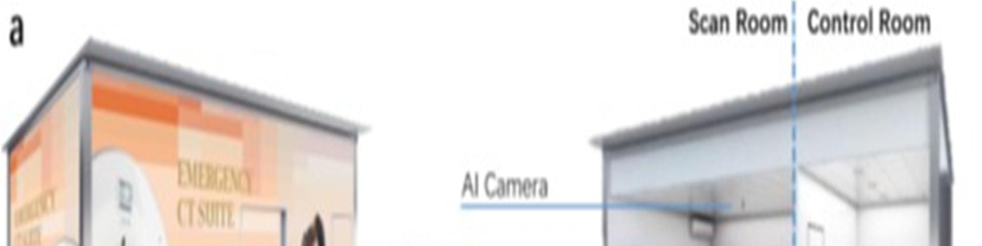
Several contemporary CT and X ray machines come with cameras for patient monitoring. These tools make it easier to conduct a contactless scanning work flow during the COVID 19 outbreak. A live video stream from the camera allows technicians in the control room to keep an eye on the patient. It is still difficult for the technician to establish the scanning characteristics, such as scan range, from just the overhead view of the camera. By recognising the patient's stance and shape from the data collected with visual sensors like RGB, Time of Flight (TOF) pressure imaging, or thermal (FIR) cameras, AI is possible to automate the process in this situation. As a result, the ideal scanning parameters can be found. The CT scans scan range, which specifies the beginning and finishing positions of the image, is one typical scanning parameter that can be calculated with AI enabled visual sensors. The anatomical joints of the person can be found in the images to determine the scan range. Estimating the 2D or 3D key point positions on the patient's body has been the topic of a lot of recent research. The neck, shoulders, elbows, ankles, wrists, and knees are common examples of these key point positions. But these key points often only represent a relatively small sampling of the entire 3D mesh in the 3D environment (that defines the digital human body). AI can also infer other crucial scanning characteristics, such as ISO centring. ISO centring is the process of aligning the subject's target body area so that its centre is visible. Overlaps with the scanner's ISO centre, resulting in the best possible imaging quality. According to studies, the radiation dosage can be decreased while retaining equivalent image quality by improving ISO centring. They apply hierarchical kinematic reasoning, in contrast to earlier work in the field, to iteratively enhance the estimation of each anatomical key point and increase the system's robustness to clutters and partial occlusions near the patient's joints.
2) Ai Streamlines Imaging Processes
For the screening and diagnosis of COVID 19, chest X rays and CT scans are frequently employed. To reduce the serious dangers of infection during the COVID 19 pandemic, it is crucial to use a contactless and automated image acquisition method. However, the traditional imaging procedure necessitates constant interaction between technicians and patients. Particularly in patient positioning, technicians first assist in posing the patient in accordance with a given protocol, such as head first versus feet first and supine versus prone in CT, after which they manually adjust the patient's position in relation to the X ray tube after visually locating the target body part on the patient. Due of the technicians near proximity to the patients throughout this process, there is a significant danger of virus exposure. In order to reduce interaction, a contactless and automated imaging approach is required.
International Journal for Research in Applied Science & Engineering Technology (IJRASET) ISSN: 2321 9653; IC Value: 45.98; SJ Impact Factor: 7.538 Volume 10 Issue VIII August 2022 Available at www.ijraset.com 1171©IJRASET: All Rights are Reserved | SJ Impact Factor 7.538 | ISRA Journal Impact Factor 7.894 |
Following acquisition, CT images will be processed and examined for screening and diagnosis reasons.
International Journal for Research in Applied Science & Engineering Technology (IJRASET) ISSN: 2321 9653; IC Value: 45.98; SJ Impact Factor: 7.538 Volume 10 Issue VIII August 2022 Available at www.ijraset.com 1172©IJRASET: All Rights are Reserved | SJ Impact Factor 7.538 | ISRA Journal Impact Factor 7.894 |
Even if one of the sensor modalities fails when using this framework with an RGB depth input sensor, the model above can still perform 3D patient body inference with the remaining data. The patient is directed to position on the patient bed after entering the scan room by visual and audible cues. Technicians can adjust the patient's attitude, if necessary, by looking through the glass and watching the live footage sent from the ceiling mounted AI camera in the scan room. The patient positioning algorithm will automatically recover the 3D pose and fully reconstructed mesh of the patient from the photos taken with the camera whenever the patient is deemed ready, either by the technician or the motion analysis algorithm in above picture. Based on the 3D mesh, the technician estimates the scan range and the 3D centre line of the patient's target body part, converts them into control signals, and then uses the optimised scanning settings to check the results. The technician can make modifications if required. As soon as this is confirmed, the patient bed will automatically slide into the CT gantry and align to the ISO centre.
Fig. 1. (a) A mobile CT platform equipped with AI empowered automated image acquisition workflow; (b) An example image captured by patient Monitoring camera of CT system; (c) Positioning and scanning of patient operated remotely by a technician.
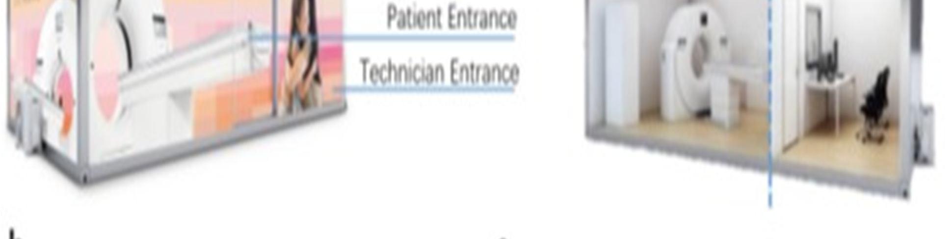

International Journal for Research in Applied Science & Engineering Technology (IJRASET) ISSN: 2321 9653; IC Value: 45.98; SJ Impact Factor: 7.538 Volume 10 Issue VIII August 2022 Available at www.ijraset.com 1173©IJRASET: All Rights are Reserved | SJ Impact Factor 7.538 | ISRA Journal Impact Factor 7.894 | III. MAIN APPLICATION OF AI IN COVID 19 PANDEMIC Fig. 2. General procedure of AI and non AI based applications that help general physicians to identify the COVID 19 symptoms. A. Early Detection And Diagnosis Of The Infection AI has the ability to quickly analyse unusual symptoms and other "red flags," alarming both patients and healthcare authorities. Faster, more economical decision making is facilitated by it. Through practical algorithms, it aids in the development of a novel diagnosis and management approach for the COVID 19 cases. With the aid of medical imaging technologies like Computed Tomography (CT) and Magnetic Resonance Imaging (MRI) scans of human body parts, AI is useful in the diagnosis of infected cases. Patientwith symptoms Ai approachbased Physician identify the possible match of covid 19 symptom with AI support Samples taken to confirm infection & decide further therapy Patients quarantined/admittedgetsStartAIbasedtreatment&monitoringRecoveryPhase Reset COVIDfor19 IsolationPositive NegativeCured Conventionalapproach (non PhysianAI)analyze the symptoms If IsolationPositivequarantiined/admittedmatchesmultiplefoundTestsampletakenPatientsgetSymptomatictreatmentstartedRecoveryPhaseResetforCovid19NegativeCured If no Nomatchesmultiplefoundtestsampletaken


C. Contact Tracing Of The Individuals
AI can provide up to date knowledge that is useful in the prevention of this disease with the aid of real time data analysis. During this crisis, it can be used to forecast the likely areas of infection, the spread of the virus, and the demand for beds and medical personnel. With the aid of older guided data over more recent data, AI is useful for preventing future viruses and disorders. It pinpoints characteristics, root causes, and mechanisms behind illness spread. This technology will be crucial in the future to combating subsequent pandemics and outbreaks. It can fight many different ailments and act as a preventive measure. Future healthcare will be more predictive and preventive thanks in large part to AI.
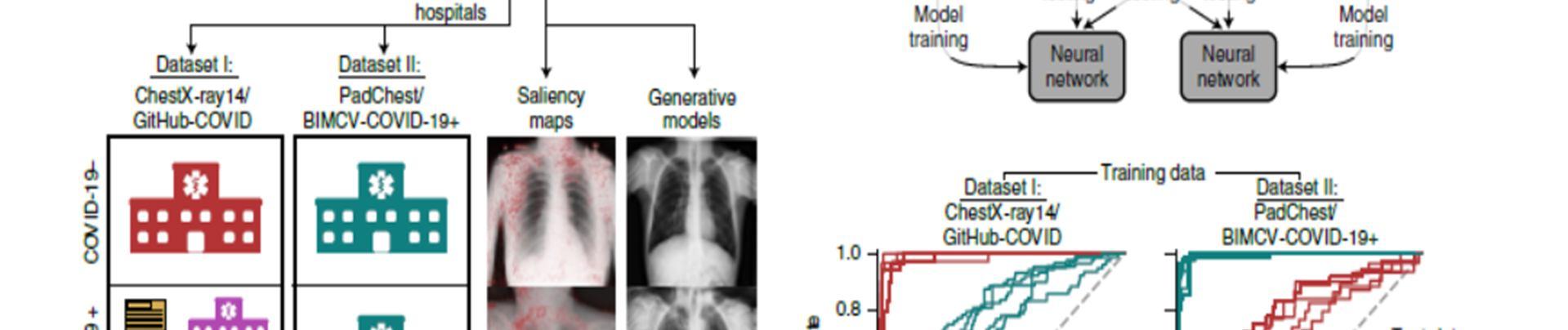
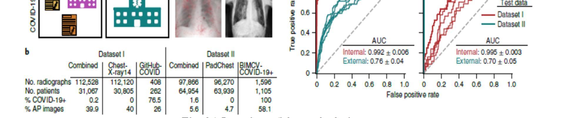
Recently, radiologists and artificial intelligence (AI) researchers reported AI algorithms that reliably identify COVID 19 in chest radiographs. It's unclear, though, how reliable these systems are. We show that recent deep learning systems to detect COVID 19 from chest radiographs rely on confounding factors rather than medical pathology, creating an alarming situation in which the systems appear accurate but fail when tested in new hospitals. This is done using cutting edge explainable AI techniques. We note that the method used to gather training data for these AI systems generates a situation that is almost ideal for AI to pick up these fictitious "shortcuts." Because training data for the identification of COVID 19 in computed tomography has also been obtained using this method of data collecting, our analysis highlights a significant issue for medical imaging activities connected to various disorders and for tomography scans, in AI for medical imaging. Additionally, we demonstrate that testing a model against external data is insufficient to guarantee that AI systems rely on pathology that is medically relevant because the unintended "shortcuts" that AI systems learn may not negatively impact performance in new Theseinstitutions.results
E. Development Of Drugs And Vaccines
AI can create a smart platform for the automatic tracking and forecasting of this virus's spread. The visual characteristics of this condition can also be extracted using a neural network, which will aid in the proper monitoring and care of those who are afflicted. It is able to provide daily updates on the patients' conditions as well as guidelines for dealing with the COVID 19 epidemic.
International Journal for Research in Applied Science & Engineering Technology (IJRASET) ISSN: 2321 9653; IC Value: 45.98; SJ Impact Factor: 7.538 Volume 10 Issue VIII August 2022 Available at www.ijraset.com 1174©IJRASET: All Rights are Reserved | SJ Impact Factor 7.538 | ISRA Journal Impact Factor 7.894 |
By locating clusters and "hot spots" and analysing the virus's level of infection, artificial intelligence (AI) can successfully infect people and keep track of them. It can forecast the progression of this illness and its likelihood of recurrence.
By analysing the COVID 19 data that is now available, AI is employed for drug research. Designing and developing drug delivery systems can benefit from it. When compared to traditional testing, which takes a long time, this technology helps to considerably speed up the procedure, which may not be possible for a human to do. It can aid in the discovery of effective medications for the treatment of COVID 19 patients. It has developed into a potent tool for developing vaccines and designing diagnostic tests. AI supports clinical trials throughout the creation of the vaccine and speeds up the process of producing vaccines and medicines.
B. Monitoring The Treatment
F. Reducing The Workload Of Healthcare Workers
The COVID 19 epidemic has put a sudden and significant increase in patient numbers on healthcare personnel, who now have an extremely heavy task. In this case, AI is employed to lighten the workload of healthcare professionals. It provides the best training to students and professionals regarding this new disease and aids in early diagnosis and early treatment using digital techniques and decision science. AI can improve patient care in the future and handle more possible problems, which will lighten the pressure on doctors.
G. Prevention Of The Disease
IV. AI CHOOSES SHORTCUTS ABOVE SIGNAL FOR RADIOGRAPHIC COVID 19 DETECTION
show that the clinical application of machine learning healthcare models should be viewed as predicated on explainable AI.
D. Projection Of Cases And Mortality
Using information about the dangers of infection and its anticipated propagation from social media and other media platforms, this technology can track and predict the virus's characteristics. It can also forecast the number of positive cases and fatalities in any area. AI can assist in identifying the most susceptible nations, populations, and regions so that appropriate steps can be taken.
V. LITERATURE SURVEY
identifying infections in X ray and CT images, allowing for later quantification. the full spectrum of COVID 19 related medical imaging and analytic methods, including image acquisition, segmentation, diagnosis, and follow up.
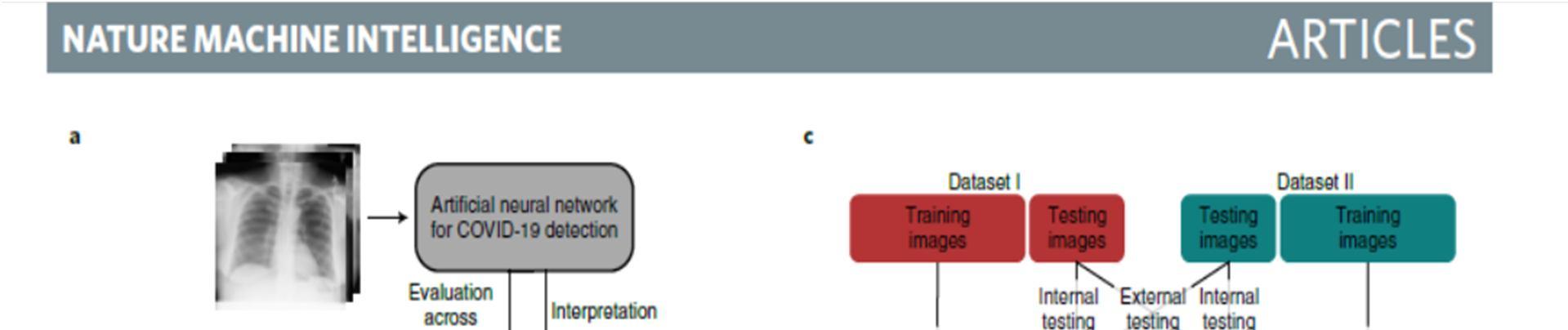
1) A neural network model is trained to detect COVID 19 using radiographs from either of two datasets, and then evaluated on both datasets to learn how performance may drop in deployment (that is, a generalization gap). Interpretability methods are then applied to infer what the model learned and which features were important for its decisions. Whereas dataset I draws radiographs from multiple hospital systems as well as cropped images from publication figures, dataset II draws radiographs from multiple hospitals from a single regional hospital system.
Fig. 3 | Overview of the study design
International Journal for Research in Applied Science & Engineering Technology (IJRASET) ISSN: 2321 9653; IC Value: 45.98; SJ Impact Factor: 7.538 Volume 10 Issue VIII August 2022 Available at www.ijraset.com 1175©IJRASET: All Rights are Reserved | SJ Impact Factor 7.538 | ISRA Journal Impact Factor 7.894 |
2) Characteristics of the datasets used in this study. c, Model evaluation scheme (top) and corresponding receiver operating characteristic (ROC) curves (bottom), which show the performance of our neural network models evaluated on both an internal test set (new, held out examples from the same data source as the training radiographs) and an external test set (radiographs from a new hospital system). Inset numbers indicate areas under the ROC curves, where a larger area corresponds to higher performance (area under the curve (AUC), mean ± standard deviation). The difference between internal and external test set performance is the generalization gap.
In this paper, since early 2020, the entire world has been dealing with the deadly and extremely contagious coronavirus disease (COVID 19), which the World Health Organization labelled a pandemic on March 11, 2020. Medical imaging, such as chest X rays and computed tomography scans, are quickly becoming one of the most important clinical diagnosis tools in the fight against COVID 19, thanks to Artificial Intelligence based solutions that enable speedy decision making and the saving of lives. The accuracy of Covid 19 diagnosis has increased thanks to AI and machine learning technologies, and the majority of commonly used deep learning techniques have been incorporated and successfully employed for COVID19 diagnosis with little data. This review provides a thorough theoretical analysis for assessing AI based techniques for COVID 19 detection from medical Thispictures.report discusses the quick reactions to COVID 19 in the medical imaging community (driven by AI). AI powered picture acquisition, for instance, can considerably assist in automating the scanning process and also reorganise the workflow with little touch to patients, offering the best protection to the imaging technicians. Additionally, AI can increase productivity by precisely
[2] Sohrabi, C. et al. World Health Organization declares global emergency: a review of the 2019 novel coronavirus (COVID 19). Int J. Surg. 76, 71 76 (2020).
[6] Yu, M. et al. Thin section chest CT imaging of coronavirus disease 2019 pneumonia: comparison between patients with mild and severe disease. Radiol. Cardiothorac. Imaging 2 . [7] Xie, X. et al. Chest CT for typical 2019 nCoV pneumonia: relationship to negative RT PCR testing. Radiology. [8] Inui, S. et al. Chest CT findings in cases from the cruise ship “Diamond Princess” with coronavirus disease 2019 (COVID 19). Radiol. Cardiothorac. Imaging. 2, p.e200110 [9] Ai, T. et al. Correlation of chest CT and RT PCR testing in coronavirus disease 2019 (COVID 19) in China: a report of 1014 Cases [10] Jin, S. et al. AI assisted CT imaging analysis for COVID 19 screening: building and deploying a medical AI system in four weeks.
[11] Review of Artificial Intelligence Techniques in Imaging Data Acquisition, Segmentation, and Diagnosis for COVID 19. Feng Shi , Jun Wang , Member, IEEE, Jun Shi , Ziyan Wu , Qian Wang, Zhenyu Tang, Kelei He , Yinghuan Shi , and Dinggang Shen , Fellow, IEEE.
VI. CONCLUSION
International Journal for Research in Applied Science & Engineering Technology (IJRASET) ISSN: 2321 9653; IC Value: 45.98; SJ Impact Factor: 7.538 Volume 10 Issue VIII August 2022 Available at www.ijraset.com 1176©IJRASET: All Rights are Reserved | SJ Impact Factor 7.538 | ISRA Journal Impact Factor 7.894 |
A sickness called COVID 19 has spread throughout the entire world. The fight against COVID 19 has benefited greatly from intelligent medical imaging. This study describes how AI offers COVID 19 applications with reliable, accurate, and effective imaging solutions. A thorough evaluation of the clinical diagnosis, innovative research, and intelligent imaging platforms is provided in COVID 19, which covers the complete pipeline of AI powered imaging applications. The usefulness of AI powered medical imaging for COVID 19 is shown using two imaging modalities, namely CT and X ray. It's important to remember that imaging only gives COVID 19 patients a partial picture of their condition. To aid with better screening, identification, and diagnosis of COVID 19, it is crucial to correlate imaging data with both clinical signs and laboratory test results. In this situation, we think AI will show that it is naturally capable of combining data from many sources to perform accurate and effective diagnosis, analysis, and follow up.
[1] Hoehl, S. et al. Evidence of SARS CoV 2 infection in returning travellers from Wuhan, China. N. Engl. J. Med (2020).
REFERENCES
The study discusses the worldwide coronavirus disease pandemic (COVID 19), which has killed millions of people and adversely impacted the livelihoods of many more. Although it is a difficult challenge for the medical community, early and quick detection of COVID 19 is essential for halting the spread of the SARS CoV 2 virus. Artificial intelligence (AI) has previously been validated in a number of scientific domains, which has motivated academics to continue working on this issue. By facilitating early detection, a number of medical imaging modalities, including computed tomography (CT), ultrasonography (US), and X ray, have significantly contributed to containing the COVID 19 outbreak. The installation of an AI system for disease detection during the COVID 19 pandemic is discussed in this paper along with several authors' methods, the importance of these research endeavours, potential difficulties, and emerging trends.
[3] He, X. et al. Temporal dynamics in viral shedding and transmissibility of COVID 19. Nat. Med. [4] Ranney, M. L., Griffeth, V. & Jha, A. K. Critical supply shortages the need for ventilators and personal protective equipment during the Covid 19 pandemic. N. Engl. J. Med [5] Fang, Y. et al. Sensitivity of chest CT for COVID 19: comparison to RT PCR. Radiology.